




Abstract
The aim of this study was to analyze the single and combined effects of N-acetyl cysteine (NAC) and doxycycline (Dox) on the inflammatory and angiogenic factors in the rat model of alkali-burned cornea. Rats were treated with a single and combined 0.5% NAC and 12.5 μg/mL Dox eye drops and evaluated on days 3, 7, and 28. In the corneas of various groups, the activity of Catalase (CAT), superoxide dismutase (SOD), and glutathione peroxidase (GPx) enzymes was assessed. The expression of inflammatory factors (TNF-α, Rel-a, and CXCL-1) and angiogenic factors (VEGF-a, MMP2, and MMP9) was measured using real-time polymerase chain reaction. The antioxidant enzyme activities decreased substantially 3 days after injury with sodium hydroxide (NaOH). NAC and combined NAC+ Dox topical treatments increased the SOD enzyme activity on day 28 (P < 0.05). The expression of TNF-α and Rel-a genes following single and combined treatment of NAC and Dox decreased significantly on days 7 and 28 (P < 0.05). The mRNA level of angiogenic factors and corneal neovascularization (CNV) level declined in NaOH-injured rats treated with Dox (P < 0.05). The topical treatment of Dox could attenuate inflammation and CNV complications. However, NAC treatment may not reduce the expression of angiogenic genes.
Introduction
Chemical injury is a common cause of ocular diseases and has a high potential to progress into blindness. Alkali burn injury accounts for 72%–80% of chemical corneal injury happening in the factories, laboratories, and household detergents (Singh and others 2013; Shahriary and others 2021). Corneal alkali injury is accompanied by extensive eye trauma and damage to the cornea and anterior segment, with a poor prognosis for rehabilitation (Cejka and others 2017). Sodium hydroxide (NaOH) toxicity may be the result of direct damage to cellular components by alkylation or production of intracellular reactive oxygen species (ROS) (Cejka and Cejkova 2015). Accordingly, ROS damage interferes with DNA-based oxidation (inhibits cell proliferation and repair), lipid peroxidation, and protein oxidation through the function and structure alteration.
The acute phase of corneal alkali injury is characterized by the recruitment of inflammatory cells into the cornea. In chronic inflammation, the release of proteolytic enzymes by immune cells and corneal neovascularization (CNV) damage the normal structure of the cornea (Spiteri and others 2016). CNV is the main complication of corneal alkali injury-causing corneal edema or even blindness. Typically, CNV manifests as a growing blood vessel in the cornea (Li and Zhao 2016). Therapeutic strategies for the corneal alkali injury include steroids or nonsteroidal anti-inflammatory drugs, antiangiogenic agents, and immunosuppressive drugs that cannot extensively prevent CNV and inflammation (Sulaiman and others 2016).
N-acetyl cysteine (NAC), a thiol-assisted precursor of the amino acid cysteine, taps ROS by raising the levels of glutathione in the mitochondria and cytosol (Kretzmann and others 2012). The concentration of glutathione is decreased during oxidative stress and increased xenobiotics and peroxide compounds. NAC is converted to cysteine in the body, which is the substrate of glutamate–cysteine antiporter (Odewumi and others 2016). NAC is FDA approved for the treatment of acetaminophen poisoning, although it is commonly used in internal medicine for the treatment of pulmonary fibrosidopathies (Nouri and others 2017).
Doxycycline (Dox) is a tetracycline antibiotic with extensive antibacterial and anti-inflammatory properties (Zhang and others 2014). Treatment with Dox in the corneal allograft leads to the survival of transplantation in accordance with neovascularization and inflammation reduction. Studies have shown that tetracycline antibiotics have the potential to inhibit matrix metalloproteinase (MMP) enzyme activity and collagen degradation (Yi and Zou 2019; Sabzevare and others 2021).
In this study, the single and combined effects of NAC and Dox eye drops were analyzed in rat models of corneal alkali injury. The activity of antioxidant enzymes, as well as the expression of inflammatory and angiogenic factors, was evaluated over 28 days after NaOH-induced corneal injury. Furthermore, clinical and pathophysiological examinations were done on days 3, 7, and 28.
Materials and Methods
Animal models and experimental design
In this investigation, 90 healthy male Sprague-Dawley rats weighing 190–240 g were utilized. The rats were kept at 40%–60% humidity, 24°C temperature, and 12-h light/12-h dark cycle. This experiment was approved by the Biomedical Research Ethics Committee of AJA University of Medical Sciences (IR.AJAUMS.REC.1400.037).
All rats were randomly assigned to 1 of 5 groups, each group with 18 rats: Group 1 (Healthy group): no NaOH injury as the vehicle-treated group, and left eyes were treated with normal saline drop; Group 2 (NaOH group): left eyes were exposed to NaOH on day 0; Group 3 (Dox+ NaOH group): left eyes were treated with NAC drop following NaOH-induced corneal injury; Group 4 (NAC+ NaOH group): left eyes were treated with Dox drop following NaOH-induced corneal injury; and Group5: (Dox+NAC+ NaOH group): left eyes were treated with Dox and NAC drop following NaOH-induced corneal injury. The NaOH-induced corneal injury was set on day 0. Six rats from each group were euthanized 3, 7, and 28 days following corneal injury and scarified for the next examinations.
In vivo animal model of cornea alkali injury
To avoid corneal infection, all rats received 0.3% tobramycin eye drops before the start of the experiment (Yi and Zou 2019). Rats belonging to the treatment groups (groups 2 to 5) were anesthetized on day 0 with 35 mg/kg body weight ketamine hydrochloride 10% and xylazine 2% mixture (Alfasan Company, Netherlands). On day 0, the left eye of the rats was injured on day 0 according to the method in a similar study (Xu 2017). A paper disc (circular 3.0 mm diameter) was immersed in NaOH 0.1 N and was then placed in the center of the rat cornea and kept open for 30 s. Next, eyes were washed with normal saline 3 times. NaOH injury was determined by slit-lamp imaging. This method was also used to determine the angiogenesis rate.
The left eyes of the NAC+ NaOH group were treated twice daily with 1 drop of 0.5% NAC (Exir pharmaceutical Company, Iran) dissolved in normal saline solution at a pH of 7.4 (Hongyok and others 2009). The left eyes of the Dox+ NaOH group were treated with 1 drop of pure liquid Dox (12.5 μg/mL) (Kimiadoxy 50R, Iran) applied twice per day (Gordon and others 2010). To avoid a potential interaction between NAC and Dox, the left eyes of the rats in group 5 (Dox+NAC+ NaOH) were treated separately with 1 drop of Dox (12.5 g/mL) twice per day and 1 drop of 0.5% NAC twice per day. The right eye was left unaffected and used as an internal control.
Rats were scarified after being anesthetized with a combination of 35 mg/kg body weight ketamine hydrochloride and xylazine. For the next tests, the cornea was quickly separated. Three tissues from each group were stored in 10% buffered formalin for histological evaluation, and 3 tissues were utilized for molecular and biochemical analyses.
Histological analyses
The cornea was fixed in 10% buffered formalin for 24 h. The samples were then coated with paraffin and cut into 5 m-thick sections. The thickness of the cornea, the number of polymorph nuclear cells infiltrated into the cornea, and the degree of neovascularization were assessed using hematoxylin and eosin (H&E) for histopathological examination.
The neovascularization initiated from dilated limbal vessels was based on the extent of centripetal invasion of vessels in the corneal regions. On days 3, 7, and 28, the slit lamp biomicroscope was scored from 0 to 3 for each cornea based on a similar study. (0: no neovessels; 1: neovessels at the corneal limbus; 2: neovessels spanning the corneal limbus and approaching the corneal center, and 3: neovessels spanning the corneal center). Vessel branches not penetrated into the corneal stroma were not counted for neovascularization.
Real-time polymerase chain reaction
The corneal tissues of each group were split into 2 equal portions. One portion was homogenized in 1 mL RNX-Plus reagent (Cat. No. EX6101; SinaClon BioScience) to extract RNA, and the other was utilized for biochemical studies. Total RNA purity and quantity were determined using a Thermo Scientific™ NanoDrop 2000 at 260/280 and 260/230 ratios, and cDNA was produced using a Revert-Aid First Strand cDNA Synthesis Kit (YTA, Tehran, Iran) based on the manufacturer's instructions. The primer sequences are listed in the Table 1.
The Sequence of Primers
Biochemical examination
Three corneal tissues from each group were kept at −80°C and homogenized in cold phosphate-buffered saline (100 mM, pH 7.4) at 4,000–6,000 rpm. The supernatant was then collected and transferred to a clean tube to measure the Catalase (CAT), superoxide dismutase (SOD), and glutathione peroxidase (GPx) activities by calorimetrically enzymatic assay kits (ZellBio GmbH, Ulm, Germany). The sensitivity of these kits was 0.5 U/mg.
Statistical analysis
All of the data were normal and presented as mean ± standard deviation. The 2-way ANOVA test was used to statistically evaluate the data. On days 3, 7, and 28, the data of the NaOH group (group 2) were compared to those of the other groups. For data analysis, GraphPad Prism software, version 6 (GraphPad Software, San Diego, CA) was utilized. The statistical significance was set at P ≤ 0.05.
Results
Alkali burn decreases the antioxidant defense system of the cornea
To evaluate the antioxidant defense system in the alkali-burned cornea, the CAT, SOD, and GPx activities were assessed. The antioxidant enzyme activity was measured in the single and combined topical treatment of NAC and Dox 3, 7, and 28 days after injury. The administration of NaOH significantly decreased the antioxidant enzymes by one-fourth on day 3 (P < 0.05).
The enzyme activities remained at a low range in all groups 3 and 7 days following corneal alkali burn (Fig. 1). Interestingly, the SOD activity level was significantly enhanced to 66.975 and 79.14 U/mg on day 28 in NAC (group 4) and combined NAC+ Dox treatment (group 5), respectively (P < 0.05); however, injury was not entirely recovered in this period. The increase of CAT and GPx activities on day 28 showed no statistically significant difference (P > 0.05; Fig. 1A, B).
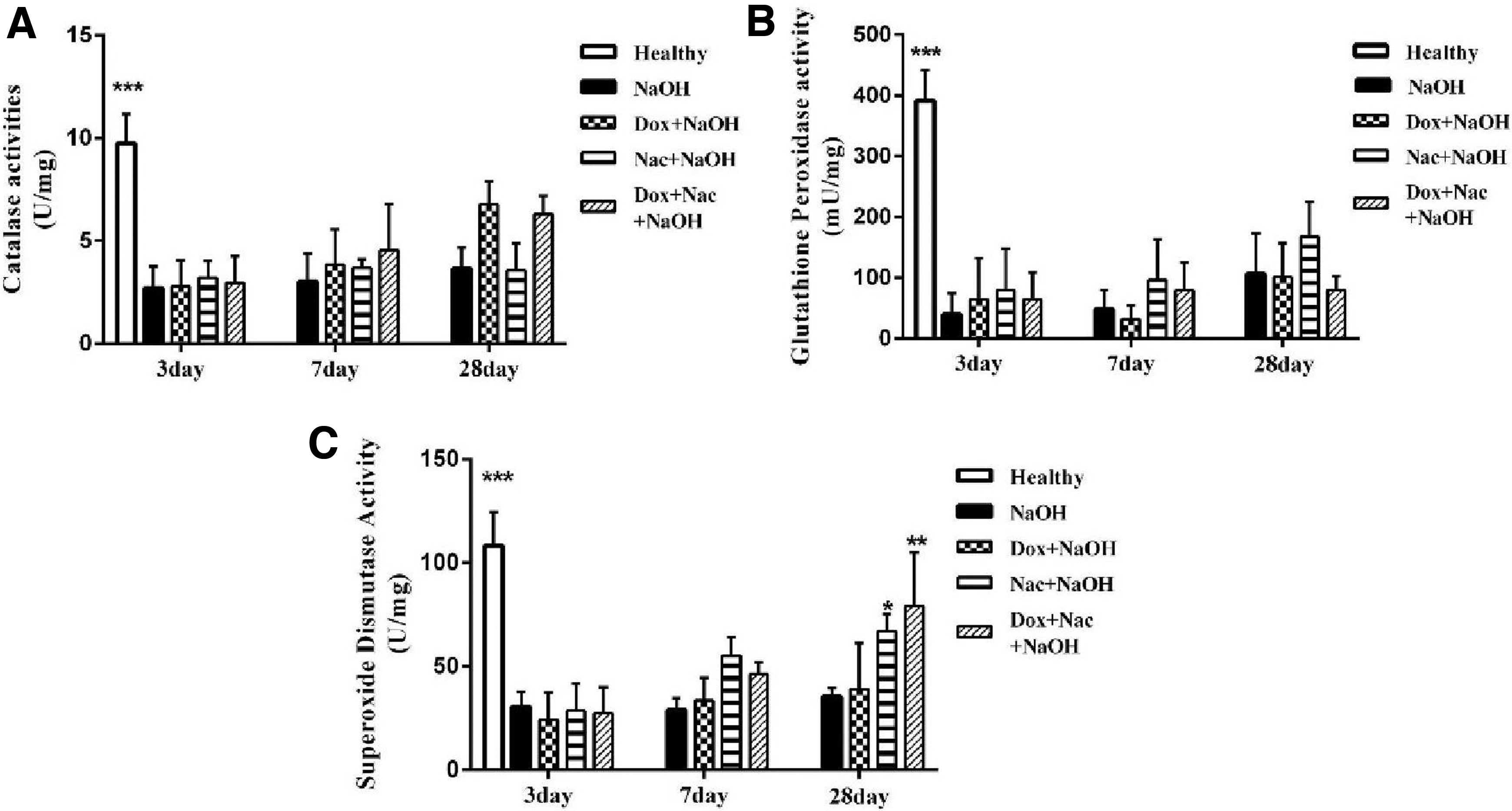
The activity of antioxidant enzymes in the cornea 3, 7, and 28 days following NaOH-induced injury. The SOD activity
NAC and Dox reduced TNF-α and Rel-a gene expression
The expression of tnf-α gene in the alkali-burned cornea (group 2) was evaluated by real-time polymerase chain reaction (RT-PCR) on days 3, 7, and 28 separately. In comparison to the healthy group (group 1), the mRNA level of TNF-α had a significant increase in the cornea of the NaOH group (group 2) (P < 0.05; Fig. 2A). The tnf-α gene expression decreased significantly 7 and 28 days following single and combined NAC and Dox treatment (groups 3, 4, and 5) (P < 0.05).

TNF-α and Rel-a mRNA levels were evaluated in the alkali-burned cornea by real-time PCR on days 3, 7, and 28. Compared with the alkali-burned group, TNF-α
The Rel-a gene expression also increased in the alkali-burned cornea (group 2) compared to the healthy group on day 3 (P < 0.05; Fig. 2B). Interestingly, the Rel-a gene expression decreased after single and combined NAC and Dox treatment on days 7 and 28, which was similar to the tnf-α gene expression (P < 0.05; Fig. 2B). However, the comparison of rel-a gene expression on day 3 in treatment groups did not indicate any statistically significant difference (Fig. 2B). Therefore, the topical NAC and Dox treatment could attenuate the expression of tnf-α and rel-a genes.
Dox decreased chemotaxis and angiogenesis factors on days 7 and 28
To determine the effects of NAC and Dox on the chemo-attractive factor, CXCL-1 gene expression was evaluated in the alkali-burned cornea. The CXCL-1 gene was overexpressed in the cornea of rats injured with NaOH (group 2) in comparison to the control group on day 3 (P < 0.05; Fig. 3A). The combined NAC and Dox treatment on days 7 and 28 and Dox on day 28 declined the CXCL-1 gene expression (P < 0.05).
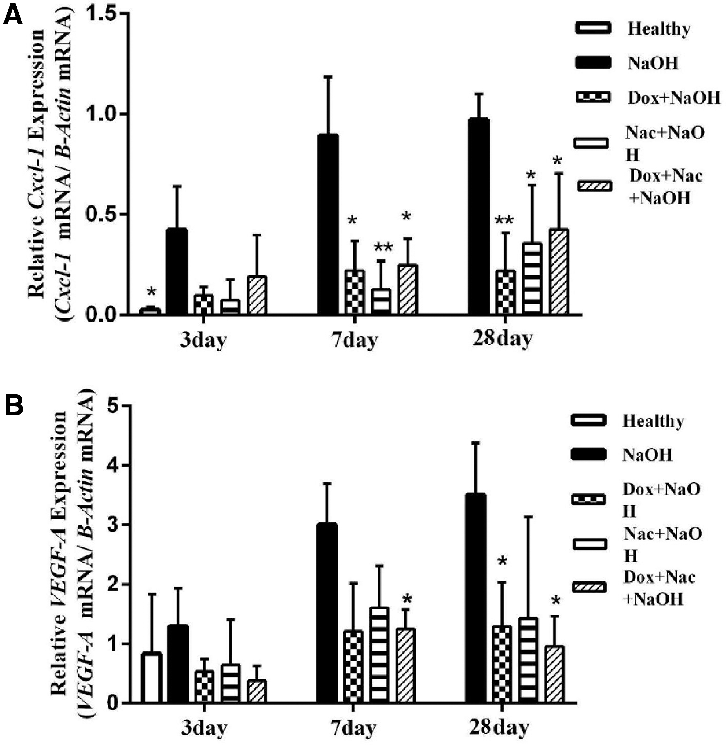
Cxcl-1 and vegf-a gene expression was analyzed on days 3, 7, and 28 in the cornea of rats exposed to NaOH.
Figure 3B showed that the VEGF-A, as the main promoting angiogenesis factor, was overexpressed in the NaOH-injured cornea rather than in the healthy group (group1) (P < 0.05). The comparison of VEGF-a gene expression did not show any statistically significant difference between group 2 (injured with NaOH) and group 4 (treated with NAC drop) (P > 0.05). There was a significant decrease in the VEGF-A mRNA expression after combined NAC and Dox treatment on days 7 and 28 and Dox treatment on day 28 (P < 0.05; Fig. 3A).
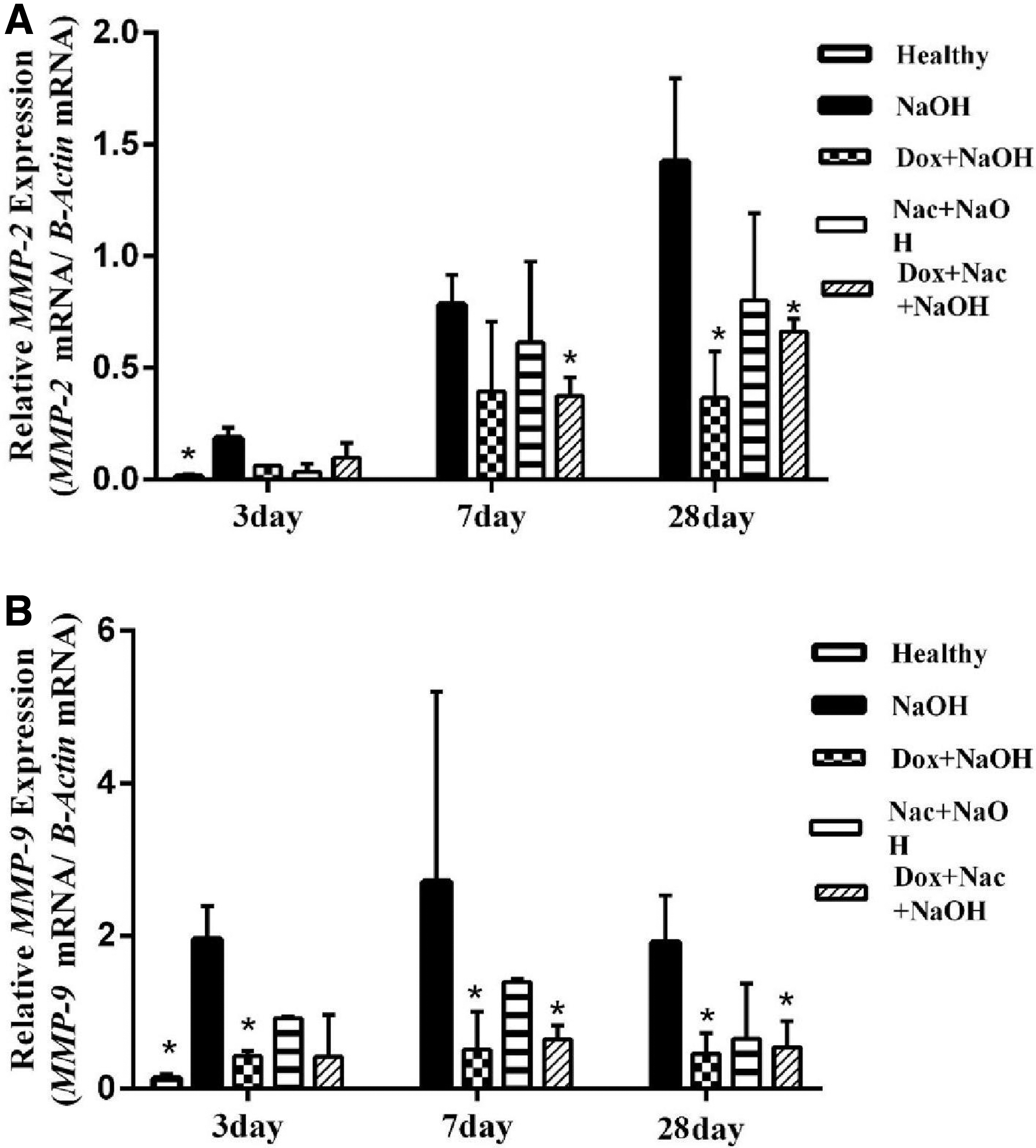
Based on the RT-PCR evaluation of the alkali-burned cornea, the mean expression of MMP2 and MMP9 genes seemed to be higher in group 2 than in the healthy samples (Fig. 4). Similar to VEGF-a gene expression, the mRNA level of MMP2 and MMP9 was downregulated in the NaOH-injured rats treated with Dox and Dox+ NAC (groups 3 and 5) on day 7 (P < 0.05). Interestingly, NAC treatment (group 4) after 7 and 28 days could not statistically reduce the mRNA expression of MMP2 and MMP9 (P > 0.05). Therefore, it indicated that the reduction of VEGF-a, MMP2, and MMP9 expression might not be affected by the topical NAC treatment.
Dox altered the infiltration of inflammatory cells and CNV
Using the slides obtained by H&E staining and examining them under a light microscope, the migration rate of inflammatory cells, the structure of corneal epithelial cells, and the CNV rate on days 3, 7, and 28 were reported (Fig. 5). The migration of inflammatory cells to the stroma and anterior chamber was observed 3 and 7 days following NaOH-induced corneal injury. Moreover, stromal edema and squamous epithelial disruption were more visible in the alkali-burned group than in the healthy group.
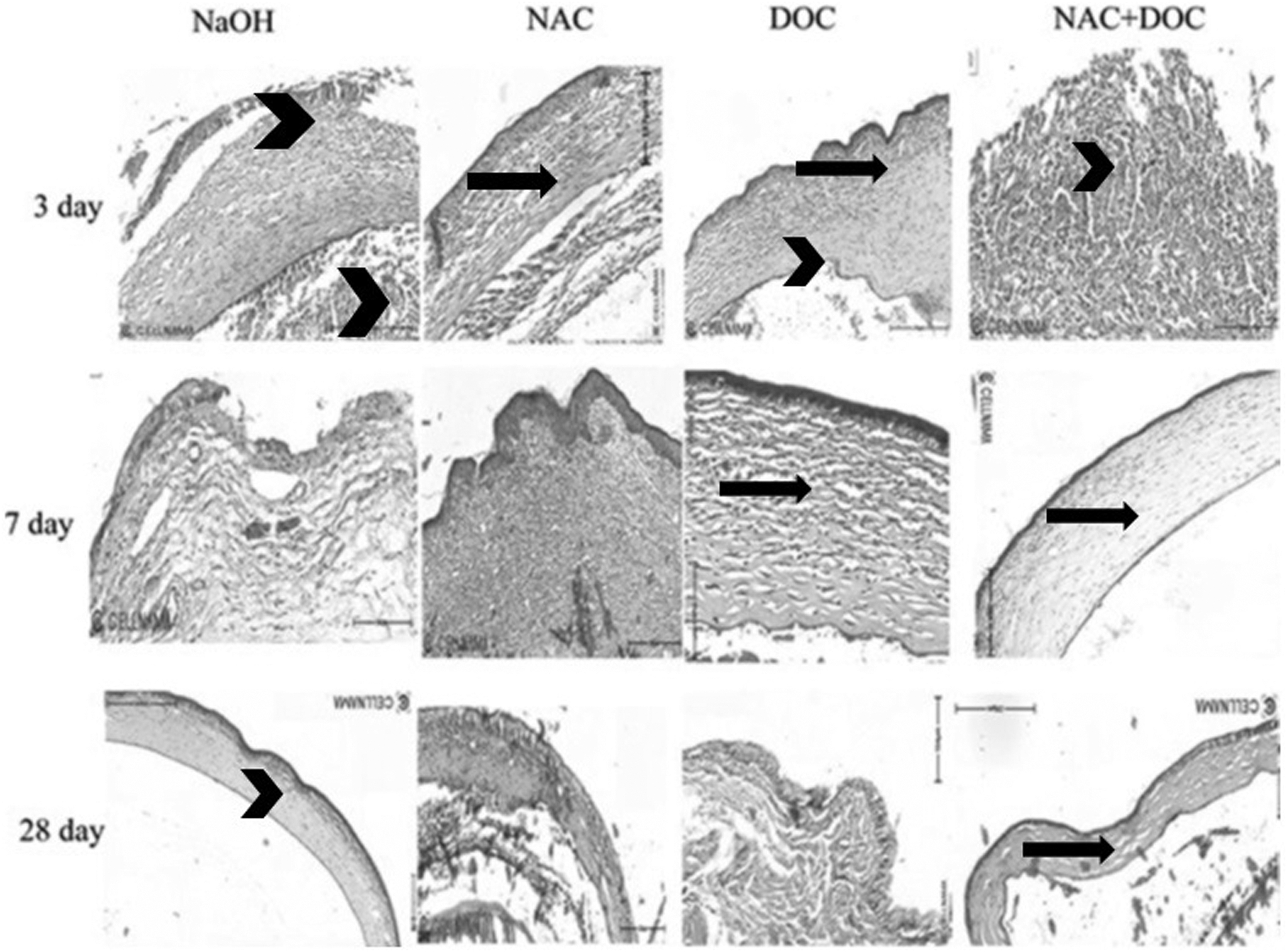
H&E staining evaluation in the corneal tissues after NaOH-induced injury. The migration of inflammatory cells (arrowhead) to the stroma and anterior chamber was observed for a short time following injury. Dox and the combined NAC and Dox treatments reduced the level of CNV (arrow) in the long period (magnification 20 × for all images). CNV, corneal neovascularization; H&E, hematoxylin and eosin.
No significant change was observed in the corneal structure and inflammatory cells in the treated groups (groups 3, 4, and 5) compared to the control group 3 days after corneal injury. On day 28, a high level of CNV and rearrangement of collagen structures were reported in the injured cornea. The migration level of inflammatory cells was decreased in all 3 treatment groups, and also the level of CNV was declined after treatment with Dox and the combination of NAC and Dox on day 28.
Discussion
Extensive use of NaOH in various industries, as well as household detergents, can injure various body organs, including the lungs, skin, and eyes. Inflammation and angiogenesis in the cornea are the 2 main complications in the eyes of patients exposed to NaOH. Despite many efforts, scientists have not yet been able to find an effective treatment to control the recurrent mechanisms in the cornea of this group of patients (Xu 2017). In this study, the single and combined effects of NAC and Dox on the injured cornea of rats exposed to NaOH 0.1 N were investigated. NaOH causes oxidative stress, which alters the configuration and function of cellular macromolecules such as proteins, lipids, and nucleic acids (Gould and others 2009).
The innate immune defense mechanism against ROS is based on the conversion of ROS to weaker compounds. The SOD enzyme converts superoxide to hydrogen peroxide and oxygen molecules. The CAT and GPx enzymes can change hydrogen peroxide to H2O and oxygen. Several in vivo and in vitro studies have shown the effect of NaOH on the function of these antioxidant enzymes (Muth and others 2004; Cejka and others 2016).
By examining the activity of these 3 enzymes, the activity of all 3 enzymes was significantly reduced on day 3 after injury in the alkali-burned cornea of different groups. Moreover, only on day 28 after topical treatment with NAC and the combination of NAC and Dox, the enzymatic activity of SOD increased significantly compared to the alkali-injured group. This recovery of enzymatic activity may be due to the antioxidant effect of the NAC compound in inactivating the electrophiles of the invading compounds and inducing the intracellular reduction signals (Samuni and others 2013). A similar effect has been observed with the use of NAC in increasing the activity of antioxidant enzymes after retinal damage to the eye (Ozdemir and others 2009).
The expression of the TNF-α and Rel-a genes as the major inflammatory pathways in the cornea of NaOH-damaged rats was examined and compared with the NAC and Dox eye drop groups by RT-PCR. Increased expression of TNF-α and Rel-a in the alkali-burned cornea indicated an increase in oxidative stress and inflammation. Our data showed a decrease in TNF-α and Rel-a expression in the groups receiving single and combined NAC and Dox eye drops in the long term. Induction of intracellular reduction signal and increase of compounds such as glutathione by NAC in the reduced state may have stopped the induction of inflammation in the damaged cornea (Samuni and others 2013).
A high level of TNF-α, as a pro-inflammatory cytokine, has been reported in alkaline-damaged corneas in BALB/c mice. The use of Infliximab antibody, which acts as an inhibitor of the TNF-α cytokine, reduced the expression of other inflammatory cytokines in the cornea of the mice in this study (Cade and others 2014). The results of the histopathological evaluation showed that the migration of inflammatory cells in the alkali-burned cornea was high on day 3. Although NAC was able to reduce the number of infiltrating neutrophils in the corneal stroma, a high number was reported in the long-term treatment compared to the healthy corneas.
TNF-α, as a subunit of NF-κB, plays an important role in inducing the expression and activation of the Rel-a gene (Zhu and others 2011). In downstream, the NF-κB transcription factor induces the expression of the CXCL-1 gene, which has a special function in invoking immune cells to the site of inflammation (Lennikov and others 2018). The CXCL-1 mRNA was overexpressed in all groups 3 days after NaOH-induced damage to the cornea and decreased gradually in different treatment groups on days 7 and 28. It is possible that decreased expression of CXCL-1 gene under the influence of NAC and Dox treatments alone does not reduce the migration of inflammatory cells to the cornea.
Lu and others (2012) reported that alkaline corneal damage in mice with knocked down tnf-αR gene (TNF-Rp55 KO) caused the lower expression of the inducible Nitric Oxide Synthase inflammatory factor and angiogenesis factors such as VEGF-A. However, the migration of neutrophils to the cornea and expression of chemokines such as C–C motif chemokine ligand 2 (CCL2) and CXCL-2 did not decrease significantly (Lu and others 2012).
Inflammatory cytokines and downstream signals in immune cells lead to CNV by producing angiogenesis-inducing factors and altering the extracellular matrix. Histopathological observations and slit lamp imaging showed a significant reduction in CNV 7 and 28 days after Dox treatment. Furthermore, VEGF-a gene expression decreased significantly in the Dox-treated group in the long term. Investigations have shown that the NF-κB transcription factor induces angiogenesis by inducing VEGF-A expression within endothelial cells and, consequently, increases proliferation (Horwitz and others 2014). Although the expression of VEGF-a gene was decreased in the group treated with NAC eye drop, this reduction was not statistically significant.
The effect of Dox on reducing VEGF-a gene expression may have been mediated by a pathway which is different from the ReLA factor. Su and others (2013) concluded that Dox inhibits cell proliferation and angiogenesis by inhibiting the Phosphatidylinositol 3-kinase/protein kinase B (PI3K/Akt) pathway and reducing VEGF-α gene expression in human umbilical vein endothelial cells. In another study, the effect of Dox on the inhibition of VEGF-A receptor (VEGFR1/2) activity was confirmed (Su and others 2011).
The expression of MMP2 and MMP9 genes was significantly reduced in groups treated with Dox on day 28 and with a combination of NAC and Dox on days 7 and 28. One study (Su and others 2013) reported the effect of Dox on reducing the expression of MMP2 and MMP9 genes by inhibiting the PI3K/Akt pathway. In addition, the use of 10% NAC in physical injury to the cornea of horses reduced the activity and expression of the MMP2 and MMP9 genes (Ollivier 2004).
MMP2 and MMP9 endopeptidase enzymes are zinc-dependent enzymes produced by the stimulation of TNF-α in the macrophage and neutrophil cells (DeSantis-Rodrigues and others 2016). These enzymes provide the conditions for angiogenesis by degrading the collagen IV of the basement membrane and adhesive molecules between endothelial cells (Barbariga and others 2019). In the alkali-burned cornea, MMP9 also plays an important role in the destruction of adhesion molecules between corneal epithelial cells (Yi and Zou 2019).
Conclusions
The progression of inflammation in the NaOH-induced corneal injury led to the induction of angiogenesis factors. NAC treatment could recover the enzymatic activity in the long term; however, NAC may not affect the reduction of CNV. Dox treatment reduced the developed CNV and angiogenesis factors, as well as inflammatory signals. Further research is needed to shed light on the recruitment pattern of inflammatory cells in the cornea.
Authors' Confirmation Statement
All listed authors are submitting the article in their own personal professional capacity and are not employees of any US-sanctioned government.
Footnotes
Author Disclosure Statement
No competing financial interests exist.
Funding Information
This research did not receive any specific grant from funding agencies in the public, commercial, or not-for-profit sectors. The expenses were supported by the corresponding author.