




Abstract
Metabolic disorders represent significant public health challenges worldwide. Emerging evidence suggests that cytokines and microRNAs (miRNAs) play crucial roles in the pathogenesis of metabolic disorders by regulating various metabolic processes, including insulin sensitivity, lipid metabolism, and inflammation. This review provides a comprehensive overview of the intricate interplay between cytokines and miRNAs in the context of metabolic disorders, including obesity, type 2 diabetes, and cardiovascular diseases. We discuss how dysregulation of cytokine–miRNA networks contributes to the development and progression of metabolic disorders and explore the therapeutic potential of targeting these interactions for disease management.
Introduction
Metabolic disorders, encompassing conditions such as obesity, type 2 diabetes (T2D), and cardiovascular disease (CVD), are major contributors to global morbidity and mortality (Clemente-Suarez et al., 2023a). The pathogenesis of these disorders involves complex interactions between genetic, environmental, and lifestyle factors. Emerging evidence suggests that dysregulation of cytokines and microRNAs (miRNAs) plays a central role in disrupting metabolic homeostasis and promoting disease progression (Marques-Rocha et al., 2015). In this review, we aim to elucidate the intricate interplay between cytokines and miRNAs in the context of metabolic disorders, highlighting their regulatory roles in key metabolic pathways and their potential as therapeutic targets.
Cytokines are small proteins (<40 kDa) secreted by various cells, including adipocytes, immune cells, and endothelial cells, which regulate immune responses, inflammation, and metabolic processes (Kany et al., 2019). Adipose tissue-derived cytokines, known as adipokines, such as adiponectin and leptin, play crucial roles in modulating insulin sensitivity, energy metabolism, and inflammation. However, dysregulation of adipokine signaling, particularly in obesity, leads to insulin resistance and chronic low-grade inflammation, contributing to the development of metabolic disorders (Kirichenko et al., 2022). Pro-inflammatory cytokines, including tumor necrosis factor-alpha (TNF-α), interleukin-6 (IL-6), and interleukin-1 beta (IL-1β), are elevated in obesity and T2D and promote insulin resistance by interfering with insulin signaling pathways and promoting adipose tissue inflammation (Zatterale et al., 2019). Understanding the role of cytokines in metabolic regulation is essential for unraveling the pathogenesis of metabolic disorders and developing targeted therapeutic interventions.
miRNAs are small noncoding RNA molecules that post-transcriptionally regulate gene expression by binding to the 3′ untranslated region (UTR) of target mRNAs, leading to mRNA degradation or translational repression (O’Brien et al., 2018). miRNAs play critical roles in various physiological processes, including cell proliferation, differentiation, apoptosis, and metabolism (Kabekkodu et al., 2018). Dysregulated miRNA expression has been implicated in the pathogenesis of metabolic disorders, including obesity, T2D, and CVD (Lopez-Pastor et al., 2020). miRNAs are involved in the regulation of key metabolic pathways, such as insulin signaling, lipid metabolism, and glucose homeostasis. Specific miRNAs, such as miR-122, miR-33a/b, and miR-155, have been shown to modulate lipid metabolism, adipogenesis, and insulin sensitivity, highlighting their importance in metabolic regulation (Afonso et al., 2021; Hu et al., 2021; Price et al., 2018; Su et al., 2019; Yaman et al., 2021; Zhang et al., 2024).
There is growing evidence suggesting crosstalk between cytokines and miRNAs in the regulation of metabolic processes and the pathogenesis of metabolic disorders (Chakraborty et al., 2020; Landrier et al., 2019). Cytokines can modulate the expression and activity of miRNAs, whereas miRNAs can target cytokine signaling pathways and inflammatory mediators, creating a complex regulatory network (Chakraborty et al., 2020). For example, TNF-α and IL-6 have been shown to regulate the expression of specific miRNAs involved in insulin signaling, adipocyte differentiation, and inflammation (Lee et al., 2013; Xu et al., 2014; Zhang et al., 2017). Conversely, miRNAs, such as miR-146a and miR-155, can target components of cytokine signaling pathways, such as Toll-like receptors (TLRs) and nuclear factor-kappa B (NF-κB), thereby modulating inflammatory responses and metabolic dysfunction (Ali et al., 2021; He et al., 2022; Nemati et al., 2021; Rasoulinejad et al., 2021). Understanding the interplay between cytokines and miRNAs provides valuable insights into the molecular mechanisms underlying metabolic disorders and may uncover novel therapeutic targets for intervention.
In conclusion, cytokines and miRNAs play critical roles in the regulation of metabolic homeostasis and the pathogenesis of metabolic disorders. The intricate interplay between cytokines and miRNAs contributes to the dysregulation of metabolic pathways, inflammation, and insulin resistance, thereby promoting the development and progression of obesity, T2D, and CVD. Elucidating the molecular mechanisms underlying cytokine–miRNA interactions may lead to the identification of novel therapeutic targets and biomarkers for metabolic disorders, offering new avenues for disease management and prevention. Further research in this field is warranted to translate these findings into effective clinical interventions for improving metabolic health.
Regulation of Obesity by Cytokines and miRNAs
Obesity, characterized by excessive adiposity and metabolic dysregulation, has become a global health epidemic with profound implications for morbidity, mortality, and health care costs (Sarma et al., 2021). The intricate interplay between cytokines and miRNAs plays a pivotal role in the pathogenesis of obesity and associated metabolic disorders (Landrier et al., 2019). Here, we provide a comprehensive overview of the regulatory roles of cytokines and miRNAs in the development and progression of obesity, highlighting key miRNAs implicated in adipogenesis, lipid metabolism, insulin signaling, and inflammation.
Adipose tissue, once considered an inert energy storage depot, is now recognized as a dynamic endocrine organ secreting a plethora of bioactive molecules, collectively termed adipokines or cytokines (Gu et al., 2023). Dysregulated secretion of adipokines contributes to the low-grade chronic inflammation observed in obesity, predisposing individuals to insulin resistance, T2D, CVD, and other metabolic complications (Kawai et al., 2021; Unamuno et al., 2018). Among the pro-inflammatory cytokines, TNF-α, IL-6, and monocyte chemoattractant protein-1 (MCP-1) are prominent contributors to adipose tissue dysfunction and systemic metabolic dysregulation (Kawai et al., 2021). These cytokines exert detrimental effects on adipocyte function, impair insulin signaling, promote lipolysis, and recruit immune cells to the adipose tissue, perpetuating a state of chronic inflammation and metabolic dysfunction.
The intricate interplay between cytokines and miRNAs constitutes a sophisticated regulatory network that governs various aspects of adipose tissue biology and energy homeostasis, ultimately influencing the development and progression of obesity. Among the key miRNAs implicated in obesity, miR-122 emerges as a central regulator, orchestrating hepatic lipid metabolism and insulin sensitivity. Dysregulation of miR-122 has been associated with aberrant lipid accumulation and insulin resistance, contributing to the pathogenesis of obesity-related metabolic disorders (Hu et al., 2021; Wang et al., 2015; Wu et al., 2017). Similarly, miR-33a/b plays a crucial role in modulating cholesterol homeostasis and fatty acid metabolism, thereby impacting lipid metabolism and adipocyte function (Davalos et al., 2011; Rayner et al., 2010; Wijesekara et al., 2012). In addition, miR-21 promotes adipocyte differentiation and lipid accumulation, facilitating adipose tissue expansion in obesity (Calo et al., 2016; Kang et al., 2013; Kim et al., 2009). Conversely, miR-155 contributes to adipose tissue inflammation and insulin resistance, exacerbating metabolic dysfunction (Johnson et al., 2018; Mahdavi et al., 2018; Zheng et al., 2019). Conversely, miR-146a acts as a suppressor of inflammation by inhibiting TNF-α-induced NF-κB activation and inflammatory cytokine expression, thus mitigating adipose tissue inflammation (Roos et al., 2016; Wu et al., 2016). Furthermore, miR-27a regulates adipogenesis and lipid droplet formation. Cold exposure reduces miR-27 levels in brown adipose tissue (BAT) and ubcutaneous white adipose tissue (SAT) and during brown adipogenesis in vitro. It targets the PRDI-BF1-RIZ1 (PR) domain containing 16 (Prdm16), peroxisome proliferator-activated receptors α (Pparα), cAMP responsive element binding protein (Creb), and partly PPARγ-coactivators-1 β (Pgc1β), inhibiting brown differentiation in cells and SAT preadipocytes, alongside its impact on peroxisome proliferator-activated receptors γ (Pparγ) and PPARγ-coactivators-1 (Pgc1α) (Sun and Trajkovski, 2014). miR-27a has also been validated as a negative regulator of adipocyte differentiation via suppressing PPARγ expression (Kim et al., 2010). Another study observed that miRNA-27a-3p, but not -5p, is a crucial mediator of human adipogenesis (Wu et al., 2021a). Microarray analysis showed miR-27b downregulation during adipogenic differentiation, whereas lipoprotein lipase (LPL) initially increased, stabilizing after 14 days. Luciferase assays confirmed miR-27b negative regulation of LPL. Overexpression and silencing of miR-27b reversed LPL regulation, affecting fat-related biomarkers and lipid droplet accumulation (Hu et al., 2018). miR-27a has also been reported to regulate sheep adipocyte differentiation by targeting the CPT1B gene (Li et al., 2021). Although miR-370 modulates adipocyte differentiation and glucose metabolism, it influences overall energy homeostasis (Zhang et al., 2021a). Moreover, miR-378 regulates adipocyte lipogenesis and insulin signaling (Huang et al., 2015; Ishida et al., 2014; Pan et al., 2014; Zhang et al., 2016a), whereas miR-29 controls adipocyte differentiation, modulating adipose tissue function (Liu et al., 2019; Wu et al., 2021b; Zhu et al., 2017). Furthermore, miR-34a negatively impacts adipocyte function and insulin sensitivity (Cornejo et al., 2022), and miR-30 regulates adipogenesis and insulin signaling pathways, influencing adipose tissue development and metabolism (Koh et al., 2018). In addition, miR-103/107 fine-tunes insulin sensitivity and glucose metabolism (Trajkovski et al., 2011), whereas miR-519d promotes adipocyte hypertrophy and inflammation, exacerbating obesity-related pathologies (Martinelli et al., 2010). Moreover, miR-132 plays a pivotal role in regulating brown adipose tissue thermogenesis and energy expenditure (Kariba et al., 2020), whereas miR-210 impairs mitochondrial function and energy metabolism, contributing to metabolic dysregulation (Muralimanoharan et al., 2015). In addition to these miRNAs, other players such as miR-221, miR-335, and miR-802 (Meerson et al., 2013; Nakanishi et al., 2009; Ni et al., 2021; Sun et al., 2019) have been implicated in the regulation of obesity-related pathways, further highlighting the complexity of miRNA-mediated mechanisms in metabolic disorders.
The role of pro-inflammatory cytokines in obesity extends beyond their canonical functions in immune regulation to encompass intricate interactions with miRNAs, thereby shaping adipocyte function and metabolic homeostasis. Pro-inflammatory cytokines such as TNF-α, IL-6, and IL-1β have been shown to modulate miRNA expression profiles in adipocytes and immune cells, orchestrating a complex regulatory network that impacts adipokine secretion and metabolic processes. For instance, TNF-α has been reported to upregulate miR-155 expression in adipocytes, leading to impaired insulin signaling and increased adipose tissue inflammation (Karkeni et al., 2016). Similarly, IL-6 and TNF-α stimulation has been associated with elevated levels of miR-146b, promoting an obesity-related inflammatory response (Shi et al., 2014). In addition, IL-1β-mediated activation has been linked to the induction of miR-146a expression, which serves as a negative regulator of NF-κB signaling, thereby attenuating inflammatory responses in adipose tissue (Jimenez et al., 2020). Furthermore, pro-inflammatory cytokines can modulate miRNA expression in immune cells infiltrating adipose tissue, such as macrophages and T cells, contributing to the polarization of these cells toward a pro-inflammatory phenotype. For example, TNF-α-induced miR-155 expression in macrophages promotes M1 polarization and exacerbates adipose tissue inflammation (Zhang et al., 2016b). Conversely, anti-inflammatory cytokines such as transforming growth factor-beta (TGF-β) may exert opposing effects on miRNA expression profiles, dampening inflammation and promoting metabolic homeostasis (Zhao et al., 2022).
In summary, cytokines and miRNAs (Tables 1 and 2, Fig. 1) play integral roles in the regulation of adipose tissue biology, energy metabolism, and inflammation in obesity. Dysregulated cytokine and miRNA signaling contribute to adipose tissue dysfunction, chronic inflammation, insulin resistance, and metabolic dysregulation observed in obesity. Elucidating the molecular mechanisms underlying cytokine–miRNA interactions may unveil novel therapeutic targets for the management and prevention of obesity and its associated metabolic complications. Further research is warranted to elucidate the therapeutic potential of targeting cytokines and miRNAs for the treatment of obesity and related metabolic disorders.
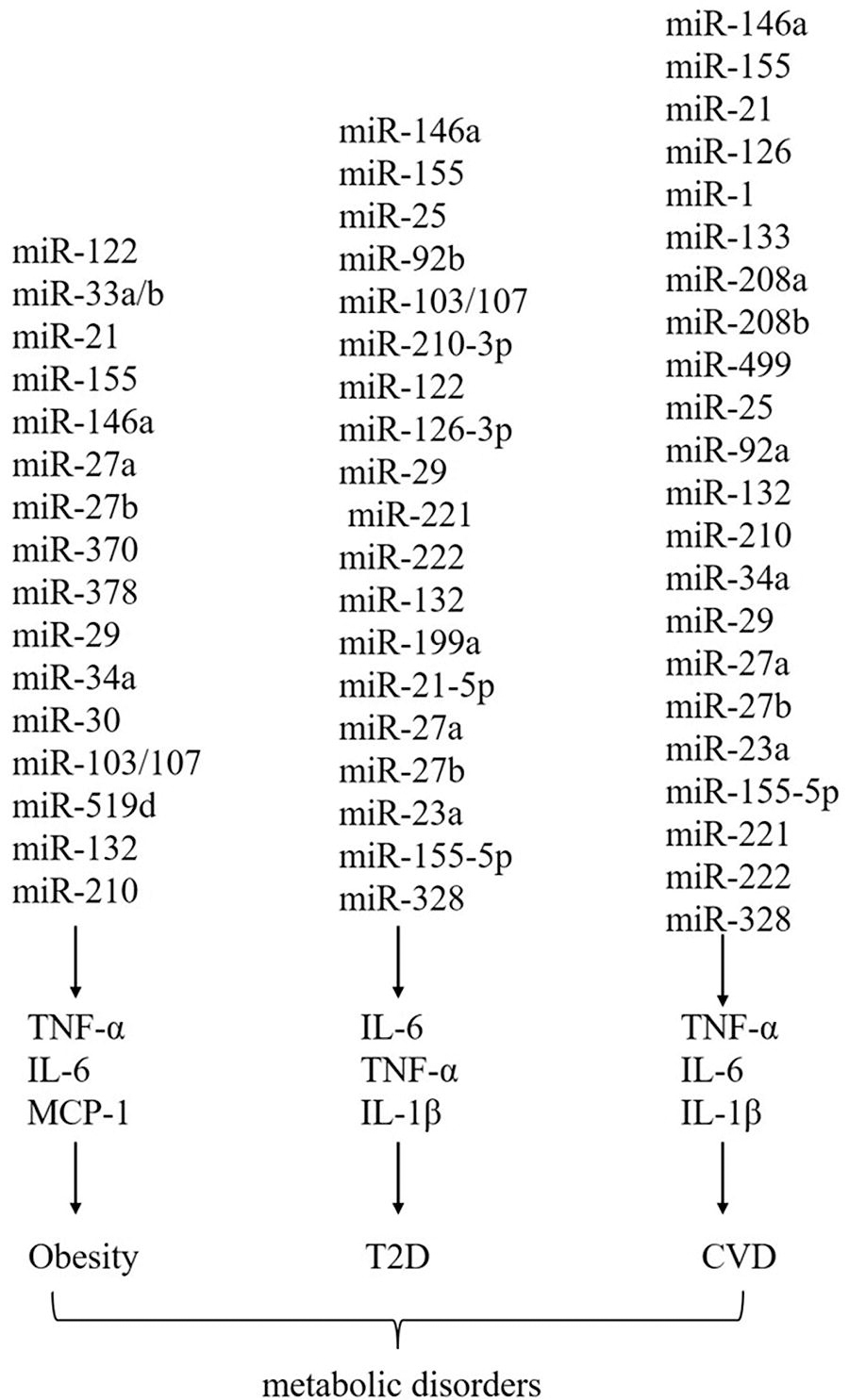
Summary of miRNAs that regulate cytokines in obesity, T2D, and CVD. CVD, cardiovascular disease; IL-6, interleukin-6; MCP-1, monocyte chemoattractant protein-1; miRNAs, microRNAs; T2D, type 2 diabetes; TNF-α, tumor necrosis factor-alpha.
Function of Cytokine and miRNAs in Obesity
GDM, gestational diabetes mellitus; IL-6, interleukin-6; miRNA, microRNA; NF-κB, nuclear factor-kappa B; T2DM, type 2 diabetes mellitus; TNF-α, tumor necrosis factor-alpha; TRAF, TNF-receptor-associated factor 3.
Function of miRNAs in Obesity
T2D, type 2 diabetes.
Regulation of T2D by Cytokines and miRNAs
T2D represents a multifactorial metabolic disorder characterized by insulin resistance, impaired glucose metabolism, and pancreatic β-cell dysfunction. Emerging evidence highlights the intricate interplay between cytokines and miRNAs in the pathogenesis and progression of T2D. Here, we delve into the multifaceted roles of cytokines and miRNAs in regulating T2D, exploring their impact on insulin sensitivity, glucose homeostasis, inflammation, and β-cell function.
In T2D, dysregulated production of inflammatory cytokines, such as IL-6, TNF-α, and IL-1β, contributes to insulin resistance and chronic inflammation (Tsalamandris et al., 2019). These cytokines exert their effects by activating various intracellular signaling pathways, leading to impaired insulin signaling and glucose metabolism (Tsalamandris et al., 2019). IL-6, for example, promotes insulin resistance by inhibiting insulin receptor substrate-1 (IRS-1) phosphorylation and disrupting insulin signaling cascades in insulin-sensitive tissues (Akbari and Hassan-Zadeh, 2018). TNF-α induces serine phosphorylation of IRS-1, impairing its interaction with the insulin receptor and attenuating insulin-stimulated glucose uptake (Rui et al., 2001; Zhang et al., 2008). IL-1β contributes to β-cell dysfunction and apoptosis, further exacerbating insulin secretion defects in T2D (Ghiasi et al., 2018). Collectively, these inflammatory cytokines create a pro-inflammatory milieu that perpetuates insulin resistance, glucose intolerance, and β-cell dysfunction in T2D.
The interplay between inflammatory cytokines and miRNAs orchestrates a sophisticated regulatory network profoundly influencing insulin sensitivity, glucose homeostasis, inflammation, and β-cell function in the context of T2D. Inflammatory cytokines, including TNF-α, IL-6, and IL-1β, exhibit the capacity to directly modulate miRNA expression by activating specific transcription factors or signaling pathways governing miRNA biogenesis and maturation. This direct regulation occurs through various mechanisms, such as the activation of NF-κB or mitogen-activated protein kinase (MAPK) signaling pathways, which, in turn, control the expression of key enzymes involved in miRNA processing, including Drosha, Dicer, and Argonaute proteins. Conversely, miRNAs reciprocally influence the expression of inflammatory cytokines and their downstream effectors, thereby exerting a regulatory role in inflammatory responses in T2D (Vezza et al., 2021). For instance, miR-146a and miR-155 have emerged as pivotal negative regulators of TNF-α, IL-6, and other pro-inflammatory cytokines, attenuating inflammation and insulin resistance in T2D (Baldeon et al., 2014). These miRNAs typically target the 3′ UTRs of mRNAs encoding inflammatory cytokines, leading to mRNA degradation or translational repression, thereby dampening the inflammatory cascade. In contrast, the dysregulation of miRNAs in response to inflammatory stimuli can further exacerbate metabolic dysfunction in T2D. Inflammatory cytokines such as TNF-α and IL-1β can induce the expression of specific miRNAs implicated in insulin resistance and β-cell dysfunction, thus establishing a feedback loop that perpetuates metabolic dysregulation in T2D. Previous studies have revealed a crucial β cell-macrophage communication pathway facilitated by the miRNA-29-TNF-receptor-associated factor 3 (TRAF3) axis. Transgenic miR-29a/b/c mice exhibit glucose intolerance and insulin resistance on a high-fat diet, primarily mediated by macrophages. Mechanistically, miR-29 promotes monocyte and macrophage recruitment and activation via TRAF3-dependent exosome release (Corral-Fernandez et al., 2013; Jankauskas et al., 2021). miR-25 and miR-92b have also been validated to regulate insulin biosynthesis and pancreatic β-cell apoptosis (Shen et al., 2022). miR-103/107 targets key components of insulin signaling pathways or β-cell function, thereby contributing to the development and progression of T2D (Trajkovski et al., 2011). miR-210-3p, a key player in gestational diabetes mellitus (GDM), is upregulated in GDM pancreases. It induces apoptosis and impairs pancreatic β-cell function by targeting Dtx1. Downregulation of Dtx1 increases insulin expression, improving β-cell function. This highlights miR-210-3p as a potential therapeutic target for GDM management (Cao et al., 2022). Moreover, in a population-based, case–control study, a direct correlation was observed between IL-6, TNF-α, and Homeostatic Model Assessment for Insulin Resistance (HOMA-IR) with elevated expression of pro-inflammatory miRNA-122, whereas anti-inflammatory miRNAs, miRNA-126-3p, and miRNA-146a showed an inverse association. This suggests a regulatory role of these miRNAs in modulating inflammation and insulin resistance in patients with type 2 diabetes mellitus (T2DM), highlighting their potential as biomarkers and therapeutic targets (Zeinali et al., 2021).
Therefore, unraveling the intricate crosstalk between inflammatory cytokines and miRNAs (Tables 3 and 4, Fig. 1) holds immense potential for elucidating the underlying mechanisms driving T2D pathophysiology and identifying novel therapeutic targets for the management of the disease. Further research efforts aimed at deciphering the precise molecular mechanisms governing these interactions are warranted to facilitate the development of targeted therapeutic interventions aimed at mitigating T2D and its associated complications.
Function of Cytokine and miRNAs in T2D
Function of miRNAs in T2D
Regulation of CVD by Cytokines and miRNAs
CVD encompasses a spectrum of disorders affecting the heart and blood vessels, including coronary artery disease, myocardial infarction (MI), stroke, and heart failure. Despite significant advances in diagnosis and treatment, CVD remains a leading cause of morbidity and mortality globally (Zhou et al., 2018). Emerging evidence suggests that dysregulation of cytokines and miRNAs plays a pivotal role in the pathogenesis of CVD (Sessa et al., 2023). Cytokines, as key mediators of inflammation, orchestrate immune responses and vascular remodeling processes critical in CVD development (Amin et al., 2020). Conversely, miRNAs, small noncoding RNAs, regulate gene expression post-transcriptionally and have emerged as critical regulators of cardiovascular homeostasis (Sessa et al., 2023). Here, we elucidate the intricate interplay between cytokines and miRNAs in the context of CVD, shedding light on their potential as diagnostic biomarkers and therapeutic targets.
Pro-inflammatory cytokines such as TNF-α, IL-6, and IL-1β play a central role in promoting endothelial dysfunction, vascular inflammation, and atherosclerosis (Amin et al., 2020). TNF-α, for instance, contributes to endothelial activation by upregulating adhesion molecules and promoting the release of chemokines, facilitating leukocyte recruitment and adhesion to the vascular endothelium (Cheng et al., 2010). IL-6, a pleiotropic cytokine, promotes inflammation and thrombosis by inducing the hepatic synthesis of acute-phase proteins such as C-reactive protein (Narazaki and Kishimoto, 2018). IL-1β, in contrast, promotes endothelial dysfunction and vascular smooth muscle cell proliferation, contributing to plaque destabilization and thrombosis (Henein et al., 2022). Moreover, dysregulated cytokine signaling has been implicated in the pathogenesis of MI and heart failure, where sustained inflammation exacerbates myocardial injury and remodeling processes (Frangogiannis, 2014).
The intricate interplay between cytokines and miRNAs orchestrates various aspects of CVD, influencing inflammation, endothelial function, angiogenesis, cardiac remodeling, hypertrophy, and fibrosis. Among the myriad miRNAs involved, miR-146a stands out for its role in suppressing inflammation by targeting NF-κB signaling and mitigating inflammatory responses implicated in CVD pathogenesis (Hou et al., 2021). Similarly, miR-155 regulates endothelial cell function and inflammation, pivotal processes in vascular homeostasis and CVD progression (Bruen et al., 2019; Liang et al., 2020; Ye et al., 2016). Meanwhile, miR-21 emerges as a key player in promoting angiogenesis while modulating cardiac hypertrophy, offering dual benefits in vascular repair and cardiac function regulation (Bejerano et al., 2018). Enhancing endothelial cell function and angiogenesis, miR-126 holds promise for vascular repair and regeneration in CVD (Cao et al., 2020). In contrast, miR-1 and miR-133 exert nuanced effects on cardiac remodeling and contractility, crucial for maintaining cardiac function (Yang et al., 2024). Moreover, miR-208a and miR-208b control cardiac hypertrophy and fibrosis, influencing the structural integrity of the heart (Callis et al., 2009; Zhang et al., 2022). Meanwhile, miR-499 influences cardiomyocyte differentiation and function, adding complexity to cardiac development and function (Li et al., 2013; Sluijter et al., 2010). In cardiac remodeling and fibrosis, miR-29 plays a critical role by modulating extracellular matrix remodeling processes (Abonnenc et al., 2013; Cheng et al., 2013). In addition, miR-92a and miR-132 contribute to angiogenesis and endothelial function, crucial for vascular health (Bonauer et al., 2009; Xu et al., 2021). miR-210, known for its role in hypoxia response, also influences angiogenesis, particularly under ischemic conditions in CVD (Guan et al., 2019). Conversely, miR-34a promotes cardiac apoptosis and fibrosis, exacerbating myocardial damage in CVD (Li et al., 2019; Su et al., 2018; Zhang et al., 2021b). Moreover, miR-126-3p enhances endothelial cell function and vascular repair, providing potential therapeutic avenues for vascular regeneration in CVD (Alique et al., 2019; Cao et al., 2020; Jansen et al., 2017). Furthermore, miR-199a and miR-21-5p modulate angiogenesis and vascular smooth muscle cell function, affecting vascular integrity and tone (Cao et al., 2021; Ke et al., 2022; Liao et al., 2021; Wang et al., 2020). In addition, miR-27a and miR-27b impact lipid metabolism and inflammation, linking metabolic disorders with CVD risk (Chen et al., 2012). Furthermore, miR-133, miR-23a, and miR-155-5p regulate endothelial function and inflammation, contributing to vascular dysfunction in CVD (Cao et al., 2016; Guo et al., 2020; Li et al., 2016; Oikawa et al., 2018; Qiao et al., 2020). Notably, miR-221 and miR-222 influence vascular smooth muscle cell proliferation and migration, critical processes in atherosclerosis, and vascular remodeling (Wang et al., 2016). Finally, miR-328 modulates cardiac hypertrophy and fibrosis, further underscoring the multifaceted roles of miRNAs in CVD pathophysiology (Du et al., 2016; Zhao et al., 2018). Overall, understanding the intricate regulatory roles of cytokine-related miRNAs in CVD provides insights into potential therapeutic targets and strategies for mitigating CVD burden.
Overall, the comprehensive understanding of cytokine-related miRNAs (Tables 5 and 6, Fig. 1) and their regulatory roles in CVD offers promising avenues for developing targeted therapeutics and precision medicine approaches to mitigate the burden of cardiovascular diseases. Future research efforts aimed at elucidating the precise mechanisms of action of these miRNAs and their interactions with cytokines in the CVD pathophysiology are warranted to translate these findings into clinical applications and improve patient outcomes in the realm of cardiovascular medicine.
miRNAs and Their Function in CVD
CVD, cardiovascular disease.
miRNAs and Their Function in CVD
Challenges and the Potential Limits of Current Understanding
Significant strides have been made in unraveling the intricate relationship between cytokines and miRNAs in metabolic disorders such as obesity, T2D, and CVD. However, numerous challenges and limitations persist, impeding the translation of research findings into clinical practice and the development of effective therapeutic interventions. The complexity of interactions between cytokines and miRNAs poses a major obstacle, with interconnected and dynamic regulatory networks making it difficult to discern the precise mechanisms underlying disease pathogenesis. Moreover, the crosstalk between various cell types, tissues, and organ systems further complicates our understanding of these interactions.
Metabolic disorders exhibit significant heterogeneity among patient populations, driven by diverse genetic backgrounds, environmental exposures, and lifestyle factors (Barroso and McCarthy, 2019). This heterogeneity makes it challenging to identify biomarkers and therapeutic targets applicable universally across all patients. In addition, despite the proposal of several cytokines, such as TNF-α, IL-6, IL-1β, IL-10, IL-8, IL-18, MCP-1, leptin, resistin, and adiponectin (Baldeon et al., 2014; Clemente-Suarez et al., 2023b), as well as miRNAs, including miR-146a, miR-155, miR-21, miR-126, miR-1, miR-133, miR-208a, miR-208b, miR-499, miR-25, miR-92a, miR-132, miR-210, miR-34a, miR-29, miR-126-3p, miR-199a, miR-21-5p, miR-27a, miR-27b, miR-23a, miR-155-5p, miR-221, miR-222, and miR-328, as potential biomarkers for metabolic disorders (Soplinska et al., 2020), their clinical utility remains limited because of challenges in validation and standardization. Variability in assay techniques, sample handling, and data interpretation presents hurdles in establishing robust biomarker assays with sufficient sensitivity, specificity, and reproducibility for clinical use.
Tissue-specific effects of cytokines and miRNAs on metabolic regulation add another layer of complexity (Asirvatham et al., 2009). These molecules exhibit differential expression patterns in various tissues crucial for metabolic homeostasis. Dissecting tissue-specific effects requires advanced molecular and computational techniques, further complicating therapeutic target identification. Moreover, translating preclinical findings into clinically effective interventions is hindered by regulatory approval processes and ethical considerations surrounding emerging technologies such as gene editing and RNA-based therapeutics. Addressing these challenges demands interdisciplinary collaboration, technological innovation, robust validation studies, and careful consideration of ethical and regulatory issues.
Despite these hurdles, continued research in this field holds promise for advancing our understanding of metabolic disorders and improving patient outcomes.
Footnotes
Authors’ Contributions
X.X.: Conception and design. L.Z. and H.C.: Data acquisition, analysis, and interpretation. L.Z. and X.X.: Drafting the article. All authors approved the final version of the article.
Availability of Data and Materials
The datasets used and/or analyzed during the current study are available from the corresponding author upon reasonable request.
Author Disclosure Statement
The authors declared that there was no conflict of interest in the study.
Funding Information
This study received no funding.