
Abstract
Purpose:
To evaluate the effect of donepezil hydrochloride, an agent for the treatment of Alzheimer’s disease (AD), on the cerebral and optic nerve head (ONH) blood flow, visual field defect in normal-tension glaucoma (NTG) patients with decreased cerebral blood flow (CBF) that demonstrates an AD-like perfusion pattern.
Methods:
The subjects were 5 NTG patients who exhibited AD-like decreased CBF upon 123I-iodoamphetamine single photon emission computed tomography (123I-IMP SPECT). Donepezil hydrochloride (5 mg/day) was prescribed for each patient during a period of 12 months. Intraocular pressure (IOP), mean deviation (MD) of the Humphrey visual field, ONH blood flow determined by a laser speckle flowgraphy, and regional CBF (rCBF) determined by 123I-IMP SPECT were measured before and every 6 months during the treatment.
Results:
MD, ONH blood flow, and rCBF were improved significantly after 6 months of the treatment, although IOP did not change significantly. No deterioration of NTG morbidity was found in any of the measured parameters after 12 months of the treatment.
Conclusions:
Oral administration of donepezil hydrochloride in NTG patients might prevent deterioration of visual field defect, ONH blood flow, and rCBF in the temporal, parietal, and posterior lobes. This pilot study suggested the possibility that donepezil hydrochloride might ameliorate glaucomatous optic neuropathy in NTG.
Introduction
A
A number of reports have documented the relationship between glaucoma and AD.2–4 A recent study has shown that the anti-AD drug donepezil, a potent acetylcholinesterase (AChE) inhibitor, exerts a neuroprotective effect on retinal ganglion cells (RGCs) in rats both in vivo and in vitro.5 In the previous study,6 we found that 22.6% of NTG patients exhibited an AD-like cerebral perfusion pattern.
On the basis of these apparent similarities between AD and NTG, we hypothesized that the neurodegenerative processes in these 2 diseases might have a similar mechanism, and therefore that an anti-AD drug might be effective for treatment of glaucomatous optic neuropathy in NTG patients.
In this study, therefore, we evaluated the feasibility of donepezil for the treatment of NTG patients with decreased regional cerebral blood flow (rCBF) and an AD-like perfusion pattern.
Methods
123I-iodoamphetamine (FUJIFILM RI Pharma Co., Ltd., Tokyo, Japan) single photon emission computed tomography (123I-IMP SPECT) was carried out on 64 NTG outpatients of the Department of Ophthalmology, Osaka Medical College. The diagnostic criteria for NTG were: normal open angle; peak intraocular pressure (IOP) ≤21 mm Hg at all times, including a 24-h fluctuation; presence of typical glaucomatous visual field defects associated with glaucomatous optic nerve change; and no ocular or systemic disease responsible for optic nerve damage. The data of 123I-IMP SPECT were normalized by autographic protocol,7 and a visually qualitative inspection was performed with 3-dimensional image by Brain Guide software package (Advanced Biologic Corp., Toronto, Canada). As the results of the inspection, 23 of these patients who showed a decrease of regional cerebral blood flow (rCBF) in the area ranging from the parietal lobe to the temporal lobe were defined as showing an AD-like pattern (Fig. 1). Detection of AD-like cerebral perfusion pattern was shown to be helpful in differential diagnosis between early-stage AD and other types of dementia.8–10 The subjects showing AD-like pattern do not necessarily have AD. Practically all of the patients showing AD-like cerebral perfusion pattern in the current study were not diagnosed AD at least when SPECT was carried out. Seven of them (aged 65–78 years; 5 males and 2 females) gave informed consent for treatment with donepezil and were admitted to the study. Three of them had been previously untreated, 3 others had been receiving latanoprost, and the remaining 1 had received both timolol and latanoprost. This study followed the tenets of the Declaration of Helsinki, and was approved by the institutional review board of Osaka Medical College.
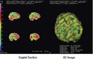
A representative example of Alzheimer’s disease (AD)-like cerebral perfusion pattern in a patient with normal-tension glaucoma (NTG). A decrease of regional cerebral blood flow (rCBF) in the area ranging from the parietal lobe to the temporal lobe is shown.
Donepezil hydrochloride (Aricept®; Eisai Co., Ltd., Tokyo, Japan; 5 mg/day) was prescribed for these 7 patients for 12 months. The prescription prior to this study was continued in each patient. Intraocular pressure (IOP), mean deviation (MD) of the Humphrey visual field test (program 30-2) were measured before and every 6 months (every 3 months for some of them) after the start of treatment. All the patients were relatively trained for the visual field test since numbers (mean ± SD) of the tests performed before entering this study for each patient was 3.0 ± 1.9. To evaluate the changes of optic nerve head (ONH) blood flow, a laser speckle flowgraphy was also performed every 6 months. A laser speckle flowgraphy was developed in Japan and has been widely used to analyze the capillary blood flow in the retina, choroid, and ONH.11 In this study, ONH square blur rate (SBR), a quantitative index of tissue blood flow was measured before and every 6 months after the start of treatment. The rCBF in the temporal, parietal, and posterior lobes was also measured by 123I-IMP SPECT at the same time points. The rCBF was automatically calculated using the 3-dimensional stereotaxic region of interest templates for quantitative evaluation (Daiichi R.L. Co., Ltd., Japan). Then, the ratio of each rCBF relative to that in the cerebellum was calculated. We also investigated adverse effects and subjective symptoms during the treatment.
In each case, data for 1 randomly selected eye were subjected to statistical analysis. Bonferroni test, following Kruskal–Wallis test, was used in the comparison between the data before and 6 or 12 months after the start of the treatment. Differences at P < 0.05 were regarded as significant.
Results
One patient requested to discontinue donepezil therapy 6 months after the start of the treatment, although adverse effects were absent. Another patient had so much difficulty in maintaining fixation during the Humphrey visual field test that his data were considered unreliable. Consequently, since the data for these 2 cases were excluded from the analysis, 5 patients were left. As the parameters regarding fixation during visual field tests for other 5 patients were substantially good enough through the study, the results for their visual fields were regarded to be reliable. The background of these 5 patients was shown in Table 1. One out of 5 patients had not received any glaucoma treatment, and other 4 patients had not changed glaucoma therapy (3 of them: latanoprost, another: timolol and latanoprost) throughout the donepezil treatment. As an adverse effect of donepezil, 1 patient complained of loose bowels. As a subjective symptom, 2 patients experienced visual improvement for newspaper reading.
B
Abbreviations: HVFT, Humphrey visual field test; MD, mean deviation; PSD, pattern standard deviation; Lat, latanoprost; Tim, timolol.
During donepezil therapy, IOP did not change significantly (data not shown). Significant improvement of MD values was observed after 6 months of the treatment (Figs. 2 and 3). In 2 of the 5 cases, improvements of more than 2 dB (compared with pretreatment) were noted at 12 months. In 3 cases, MD did not change by more than 2 dB throughout the study. For the change of ONH blood flow, SBR was increased significantly after 6 months of the treatment (Fig. 4). At 12 months, SBR was increased by >10% in 2 cases, and decreased by >10% in 1 case compared with the pretreatment value. The rCBF in the temporal region was increased significantly after 6 months of treatment (Figs. 5 and 6). At 12 months, rCBF was stable in 4 cases, and decreased in only one case by <10% relative to the pretreatment value. The rCBF in the posterior and parietal regions showed a similar change (data not shown).
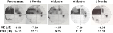
A representative example of changes in Humphrey visual fields of an normal-tension glaucoma (NTG) patient (Case A) during donepezil treatment. The visual field defect was improved gradually with its maximum at 6 months, but deteriorated to be similar as the pretreatment level at 12 months.
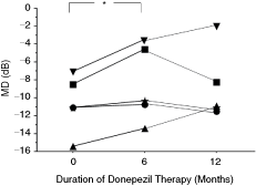
Mean deviation (MD) changes for each normal-tension glaucoma (NTG) patient (▪: Case A, ▴: Case B, ▾: Case C, ★: Case D, •: Case E) during donepezil treatment. MD was improved significantly after 6 months of the treatment (∗P < 0.05, Bonferroni test).

Changes in square blur rate (SBR) of optic nerve head (ONH) for each normal-tension glaucoma (NTG) patient (each symbol represents the same case as in Fig. 3) during donepezil treatment. SBR was increased significantly after 6 months of the treatment (∗P < 0.05, Bonferroni test).
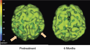
A representative example of the change in cerebral perfusion of an normal-tension glaucoma (NTG) patient (Case A) during donepezil treatment. The cerebral perfusion, which was decreased especially in the temporal lobe (arrows), was improved after 6 months of donepezil treatment.

Regional cerebral blood flow (rCBF) changes in the temporal lobe for each normal-tension glaucoma (NTG) patient (each symbol represents the same case as in Fig. 3) during donepezil treatment. rCBF in the temporal lobe was increased significantly after 6 months of the treatment (∗P < 0.05, Bonferroni test).
Discussion
In our present study, oral administration of donepezil for 12 months seemed to have probably favorable effects on NTG patients with an AD-like cerebral perfusion pattern. Although the interpretation of the results must be carefully done since the number of subjects was not so large and there was no control group in this study, we could evaluate it at least as a pilot study because all of the patients were trained for the visual field test and the reliability of results obtained by the laser speckle flowgraphy was already verified.11 For the effect of other glaucoma medications on the ONH blood flow, we reported elsewhere12 that monotherapy of latanoprost and the combined therapy of latanoprost–timolol, used in the current study, had no significant effects. All of the MD values in the visual field test, ONH blood flow, and rCBF were significantly improved at 6 months and none of the patients showed any deterioration of MD values at 12 months. At least 4 out of the 5 cases showed preservation of both ONH blood flow and rCBF at 12 months.
Donepezil hydrochloride, a selective AChE inhibitor, is widely used for the treatment of mild to moderate AD. The main clinical effect of donepezil hydrochloride is an improvement of cognitive function in AD patients by increasing the ACh level at synapses. Long-term studies of the efficacy of donepezil hydrochloride over the past few years have shown that its maximum cognitive improvement effect, assessed on the basis of Mini-Mental State Examination scores, is exerted between 3 and 6 months after the start of therapy, after which the effects are maintained or decline slowly below the baseline between 9 and 12 months.13–19 In the present study, the maximum improvements in visual field, ONH, and cerebral circulation were obtained at 6 months after the start of treatment. These results suggest that the period of effectiveness by donepezil hydrochloride for NTG patients might be limited to some extent.
Another important action of donepezil hydrochloride that has recently been revealed is neuroprotection in the central nervous system. Experiments using rat cortical neurons have demonstrated its neuroprotective effects against glutamate or N-methyl-
In addition to the neuroprotection mentioned earlier, some studies have suggested that donepezil hydrochloride has favorable effects on brain perfusion, maintaining regional perfusion during 1 year of therapy in AD patients.24,25 The rCBF preservation could be the consequence of different actions of donepezil that lead to increased bioavailability of ACh in the cholinergic synaptic cleft, thus allowing an increased postsynaptic stimulation. Cholinergic stimulation has a direct effect on cerebral vessels, thus producing a metabolism-independent rCBF increase.26 In addition, rCBF preservation can derive from metabolic activation within cholinergic neuronal pathways, in a manner similar to the glutamatergic synapses, where more efficient neurotransmission has been shown to increase the metabolism and blood flow of the neuron–astrocyte complex.27 On the other hand, pilocarpine, a cholinergic agent, was shown to relax ciliary artery, important to ONH blood flow, through nitric oxide synthesis in the endothelium.28 The actions of donepezil as a AChE inhibitor, mentioned earlier, probably support our present results including the alteration of rCBF and ONH blood flow in NTG patients, though the exact mechanism how donepezil changes the blood flow remains to be investigated. As we previously mentioned, the period of effectiveness by donepezil might be limited, the exact reason why the increased blood flow returned to the initial level in the current study is also to be revealed in the future.
Neuroprotection is now expected to be one of the most important approaches for preventing the optic nerve degeneration that results from progressive glaucoma, especially in NTG. Calcium channel blockers, used in the management of cardiovascular diseases, have also been investigated as neuroprotective agents for glaucoma, exerting a vasodilative action, and producing favorable effects in NTG patients. Recently, Koseki and colleagues reported that oral administration of nilvadipine slowed the inhibition against visual field progression and increased the ocular circulation in open-angle glaucoma patients with low-normal pressure.29 There appears to be some similarities between calcium channel blockers and donepezil because both agents exert neuroprotective effects on NTG, probably at least partly through improvement of ocular circulation.
The mechanism responsible for the neuroprotective effect of donepezil is still unclear. In experiments using rat cortical neurons, Takada and colleagues showed that the neuroprotection afforded by donepezil was prevented by methyllycaconitine (MLA), an α7-selective nicotinic AChR (nAChR) antagonist, but not by scopolamine, a muscarinic AChR (mAChR) antagonist.20 These results are consistent with Arias observation that the neuroprotective effect of donepezil was reversed by MLA in human neuroblastoma cells as well,23 and that donepezil seems to act neuroprotectively with nAChR. On the other hand, Miki and colleagues reported that both mecamylamine, a nAChR antagonist, and scopolamine did not affect the neuroprotective effect of donepezil on RGCs in vitro. This suggests that activation with mAChR or nAChR would not be the main mechanism of neuroprotection in RGCs.5 A recent report by Narimatsu and colleagues also revealed that donepezil may improve cognitive function in mice by increasing the hippocampal production of IGF-I through sensory neuron stimulation.30 The currently proposed neuroprotective mechanism is still tentative, and remains to be clarified in the near future.
In conclusion, a pilot study of 1-year donepezil therapy for NTG patients with an AD-like cerebral perfusion pattern showed a possibility of inducing improvement in the visual field, ONH circulation, and rCBF at least temporally and preventing deterioration of them. The current study might be a beginning of an important breakthrough for NTG therapy, though it should be further verified in larger numbers of NTG patients.
Footnotes
Acknowledgment
This study was partly supported by Osaka Eye Bank.
Author Disclosure Statement
No competing financial interests exist.