




Abstract
Abstract
Purpose:
The aim of this study was to investigate the suppressing effects of systemically administered ursodeoxycholic acid (UDCA) and tauroursodeoxycholic acid (TUDCA) on choroidal neovascularization (CNV) in a laser-treated rat model.
Methods:
CNV was induced by argon laser photocoagulation in the right eye of each animal. UDCA 500 mg/kg, TUDCA 100 mg/kg, or vehicle was intraperitoneally injected at 24 h before and daily after laser treatment. Fourteen days after laser treatment, fluorescein angiography was performed to evaluate leakage from CNV and eyes were enucleated for histologic evaluation. Vascular endothelial growth factor (VEGF) levels in the retina were measured using enzyme-linked immunosorbent assay at 3 days after laser treatment and were compared between the UDCA, TUDCA, and control groups.
Results:
The proportion of CNV lesions showing clinically significant fluorescein leakage was lower in the UDCA and TUDCA groups (42%, P = 0.0124; and 46%, P = 0.0292) than in the control group (67%). CNV lesion dimensions including CNV area and CNV/choroid thickness ratio were also significantly reduced in the UDCA and TUDCA groups (7,664 ± 630 μm2, P < 0.001 and 8,558 ± 570 μm2, P < 0.001; 2.35 ± 0.16, P = 0.026 and 2.27 ± 0.15, P = 0.003) compared with the control group (12,147 ± 661 μm2 and 3.10 ± 0.27). The VEGF level in the retina after laser treatment was lower in the TUDCA group than that in the control group (9.0 ± 2.7 pg/mg vs. 29.4 ± 8.2 pg/mg, P = 0.032), whereas the UDCA group showed no difference.
Conclusions:
The systemic administration of UDCA and TUDCA suppressed laser-induced CNV formation in rats, which might be associated with anti-inflammatory action. The result indicates that UDCA and TUDCA are potential candidate drugs for the treatment of many CNV-related retinal diseases, including age-related macular degeneration.
Introduction
Ursodeoxycholic acid (UDCA) and its conjugate form tauroursodeoxycholic acid (TUDCA) are known to have antiapoptotic effects in many diseases such as myocardial infarction, 7 hepatitis, 8 cholestasis, 9 and Huntington's disease. 10 UDCA inhibits the classical mitochondrial pathways of apoptosis, regulates apoptosis-related genes, 11 and diminishes Fas-ligand-induced apoptosis in animal hepatocytes. 12 Recently, the systemic administration of TUDCA into animals was found to profoundly suppress the apoptosis of photoreceptors and preserve photoreceptor function in the hereditary and environmental mouse models of human retinal degeneration.13,14 Apoptosis of retinal pigment epithelial cells and photoreceptors is known to be one of the important mechanisms of AMD. 15 In addition, apoptosis appears to be involved in the early development of CNV, which is the hallmark of exudative AMD. 5
TUDCA has been shown to have anti-inflammatory activity by reducing endoplasmic reticulum (ER) stress and associating inflammatory responses.10,16 UDCA and its conjugate derivatives were also reported to have antiangiogenic activity, 17 which suggests the possibility that it can be used to inhibit CNV development in retinal diseases.
Therefore, the proven inhibitory effects of UDCA and TUDCA on apoptosis, inflammation, and angiogenesis suggest that they are possible candidate treatments for AMD. We considered the antiangiogenic activity of UDCA and TUDCA and investigated the therapeutic potentials on CNV-related retinal diseases, including exudative AMD.
In this study, we used a laser-induced CNV model to demonstrate the suppressing effects of UDCA and TUDCA on CNV and vascular endothelial growth factor (VEGF) expression.
Methods
Animals
Male adult Brown Norwegian rats (SLC, Inc.) were used in this study. All animal procedures were approved by the Institutional Animal Care and Use Committee of the Seoul National University Hospital and adhered to the Association for Research in Vision and Ophthalmology statement for the Use of Animals in Ophthalmic and Vision Research.
Laser-induced CNV model
After anesthesia and pupillary dilatation, rats were positioned on a Mayo stand in front of a laser delivery system (Coherent PC-920 Argon Ion Laser System; Coherent Medical Laser). CNV was induced by 512 nm argon laser photocoagulation, which was performed on the right eye using a spot size of 100 μm, at 150 mW, over 100 ms. Six lesions about 2–3 disc diameters from the optic nerve head were created. The presence of a bubble at the time of laser delivery was regarded as an indication of rupture of Bruch's membrane and as the presence of an injury sufficient to induce CNV. Eyes showing massive hemorrhage after laser treatment were excluded.
Angiographic and histologic analysis of CNV
Fluorescein angiography
Fluorescein angiography (FA) was performed in anesthetized animals, using a digital fundus camera (model CF-60UVi; Cannon, Inc.), at 14 days after CNV induction. Initially, 10% fluorescein (0.2 mL) was administered intraperitoneally, and a slit cover glass was placed on the cornea to improve visualization. Lesions were graded as previously described, 18 and the allocated grades were as follows: Grade 0, no hyperfluorescence in the early and late phases; Grade 1, hyperfluorescence without leakage in the early or late phase; Grade 2A, hyperfluorescence in the early or middle phase and late leakage. The late leakage does not extend beyond the treated areas; Grade 2B, bright hyperfluorescence in the middle phase and late leakage beyond treated areas (Fig. 1). Grade 2B lesions were regarded as being clinically significant. 18 We excluded Grade 0 lesions from the analysis because it was considered that these lesions could have been caused by insufficient laser photocoagulation.
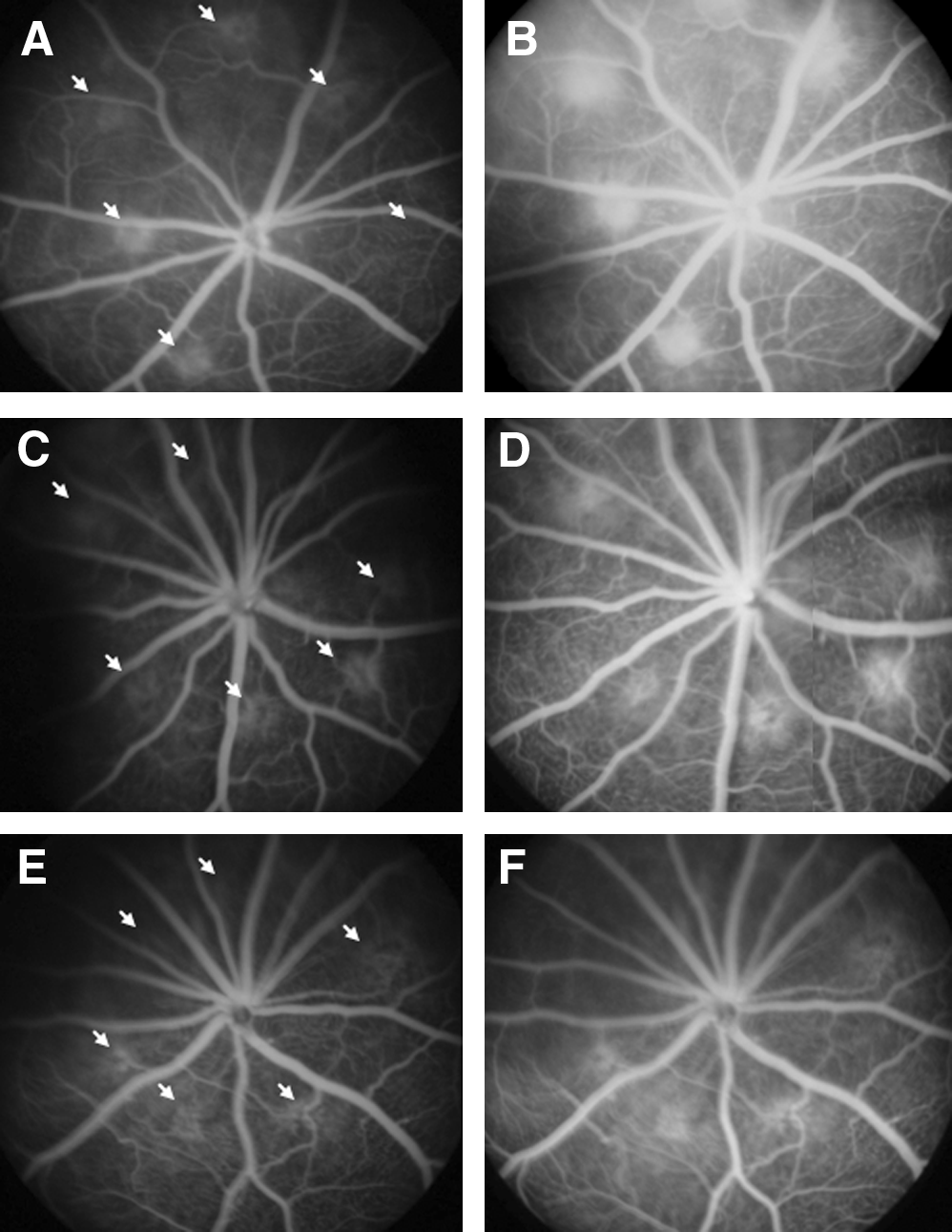
The typical fluorescein angiographic images of early (
Histology
Eyes were enucleated after the completion of FA. Vertical sections were obtained from each eye by dissecting the cornea and optic nerve. Again sections were dissected from the optic nerve plane to more peripheral planes and the presence of CNV was investigated in steps of 16 μm from the optic nerve head. CNV was identified as a fibrotic mass between the retina and choroid with vessels and endothelial cells inside it. The margin of CNV was easily discerned from surrounding tissues as the outer border of CNV contacted choroidal melanin pigments and the inner border contacted retinal tissues. When CNV was found, all neighboring slides showing the same CNV were reviewed and the slide with the largest lesion diameter was chosen for histologic evaluation. The dimensions of CNV lesions were measured on digitized histologic images, using Image-Pro plus software (Media Cybernetics, Inc.). The following parameters were recorded: CNV area (mm2), CNV/choroid thickness (X/Y) ratio, and largest CNV diameter. The CNV area was measured from the area within the drawn margin of CNV in digitized images. X/Y ratio was calculated using the maximum distance from the bottom of the pigmented choroidal layer to the top of the neovascular membrane (X), and the thickness of the intact pigmented choroid adjacent to the lesion (Y). 19
Thirty-nine animals were subjected to angiographic and histologic analyses to investigate the effects of systemic UDCA (Ursa; Daewoong Pharmaceutical Co.) and TUDCA (Taurolite; Bruschettini, Inc.).
We chose a systemic dose of 500 mg/kg for UDCA and of 100 mg/kg for TUDCA. The dose decision was based on the experiments by Boatright et al. 13 and our preliminary experiments using rats (unpublished data). In our preliminary experiments using rats and the laser-induced CNV model, UDCA 500 mg/kg and TUDCA 100 mg/kg showed a suppressing effect on CNV and also showed similar efficacy on CNV compared with UDCA 1,000 mg/kg and TUDCA 500 mg/kg. In the present study, 39 animals were allocated to 3 groups, namely, the UDCA, TUDCA, and control groups (n = 13 in each group). Controls were administered vehicle (0.15 M NaCO3). Drugs and vehicle were administered intraperitoneally daily from 24 h before to 14 days after laser induction. Fourteen days after CNV induction, FA and enucleation were performed, and angiographic and histologic evaluations of CNV lesions were performed.
VEGF and enzyme-linked immunosorbent assay
Animals received intraperitoneal injections of drugs (UDCA 500 mg/kg, TUDCA 100 mg/kg, or vehicle) at 1 day and 1 h before laser CNV induction and then daily for 2 days. Eyes were enucleated at 3 days after laser induction. Detailed methods of the preparation of enucleated eyes and the enzyme-linked immunosorbent assay (ELISA) are the same as that in the report by Zambarakji et al. 20 Cornea and lens were removed, and the retina was separated from the choroid and sclera. Retina was homogenized in cell lysis buffer (Protein Extraction Solution; Intron Biotechnology, Inc.) and incubated for 30 min on ice. The lysate obtained was centrifuged at 13,000 rpm for 5 min at 4°C, and the supernatant was transferred to a fresh tube to determine VEGF levels by using an ELISA kit (Rat VEGF Immunoassay kit; R&D Systems, Inc.). Protein concentrations were measured using a commercial assay kit (BCA Protein Assay kit; Pierce Biotechnology, Inc.). Choroid and sclera were prepared in the same way as retina.
Statistical analysis
The data were expressed as mean ± standard error of the mean. Scores of angiographic leakage from CNV lesions were compared between groups using chi-square test. Histologic dimensions of CNV lesion and VEGF levels in the retina were compared between groups using the Mann–Whitney U test. P < 0.05 was considered statistically significant. Statistical analysis was performed using SPSS v.15.0 software (SPSS, Inc.).
Results
Angiographic leakage from CNV lesions
Results of the angiographic evaluations of CNV lesions in the 3 groups (n = 13 eyes and 78 total lesions in each group) are shown in Table 1 and Fig. 2. Figure 2 demonstrates the proportion of CNV lesions showing clinically significant fluorescein leakage (Grade 2B) in each group. The UDCA and TUDCA groups had significantly lower incidences of clinically significant fluorescein leakage (42% and 46%) than the control group (67%) (P = 0.0124 for the UDCA group and P = 0.0292 for the TUDCA group by chi-square test; Table 1).

The angiographic leakage from choroidal neovascularization (CNV) lesions at 14 days after laser induction, which were graded from fluorescein fundus angiography. The bars depict percentages of CNV lesions showing significant fluorescein leakage (Grade 2B) out of the induced CNV lesions (Grades 1 and 2A) in the ursodeoxycholic acid (UDCA) (500 mg/kg), tauroursodeoxycholic acid (TUDCA) (100 mg/kg), and control groups. The UDCA and TUDCA groups had significantly lower proportions of Grade 2B lesions than the control group (P = 0.0124 and 0.0292 by chi-square test).
Six chorioretinal lesions were induced per eye with the laser. CNV was identified and graded on the angiography at 14 days after laser treatment. Animals were treated with daily intraperitoneal injection of vehicle (0.15 M NaCO3) in the control group, UDCA 500 mg/kg in the UDCA group, and TUDCA 100 mg/kg in the TUDCA group.
P values were calculated using chi-square test.
Abbreviations: CNV, choroidal neovascularization; CI, confidence interval; UDCA, ursodeoxycholic acid; TUDCA, tauroursodeoxycholic acid.
Histologic dimensions of CNV lesions
Figure 3 shows the typical CNV lesions in the 3 groups on histologic sections of eyeballs (hematoxylin–eosin staining). The comparison of histologic dimensions of CNV lesions in the 3 groups are illustrated in Fig. 4. Mean CNV areas were significantly lower in the UDCA (7,664 ± 630 μm2, P < 0.001) and TUDCA (8,558 ± 570 μm2, P < 0.001) groups than in the control group (12,147 ± 661 μm2, Fig. 4A). Further, mean CNV/choroid (X/Y) ratios were significantly lower in the UDCA (2.35 ± 0.16, P = 0.026) and TUDCA (2.27 ± 0.15, P = 0.003) groups than in the controls (3.10 ± 0.27; Fig. 4B), and the mean values of largest CNV lesion diameter were smaller in the UDCA (237.2 ± 13.7 μm, P = 0.007) and TUDCA (268.5 ± 9.1 μm, P = 0.107) groups than in controls (302.8 ± 14.4 μm; Fig. 4C).

Histologic analysis of the rat eyes with choroidal neovascularization (CNV) lesions at 14 days after laser induction (hematoxylin–eosin staining). (
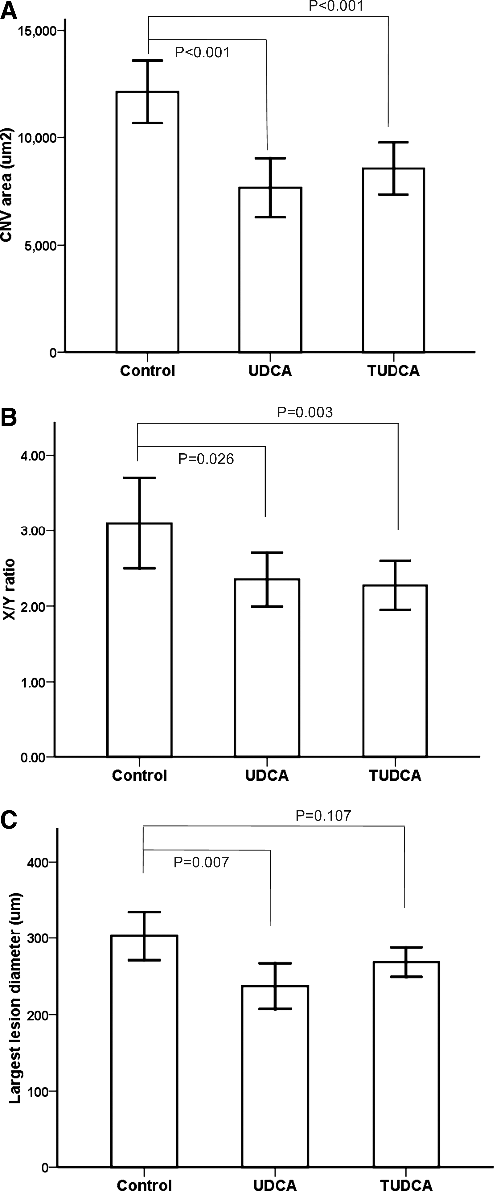
Graphs showing the histologic dimensions of choroidal neovascularization (CNV) in the control, ursodeoxycholic acid (UDCA), and tauroursodeoxycholic acid (TUDCA) groups. CNV areas (
Quantification of VEGF
Figure 5 summarizes VEGF measurements in the isolated retinal lysates at 3 days after CNV induction. Mean normalized VEGF levels of retina in the TUDCA group (9.0 ± 2.7 pg/mg total protein, n = 5, P = 0.032 by Mann–Whitney U test) were significantly lower than that in the control group (29.4 ± 8.2 pg/mg, n = 4), whereas the VEGF levels of the UDCA group (37.6 ± 6.4 pg/mg, n = 6) showed no difference compared with the control group (P = 0.190 by Mann–Whitney U test). Mean normalized VEGF levels of the choroid-sclera tissue showed no difference between drug-treated and control groups (control: 80.5 ± 24.3 pg/mg total protein; UDCA: 74.4 ± 15.5 pg/mg, P = 0.762; and TUDCA: 95.4 ± 18.5 pg/mg, P = 0.486 by Mann–Whitney U test).
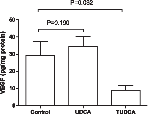
Mean vascular endothelial growth factor (VEGF) levels (standard error bars) in retinal lysate normalized for total proteins in the vehicle-treated control group (n = 4), the ursodeoxycholic acid (UDCA) (n = 6)-treated group, and the tauroursodeoxycholic acid (TUDCA) (n = 5)-treated group. Eyes were enucleated at 3 days after choroidal neovascularization (CNV) induction. P values were calculated using the Mann–Whitney U test.
Discussion
In the present study, rats treated with systemic UDCA or TUDCA before and after CNV induction showed less fluorescein leakage from CNV and reduced CNV lesion sizes. The reduced fluorescein leakage observed in the UDCA and TUDCA groups also indicates smaller induced CNV membranes than the control group. Further, our VEGF quantification result suggests that the systemic administration of TUDCA is associated with suppression of early VEGF elevation in the retina after laser injury, and that this inhibition of VEGF upregulation is associated with the observed reduction in CNV size and vascularity.
Although the pathogenesis of CNV development in exudative AMD is not fully understood, inflammation, apoptosis, and neurodegeneration are considered to be primary contributors. 21 Recent studies22,23 suggest that ER stress and protein misfolding are implicated in a variety of neurodegenerative diseases that share pathological features with AMD. Proteome analyses of the human and murine retinas show that stress-induced protein misfolding and decreased chaperoning are associated with age-related retinal degeneration.24,25 Therefore, these findings indicate that the pathogenesis of AMD may be mediated, at least in part, via ER stress and protein misfolding. 26 TUDCA, an endogenous bile acid and putative chemical chaperone, has also been shown to reduce ER stress and inflammatory responses in models of diabetes and liver disease, and to have cytoprotective and antiapoptotic effects in animal models of Huntington disease, Parkinson's disease, and stroke.10,13,16 Boatright et al. 13 reported that the systemic administration of TUDCA in a mouse retinal degeneration (rd10) model and in a mouse light-induced retinal degeneration model can suppress the apoptosis of photoreceptors and preserve their functions and morphologies.
In the present study, we also found that the inhibitory effects of TUDCA on CNV are probably related to the reduced VEGF expression. Previously, Suh et al. demonstrated the antiangiogenic activity of a conjugated form of UDCA using a chick embryo chorioallantoic membrane assay. 17 The abilities of UDCA and TUDCA to reduce ER stress and inflammatory response indicate that these agents could also inhibit inflammatory response in chorioretinal injury during and after the laser induction of CNV. As VEGF is also known to be one of the proinflmmatory factors, 27 the inhibition of inflammatory response might suppress VEGF upregulation in the retina after laser treatment and ultimately could suppress CNV induction.
VEGF is known to be the most important mediator of CNV in exudative AMD in humans, 3 and a number of clinical studies have revealed that anti-VEGF agents suppress CNV and improve vision in exudative AMD. 3 VEGF is also known to be an important mediator of CNV resulting from retinal diseases other than AMD, such as choroiditis, pathologic myopia, and angioid streak. 28 Therefore, the suppression of local inflammation and upregulation of VEGF by systemic TUDCA suggest that this drug could be helpful for preventing and suppressing CNV induced by various retinal diseases including AMD.
It is of note that these 2 similar drugs showed different responses in terms of retinal VEGF levels. The lack of difference in the retinal VEGF levels between the UDCA group and control group indicates that mechanisms other than VEGF might be involved in the suppression of CNV by UDCA. Recently, systemic administration of anti-VEGF therapy was reported to have nephrotoxicity, which was characterized by thrombotic microangiopathy and loss of podocytes resulting in renal failure.29,30 It was hypothesized that anti-VEGF antibody might cause an alteration in VEGF expression in the kidney, leading to endothelial cell dysfunction and glomerular disease. 31 Therefore, the VEGF-independent suppression of CNV by UDCA could be an important finding and may be an additional advantage as an antiangiogenic agent.
The laser-induced CNV model, which was used in our study, is not suitable for studying early or nonexudative AMD. However, this model may be the best available animal model to study CNV and CNV-related retinal diseases including exudative AMD, although there is no perfect animal model to study CNV that resembles human CNV.20,32,33 The mechanism of CNV development from AMD has not yet been fully understood, and our laser-induced CNV model reflects only parts of the pathogenesis of exudative AMD. Additional research is also necessary to reveal the exact mechanism of actions of these drugs, despite we showed the antiangiogenic effects of UDCA and TUDCA. Evaluation of the inflammatory processes, such as the recruitment of macrophages to the site of CNV, which have been histologically demonstrated near AMD lesions, 34 would better elucidate the mechanism of actions of UDCA and TUDCA on CNV.
There are several reasons why the effects of UDCA and TUDCA on CNV deserve attention. First, UDCA and TUDCA can be administered systemically via the oral route, and both have been proven to be safe and to have few side effects. Currently, the only vision-improving treatment method for exudative AMD involves the multiple intravitreal injections of anti-VEGF agents, 3 which presents the risk of endophthalmitis in 0.01%–0.16% of patients. 35 Thus, the availability of an anti-CNV drug with a good safety profile would have an enormous impact on the treatment of many retinal diseases associated with CNV. Second, if their oral administration could give additive effects to the current anti-VEGF treatments, they might reduce the frequencies of the intravitreal injections of anti-VEGF agents and reduce the economic burden and side effects related to anti-VEGF treatments.
In conclusion, the systemic administrations of UDCA and TUDCA suppressed laser-induced CNV formation in rats, which might be associated with VEGF downregulation. The results suggest that UDCA and TUDCA should be viewed as candidate drugs for the treatment of many CNV-related retinal diseases, including AMD.
Footnotes
Acknowledgments
This research was supported by a research fund from the Daewoong Pharmaceutical Company, Grant Number 0411-20070024. The authors thank Young Joo Kim for VEGF quantification by ELISA and Hyo Jin Shin for her assistance during the animal treatments.
Author Disclosure Statement
No conflicting interests exist for any author.