
Abstract
Abstract
Aim:
To study the clinical features and to identify the molecules responsible for contact-allergic reactions following ocular use of corticosteroid (CS) preparations.
Design:
Observational case series.
Methods:
We reviewed the clinical data, the patch test results, and sensitization sources in patients with a CS contact allergy, who have been patch tested in the K.U. Leuven Dermatology department during an 18-year period.
Results:
Eighteen subjects (out of 315 with CS delayed-type hypersensitivity) presented with allergic manifestations (conjunctivitis, eczema of the face, periocular skin or eyelids) of delayed-type hypersensitivity reactions to the use of CS-containing ocular preparations. The most common allergen was hydrocortisone, but most patients presented with multiple positive tests, not only to other CSs, but also to other active principles, preservatives, and vehicle components.
Conclusions:
Ophthalmic CSs, despite their anti-inflammatory and antiallergic properties, may produce contact-allergic reactions.
Introduction
ACD is said to occur in 46% to 74% of the patients suffering from conjunctivitis and/or eyelid dermatitis,4–6 the most important allergens being topical pharmaceutical products, that is, active principles, vehicle components, and preservative agents present in ophthalmic preparations.4,7–12
Topical corticosteroids (CSs), potent anti-inflammatory and immuno-modulator agents, are important pharmaceutical products for treating individuals with ACD since they reduce immunological reactions. However, in some individuals, CSs could become an antigen that triggers CS allergy. This manifests as an ACD that is unresponsive to CS or provokes a worsening of a pre-existing condition. 13
Despite the wide ocular use of CSs, only few cases of patients experiencing allergic reactions have been reported.4,7–11,14–17 For example, Alani in 1976 14 was the first to report 2 patients suspected of allergic dermato-conjunctivitis due to CSs: they were both tested with the constituents of the ointments applied and showed positive patch test reactions to betamethasone valerate and to hydrocortisone, respectively. Mathias et al.15,16 described 3 patients, suffering from chronic iridocyclitis, who developed severe conjunctival inflammation following retrobulbar injections with a methylprednisolone-acetate suspension. Lewis 17 also reported multiple contact sensitivities to eyedrop ingredients, including the drugs prednisolone. Most authors of clinical reviews mention intraocular CSs as potential causes of ACD.4,7,8,10–12
However, when problems arise on the eyes and/or eyelids, the significance of contact allergy to CSs cannot easily be assessed, since ocular preparations are not often suspected as potential causes and, hence, have rarely been patch tested.
We wanted to study the clinical features and to identify the molecules responsible for contact-allergic reactions following ocular use of CS preparations.
Methods
We reviewed the clinical data and the results of patch testing of an extended CS series in the Dermatology Department at the K.U. Leuven University Hospital during an 18-year study period, that is, from January 1, 1990, to June 30, 2008. 18 All patients attending the Contact Allergy Unit were examined clinically, and routinely patch tested with a baseline (standard) series containing (1) tixocortol pivalate 0.1% pet., a marker that detects allergic reactions to CS molecules such as hydrocortisone, methylprednisolone, and prednisolone; (2) budesonide 0.01% pet., a marker for CS such as triamcinolone acetonide and amcinonide (19) and (3) hydrocortisone 17-butyrate and prednisolone caproate both diluted in 0.1% ethanol. Moreover, those patients having been exposed to CSs were, in most cases, also tested with the respective molecules used. Patients with a proven contact allergy to a CS and some presumed to have had such a reaction, according to their history, were later tested with an extended CS series to study simultaneous positive (and cross) reactions.
Sixty-six CSs, including those from the baseline series, as well as 2 sexual hormones, that is, progesterone and testosterone, were thus tested (Table 1). All CSs (except for the 2 screening agents officially added to the baseline series 19 ) were made up in the Contact-allergy Unit by a laboratory technician; the substances used for these preparations had previously been obtained from the pharmaceutical companies marketing them. Dexamethasone Na-phosphate (introduced in November 1990) and difluprednate (Sicor, Milan, Italy; marketed in 2008 to treat ocular pain 20 ) have been included in the base line series since October 1994. The test solutions were freshly prepared every 6 to 12 months.
DMSO, dimethyl sulfoxide.
Ethanol was the vehicle used to test all CS molecules except for tixocortol pivalate and budesonide where petrolatum worked well.21,22 Due to their weak trans-epidermal penetration, hydrocortisone and cortisone acetate were diluted in an equal mixture of ethanol and dimethyl sulfoxide (DMSO). 21 Both ethanol and ethanol/DMSO were included as controls in the extended series.
The concentrations of the test solutions were defined according to the methods of Isaksson et al.23,24 (Table 1). Particularly for budesonide, lower concentrations were also tested, that is, 0.01% pet. and 0.002% eth.
The patch test materials used were van der Bend chambers (van der Bend, Briele, The Netherlands) on Mepore® (3M), covered on the upper back with Mefix®. The patches were removed after 2 days and the patch test reactions were evaluated at day 2, day 4, and in many cases also after 7 days, in accordance with the International Contact Dermatitis Recommendation Group (ICDRG) criteria. All clearly positive tests were taken into account in this study. Although, in general, dubious reactions were excluded from our analysis, weak irritant reactions to the DMSO/eth (even in the presence of hydrocortisone) could not always be ruled out with certainty.
The results were collected from a computer database containing the following information: sex, date of birth, occupation, personal and familial atopic history, localization and lesion type, use of CSs, and patch test results. The relevance of the reactions was considered on the basis of a history of known or likely contact of the affected skin area with products containing the allergen, as well as according to the follow-up of the patients in whom the dermatitis got better, for example, in the case of an underlying disease such as atopic or seborrheic dermatitis, or disappeared on discontinuing the use of the incriminated CS.
To categorize the multiple positive reactions often observed with CSs, which may be due to simultaneous or subsequent sensitization or to cross-reactivity, 25 the CSs were classified into 4 reacting groups: groups A, B, C, and D. 26 Group D has been further sub-classified into 2 subgroups: D1 and D218,27,28 (Table 2).
Results
Eighteen (5.7%) out of 315 CS allergic patients (Table 3) presented with facial, periocular, or eyelid eczema, and/or conjunctivitis, following ocular application of CSs. Their symptoms responded to treatment either poorly or not at all; decreased very rapidly when the treatment was stopped; or got worse when CS-based collyria or ointments were reapplied (Figs. 1–2 provide examples of ACD following the ocular use of dexamethasone Na-phosphate).
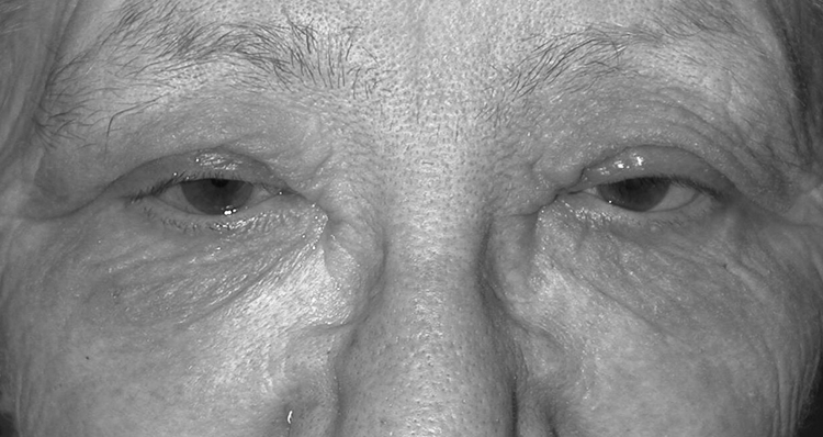
Allergic contact dermatitis from corticosteroids, in this case following the ocular use of dexamethasone (ointment) and dexamethasone Na-phosphate (collyrium), respectively: periocular edema and eczema, conjunctivitis, and worsening of the ocular pathology for which the corticosteroid was prescribed.
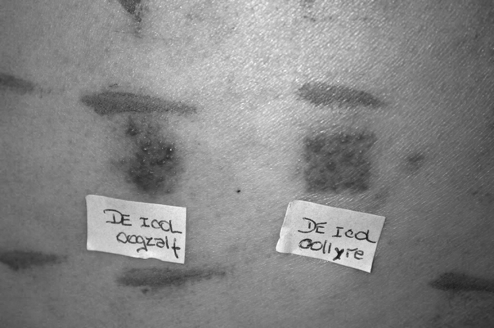
Patch test results. Positive patch tests of dexamethasone containing ointment and Na-Phosphate containing collyrium, respectively (Deicol®; Meda Pharma, Brussels, Belgium).
According to the clinical history, the patients had used ophthalmic preparations containing hydrocortisone, prednisolone, dexamethasone, fluorometholone, and/or triamcinolone actonide. All 18 patients tested positively to the group A CSs, that is, hydrocortisone, prednisone, prednisolone, methylprednisolone, and/or particularly to tixocortol pivalate, the latter being the contact allergy marker of this group.
Eight patients, all of whom had used hydrocortisone ophthalmic ointment [numbers (nos.) 1, 3, 5, 8, 11, 13, 17, and 18; Table 3], reacted exclusively to group A, particularly to hydrocortisone, and presented with cross-reactions with other CSs from the same group. Six patients (nos. 2, 4, 6, 7, 14, and 15; Table 3), who had also used ophthalmic CSs containing hydrocortisone and/or prednisolone, tested positively to group A CSs with concomitant/cross-reactions with the labile ester molecules from the D2 group, of whom 2 also tested positively to budesonide (nos. 7 and 14). Indeed, budesonide, based on the acetal function, is actually an equal mixture of the R and S diastereoisomers29,30 and is able not only to cross-react within the acetonide group (group B) but also with ester molecules (mainly of group D2). Four additional patients (nos. 9, 10, 12, and 16; Table 3) reacted to CSs from different groups. One of them (no. 9) had exclusively used collyria containing group A molecules, that is, prednisolone pivalate and fluorometholone, whereas the other 3 had used ophthalmic ointments containing dexamethasone (nos. 10, 12, and 16), dexamethasone Na-phosphate (no. 16), dexamethasone Na-metasulpho-benzoate, or triamcinolone acetonide (no. 12).
Some patients (nos. 2, 3, 4, 5, 6, 9, 10, 12, 16, and 17; Table 3) also tested positively to other allergens present in opthalmic products, such as benzalkonium chloride and thiomersal (preservative agent), polymixine B, tetracycline, neomycine, chloramphenicol, and rifamycine (local antibiotics), and wool alcohols (an excipient often used in ophtalmic ointments).
Discussion and Conclusion
Not many cases of allergic reactions due to ophthalmologic use of CSs have been reported in the literature. In this study, out of 315 patients with a CS allergy, 6% presented with ACD and/or conjunctivitis following ocular administration.
Recognizing CS allergy can be difficult as the clinical signs are neither specific nor spectacular, or display a completely atypical chronology, which is due to the anti-inflammatory properties of the CS. As mentioned by Le Coz, 31 the clinical manifestation of such reaction depends on 2 competing effects that are of variable intensity and offset in time, namely, the immunological allergic response and the pharmacological antiallergic effect.
Facial and periocular edema and/or eczema, conjunctivitis, a dry sensation, burning, itching (pruritus), or tearing are symptoms that may indicate sensitization. Worsening, rapid increase, and/or absence of response of the ocular pathology for which the CSs was prescribed should alert the clinician to suspect a contact allergy.
All patients presented with multiple positive reactions to CS, which may be due to concomitant but certainly also to cross-sensitivity.25–29,32 The potential of CSs to cross-react within and between groups could be related to structural homology and biometabolization of the CS. Foti et al. 33 suggested that the cross reactivity of CS may occur at T-lymphocyte level during antigen presentation. Molecules from group A were mainly implicated, in particular hydrocortisone, the most prescribed ophthalmic CS. Group A CSs classically cross-react with molecules from group D2, which are rapidly metabolized in the skin to group A molecules.18,26–29,34 One example of intracutaneous biometabolization is the conversion of hydrocortisone 17-butyrate (group D2) to hydrocortisone 21-butyrate, followed by an enzymatic transformation into hydrocortisone (group A). This illustrates the typical cross-reaction pattern observed between group A and group D2.
Further, besides CSs, multiple positive tests were also observed for other potential allergens present in ocular preparations, that is, topical antibiotics, antiseptics, and even vehicle components.
Conventional patch testing of ocular products as such (as is), in particular beta-blocking agents, is often unsuccessful in diagnosing ACD and the baseline standard series does not seem very useful either.35,36 The anatomological and physiological properties of eyelid skin, sometimes previously damaged by dermatitis, may be the cause of a lower threshold to contact allergy in comparison with the thick healthy skin of the back. 36 Repeated open application tests on the forearm (1 application 2 or 3 times a day during several days) is sometimes necessary to confirm sensitization.
Extensive patch testing (and sometimes additional tests) with all ingredients (including CSs) of the preparations used, are thus required to detect all relevant allergens, to advise patients about which products should be avoided, 37 as well as with regard to the use of future safe alternatives.
Footnotes
Acknowledgments
The authors thank the Foundation Saint-Luc from the Université Catholique de Louvain for supporting this research work.
Author Disclosure Statement
The authors disclose no conflict of interest in connection with the submitted article. The authors indicate no financial conflict of interest. The study and data accumulation were conducted with the approval of the Institutional Ethics Committee, Commission d'Ethique Biomédicale Hospitalo-Facultaire de l'Université Catholique de Louvain (NCT 340320084407).