




Abstract
Abstract
Purpose:
The effects of rebamipide on the number of periodic acid Schiff reagent (PAS)-positive cells in the conjunctiva, the mucin content in the cornea and conjunctiva of normal rabbits, and desiccation-induced corneal damage in vivo were examined.
Methods:
Rebamipide (0.1%–3%) was applied 6 times a day for 14 days, and the PAS-positive cell count in the bulbar conjunctiva was measured by impression cytology. The amount of conjunctival and corneal mucin-like substances was measured by Alcian blue binding. The corneal damage model was created by desiccation from air flow at room temperature. The level of corneal damage was determined by scoring the area stained with rose bengal and fluorescein dye.
Results:
Rebamipide increased the number of PAS-positive cells in the conjunctiva when instilled at concentrations of 0.3% or higher, and 1% rebamipide increased the amount of mucin-like substances of the conjunctiva and cornea. Moreover, 1% rebamipide was also found to lower the rose bengal scores of the cornea in the corneal damage model by desiccation.
Conclusions:
Rebamipide is a possible candidate drug for treatment of cornea and conjunctival epithelial damage due to its mucin-like substance increasing action, for instance, in the treatment of dry eye disease.
Introduction
There have been several reports on the importance of mucin in patients with dry eye.3,4 The level of mucin in the lacrimal fluid was reduced in these patients.5,6 The number of goblet cells involving mucin that produce and secrete mucin were also decreased.7,8 Moreover, it has been reported that gefarnate and diquafosol sodium increased the number of conjunctival periodic acid Schiff reagent (PAS)-positive cells, and a drug that acts to increase mucin is a promising option for dry eye treatment.9,10
Rebamipide is a mucosal protective and ulcer-healing agent developed in Japan and is widely prescribed in Asia. It has been reported that the use of rebamipide eye drops increased mucin levels of the conjunctiva and the cornea, and improved injuries of the epithelial conjunctiva and cornea. 11 Currently, rebamipide eye drops for dry eye is under development to obtain indication in the United States and Japan.
In the present study, we evaluated whether rebamipide has increasing effects of cornea-conjunctival mucin-like substance contents and the number of PAS-positive cells, goblet cells involving mucin, in normal rabbits. Additionally, we examined the preventive effect of rebamipide for desiccation-induced corneal damage.
Methods
Test articles
Rebamipide, 2-(4chlorobenzoylamino)-3-[2(1H)-quinolinone-4yi]-propionic acid, is a novel quinolinone derivative synthesized by Otsuka Pharmaceutical Co., Ltd. (Tokyo, Japan). Ophthalmic suspensions were formulated at concentrations of 0.1% to 3% (wt/vol). Sodium hyaluronate (0.1%, Hyalein0.1) was purchased from Santen Pharmaceutical Co., Ltd. (Osaka, Japan).
Chemicals
Sucrose, magnesium chloride, and PAS were obtained from Wako Pure Chemical Industries (Osaka, Japan). Alcian blue 8GX was obtained from Nacalai Tesque (Kyoto, Japan).
Animals
New Zealand white female rabbits (2.0–2.5 kg body weight; Kitayama Labes, Nagano, Japan) were used. The use and treatment of rabbits in all studies conformed to ARVO Standards for the Use of Animals in Ophthalmic and Vision Research.
Test article treatment
In the study investigating the effects of the dose dependency of rebamipide on PAS-positive cells and on conjunctival mucin-like substances, 0.1%–3% rebamipide or vehicle was instilled 6 times a day for 14 days with 1 additional instillation on the 15th day. In the study investigating the effects of the number of rebamipide instillations on the levels of conjunctival mucin-like substances, 1% rebamipide was instilled 2, 4, or 6 times a day for 14 days with 1 additional instillation on day 15. Rebamipide (1%) was administered 6 times a day for 7–56 days with 1 additional instillation on the day following the end of treatment in the study investigating the effects of the treatment period of rebamipide on PAS-positive cells, and was administered 4 times a day for 3–56 days with 1 additional instillation on the day following the end of treatment in the study investigating the effects of the treatment period of rebamipide on conjunctival mucin-like substances. Rebamipide was discontinued for periods of 0, 7, 14, 28, or 42 days following completion of the initial administration periods of 14 days. In the desiccation-induced corneal desiccation damage model, rebamipide (1%), sodium hyaluronate (0.1%), or saline was administered 6 times a day before creating desiccation-induced corneal damage for 14 days.
Measurement of the number of PAS-positive cells
The animals were sacrificed by intravenous injection of thiopental sodium. Immediately, cellulose acetate paper strips (3×10 mm; Millipore, Bedford, MA) were pressed on the superior bulbar conjunctival surfaces, dull side down, 2 mm away from the limbus for about 10 s and then removed. Each strip of paper carrying conjunctival epithelial cell and PAS-positive cell specimens was fixed in 95% ethyl alcohol. The specimens were stained with PAS. Photographs were taken of 4 locations selected at random under a light microscope (Olympus, Tokyo, Japan). Images of the PAS-positive cells were taken only in the regions of confirmed successful collections of conjunctival epithelial cells. The number of goblet cells in a 0.09 mm2 (0.3×0.3 mm) area at each of the 4 locations was calculated from the previously taken photographs. The average number of PAS-positive cells counted at these 4 locations was then taken as an indicator of the number of PAS-positive cells for each eye. The mean value of the number of PAS-positive cells of the left and right eyes of all animals was then taken as an indicator of the number of PAS-positive cells per eye for each animal.
Measurement of the amount of mucin-like substances
After the last administration of rebamipide or vehicle, the amount of conjunctival and corneal mucin-like substances was measured by Alcian blue binding. Rabbits were sacrificed by intravenous injection of thiopental sodium. A whole conjunctiva or cornea was removed from each rabbit. The conjunctivae were weighed, and the conjunctivae and corneas were incubated in 10 mL of a 0.1% Alcian blue solution at room temperature for 1.5 h. The conjunctivae were washed twice with 10 mL of 0.25 M sucrose for 15 and 45 min. The central area of the corneas was marked with a 10-mm-diameter trephine. The corneal epithelial cells in the marked areas were scratch-removed with a beaver scalpel. Then, the conjunctivae were incubated in 10 mL of 0.5 M MgCl2 solution at room temperature for 2 h to extract the colorant bound to the mucin layer. The corneal epithelial cells were incubated in 0.5 mL of 0.5 M MgCl2 solution. After the extract obtained from the corneas was centrifuged at 250 g for 5 min at room temperature, 0.3 mL of the supernatant was collected. The obtained extract was then purified with diethyl ether (conjunctiva: 10 mL; cornea: 0.3 mL).
The optical density of the aqueous layer of the extract was measured at 605 nm with a spectrometer (Beckman Instruments, DU-65, Fullerton, CA), and the amount of bound Alcian blue dye in the conjunctiva was represented as the optical density per weight of conjunctival tissue (OD units/g tissue), whereas the amount of bound Alcian blue dye in the cornea was represented as the optical density per cornea (OD units).
Desiccation-induced corneal damage model in vivo
Rabbits with their nictitating membrane removed before treatment were anesthetized with ketamine hydrochloride (200 mg/body, Ketalar 50; Sankyo Co., Ltd., Tokyo, Japan) and oxybuprocaine hydrochloride (2–3 drops/eye, Benoxil 0.4; Santen Pharmaceutical Co., Ltd.) before desiccation-induced corneal damage. The rabbits were placed in a fixator and speculums were applied. The rabbit eyes were dried by air blower at a constant air flow speed at 10 m/s (Sanyo Electric Co., Ltd., Osaka, Japan) at room temperature for 10 min, blown straight onto the cornea from a distance of 10 cm. The airflow was applied only to the cornea and injuries occurred 2 h after the last treatment.
Corneal damage was evaluated by gross examinations after corneal staining with a 2 μL liquid mixture of 1% rose bengal and 1% fluorescein and observed before the start of test article administration as well as immediately before and 1 day after the corneal desiccation. The degree of dye absorption was observed with a handheld slit-lamp (SL-14, magnification; ×16, Kowa, Nagoya, Japan) and sketched. The criteria for evaluation are shown below: score 0, corneal surface is left completely unstained; score 1, either 1/3 or less of the corneal surface area is stained homogeneously or 2/3 or less is stained in a punctated form; score 2, either 1/3–2/3 of the corneal surface area is stained homogeneously 2/3 or more of the corneal surface is stained in a punctated form or 1/3 or less of the corneal surface is stained homogeneously with punctated forms also recognizable; score 3, either 2/3 or more of the corneal surface area is stained homogeneously or 1/3–2/3 of the corneal surface area is stained homogeneously with punctated forms also being recognizable.
Statistical analysis
All values are expressed as mean±SE. A P-value of <0.05 was considered statistically significant. Statistical analyses were performed with SAS software version R6.12 (SAS Institute Japan Ltd., Tokyo, Japan).
Results
Effect of rebamipide at various concentrations on the number of conjunctival PAS-positive cells in normal eyes
The average conjunctival PAS-positive cell count after instillation of the vehicle, or 0.1%, 0.3%, 1%, or 3% rebamipide was 57.1±2.5, 61.1±6.9, 89.8±5.1, 90.5±3.9, and 88.9±5.2 (cells/0.09 mm2), respectively. Rebamipide at concentrations of 0.3% or higher significantly increased the number of conjunctival PAS-positive cells; the maximum cell count was shown with 0.3% rebamipide (Fig. 1B). Figure 1A shows PAS staining of filter paper containing PAS-positive cells and conjunctival epithelial cells after 15 days of treatment. The red-stained cells are PAS-positive cells and their density in the rebamipide group increased compared with that in the vehicle-treated group.
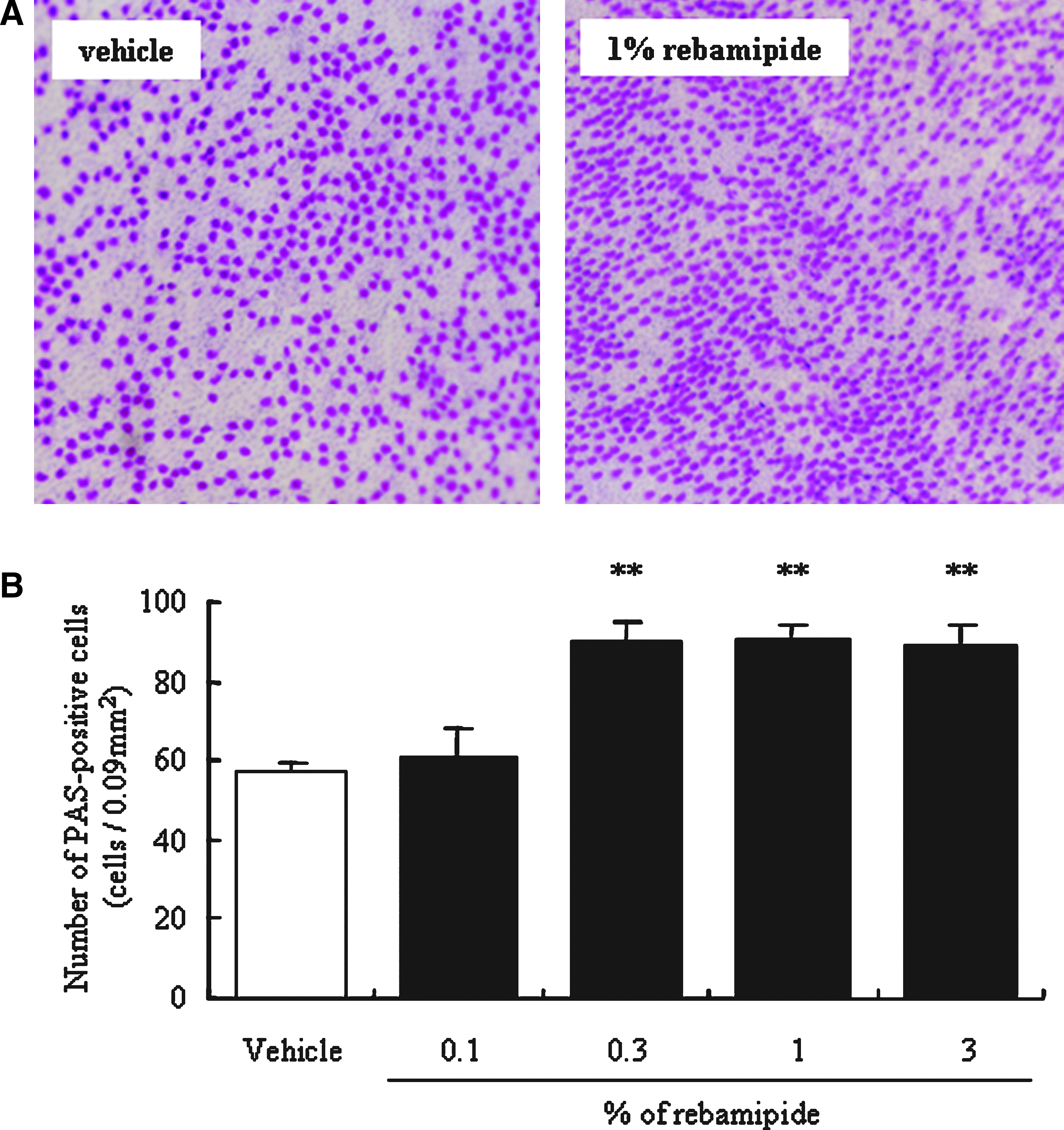
Photomicrographs
Effect of rebamipide treatment periods on the number of conjunctival PAS-positive cells
No significant increase in the number of conjunctival PAS-positive cells was observed at day 7. However, the average number of PAS-positive cells on days 14, 28, and 56 of 1% rebamipide administration was 96.0±6.1, 95.1±6.9, and 91.0±7.6 (cells/0.09 mm2), respectively, indicating a significant increase compared with untreated eyes (67.5±4.4 cells/0.09 mm2). The number of PAS-positive cells peaked on the day 14 after the start of rebamipide instillation (Fig. 2).
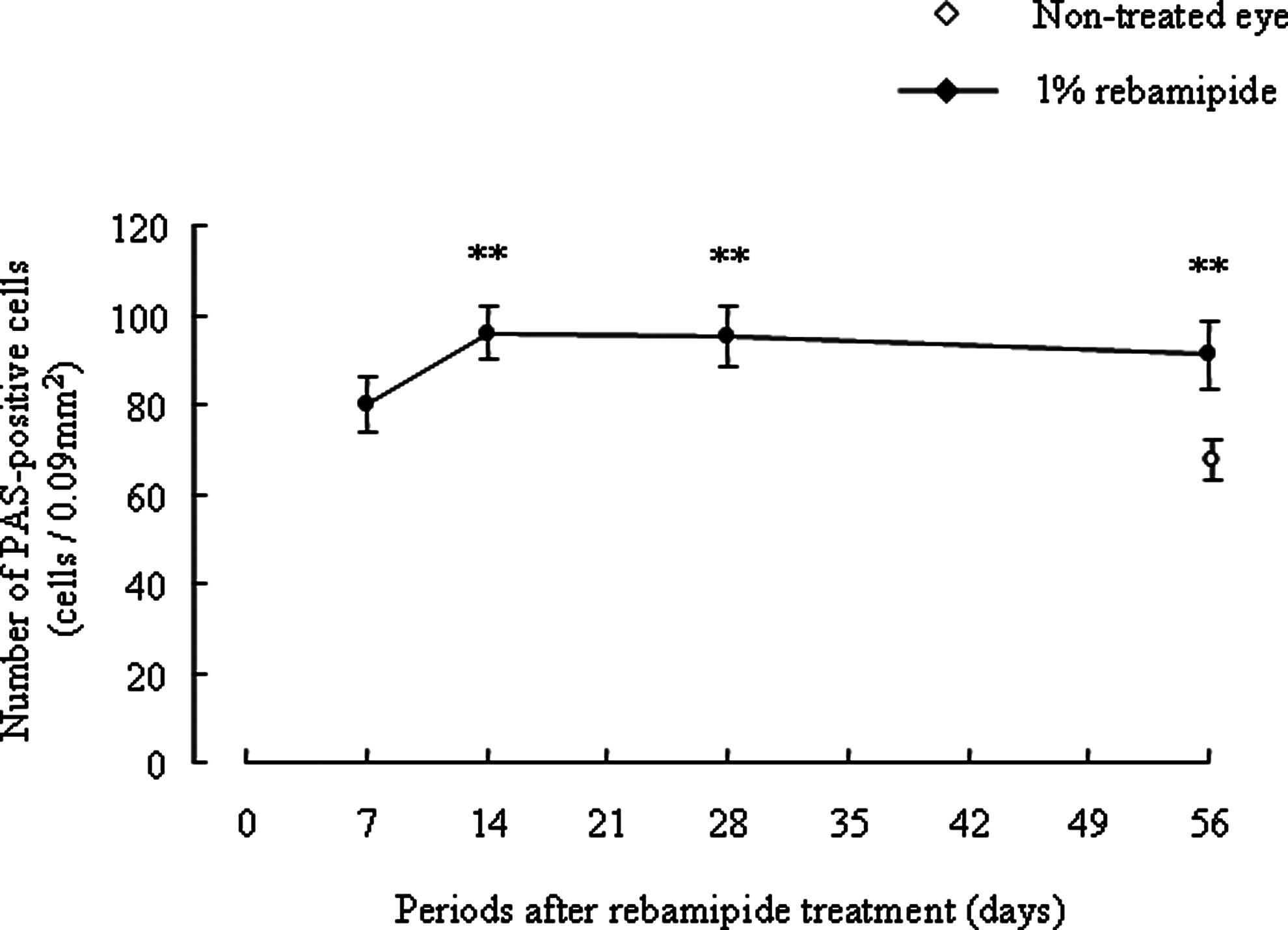
Examination of the treatment period required for development of the rebamipide-induced increase in the number of PAS-positive cells in the conjunctiva. One percent rebamipide was administered to both eyes of rabbits 6 times a day for 7 to 56 days and once on the morning after the last day of each respective period. The average number for untreated eyes was 73.0±3.8 cells/0.09 mm2. All values are expressed as mean±SE. Statistical analysis was performed by Dunnett test. There were 8 animals per group. **P<0.01 versus non-treated eye.
Continued action of rebamipide on the number of conjunctival PAS-positive cells
The mean number of PAS-positive cells immediately after termination of rebamipide instillation for 14 days was 104.2±4.8 (cells/0.09 mm2), significantly higher than the average number for untreated eyes (73.0±3.8 cells/0.09 mm2). The number of PAS-positive cells was significantly higher than that for untreated eyes on day 7 after termination of rebamipide. These levels returned to normal values approximately 27 days after termination of rebamipide (Table 1).
All values are expressed as the mean±SE. There were 8 animals per group.
P<0.01 versus untreated eye (treatment period: 0 days).
Effect of various concentrations of rebamipide on conjunctival mucin-like substances in normal eyes
The mean amount of bound Alcian blue dye was 0.115±0.005 (OD units/g tissue) in the control group eyes, as well as 0.119±0.007, 0.166±0.013, 0.259±0.015, and 0.256±0.024 (OD units/g tissue) in the eyes treated with 0.1%, 0.3%, 1%, and 3% rebamipide, respectively. A significant increase in conjunctival mucin was confirmed for rebamipide at concentrations of 1% or greater, with this action being near its maximum at a concentration of 1% (Fig. 3).
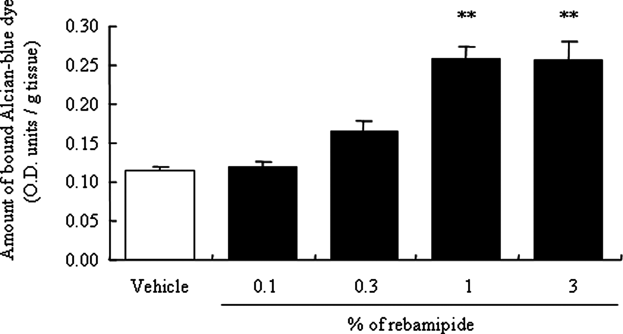
Effects of rebamipide on the amount of mucin-like substances in the conjunctiva of normal rabbits. Rebamipide or vehicle was administered to the eyes of rabbits 6 times a day for 14 days and once on the morning of day 15. All values are expressed as mean±SE. Statistical analysis was performed by Dunnett test. There were 8 animals per group. **P<0.01 versus vehicle.
Effect of the number of rebamipide instillations on conjunctival mucin-like substances in normal eyes
The mean level of bound Alcian blue on normal eyes was 0.125±0.006 (OD units/g tissue) on the day after completion of ophthalmic instillations (day 15). When 1% rebamipide was administered 2, 4, or 6 times a day, the mean amount of bound Alcian blue dye was 0.123±0.007, 0.181±0.007, and 0.181±0.009 (OD units/g tissue), respectively, with significant increases observed in cases of 4 or more instillations per day (Fig. 4).

An examination of the treatment number required for the development of the rebamipide mucin-like substance increasing effect in the conjunctiva. Rebamipide was administered to the eyes of rabbits 2 to 6 times a day for 14 days and once on the morning on the last treatment day. All values are expressed as mean±S.E. Statistical analysis was performed by Dunnett's test for comparison of 4 groups. There were 6 animals per group. **P<0.01 versus non-treated eye.
Time required for onset of rebamipide-induced conjunctival mucin-like substances increases in normal eyes
The mean level of bound Alcian blue on the conjunctiva of the 3-day, 7-day, 14-day, 28-day, and 56-day groups was 0.061±0.003, 0.064±0.005, 0.096±0.007, 0.098±0.004, and 0.097±0.006 OD units/g of conjunctival tissue, respectively. It was observed that treatments with 1% rebamipide for 14 days or longer significantly increased the level of bound Alcian blue (Fig. 5).
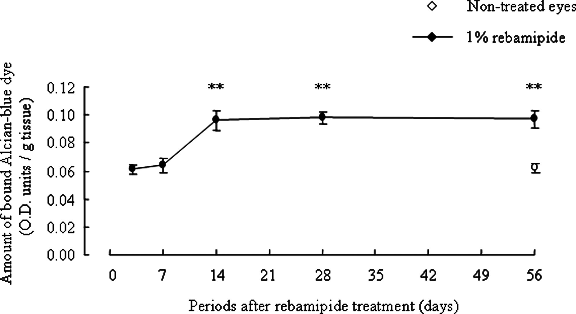
Examination of the treatment period required for development of the rebamipide-induced amount of mucin-like substances increase in the conjunctiva. One percent rebamipide was administered to both eyes of rabbits 4 times a day for 7 to 56 days and once on the morning after the last day of each respective period. The mean level of bound Alcian blue on the conjunctiva for untreated eyes was 0.062±0.003 OD units/g of conjunctival tissue. All values are expressed as mean±SE. Statistical analysis was performed by Dunnett test. There were 7–8 animals per group. **P<0.01 versus non-treated eye.
Continued action of rebamipide on conjunctival mucin-like substances in normal eyes
The mean level of bound Alcian blue dye immediately after termination of rebamipide instillation for 14 days was 0.070±0.004 (OD units/g tissue), significantly higher than the mean number for untreated eyes (0.034±0.003 OD units/g tissue). The level of bound Alcian blue dye was significantly higher than that for untreated eyes on day 7 after termination of instillation. These levels returned to normal values approximately 14 days after termination of instillation (Table 2).
All values are expressed as the mean±SE. There were 8 animals per group.
P<0.05.
P<0.01 versus untreated eye (treatment period: 0 days).
Rebamipide-induced increase in the amount of mucin-like substances in the cornea
The mean amount of bound Alcian blue dye was found to be 0.025±0.001 (OD units) in the control group eyes and 0.069±0.006 (OD units) in the eyes treated with rebamipide. A significant increase in corneal mucin levels was confirmed for 1% rebamipide (Fig. 6).
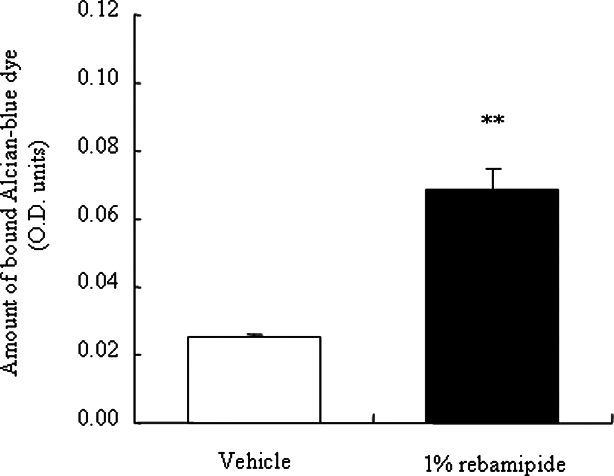
Effect of rebamipide on the amount of mucin-like substances in the cornea of normal rabbits. Rebamipide or vehicle was administered to the eyes of rabbits 6 times a day for 14 days and once on the morning of day 15. All values are expressed as mean±SE. Statistical analysis was performed by unpaired t-test. There were 10 animals per group. **P<0.01 versus vehicle.
Effects of rebamipide on desiccation-induced corneal damage
The mean rose bengal score for saline, 1% rebamipide, and 0.1% sodium hyaluronate 1 day after the injury was 2.5±0.2, 1.4±0.1, and 2.3±0.2, respectively. The mean fluorescein score for saline, 1% rebamipide, and 0.1% sodium hyaluronate 1 day after creation of the injury was 2.2±0.5, 1.2±0.3, and 1.6±0.6, respectively (Table 3). In this study, 1% rebamipide produced significantly lower rose bengal scores compared with saline and sodium hyaluronate, but no significance was found in the evaluation by fluorescein score.
All values are expressed as the mean±SE. There were 8 animals per group.
P<0.01 versus saline.
Discussion
In our present study, the effect of rebamipide in increasing the number of PAS-positive cells, goblet cells involving mucin, was evaluated by using normal eyes in the rabbit. Rebamipide significantly increased the number of PAS-positive cells at concentrations of 0.3% or higher. Dartt et al. reported that rebamipide acted on conjunctival goblet cells in several ways: promoting the number of goblet cells, inducing MUC5AC in the proliferating goblet cells, and promoting mucin secretion directly from goblet cells.12,13 These results show rebamipide's influence not only on the number of PAS-positive cells but also on the function of mucin secretion. MUC5AC is a major factor in dry eye disease and plays many important roles such as stabilization of the aqueous layer, defense of infection, and removal of foreign materials.14,15
Rebamipide increased the amounts of the conjunctival mucin-like substances in a dose-dependent manner, and significantly increased at concentrations of 1% or higher. This suggests that increasing the amounts of conjunctival mucin-like substances was related to increasing the number of conjunctival PAS-positive cells. In addition, rebamipide increased the amounts of corneal mucin-like substances. This increase of mucin-like substances by rebamipide might be caused by the production from the corneal and conjunctival epithelial cells or the conjunctival PAS-positive cells.
The corneal damage improving effect of rebamipide was evaluated using a desiccation-induced corneal damage model in vivo. In the clinical state, dry eye occurred as a result of the air flow of air-conditioning appliances. 16 The animal model in this experiment was made by producing rapid desiccation of the cornea. Dry eye animal models by desiccation have been reported previously, such as the rabbit short-term dry eye model and rat jogging board dry eye model. INS365 17 , 15(S)-HETE 18 , and gefarnate 19 were evaluated as a possible therapeutic drug to treat dry eye using the rabbit short-term dry eye model. It is important to evaluate the preventive effect of these drugs. The visual function, signs, and symptoms of dry eye patients became worse in the afternoon in comparison with the morning. 20 It is important to evaluate the preventive effect of these drugs in the desiccation-induced dry eye model. Rebamipide showed efficacy, with significant difference, by the rose bengal score compared with saline in the desiccation-induced corneal damage. The fluorescein score for rebamipide was almost half the value for saline; however, the difference was not statistically significant because of the wide deviation. On the other hand, 0.1% sodium hyaluronate did not show a difference compared with saline in each dye staining. The administration of 0.1% sodium hyaluronate had no preventive effect, because its pharmacological action is considered to be due only to its moisturizing effect,21,22 not an increasing effect on the production of mucin-like substances in the ocular surface. These results indicate that the preventive effect for desiccation-induced corneal damage may be due to increasing the amount of mucin-like substances in the ocular surface by pretreatment with rebamipide.
In conclusion, rebamipide is a possible candidate treatment for cornea and conjunctival epithelial damage due to its mucin-like substances increasing action, for instance, in the treatment of dry eye.
Footnotes
Author Disclosure Statement
H. Urashima, Y. Takeji, T. Okamoto, S. Fujisawa, and H. Shinohara were employed at Otsuka Pharmaceutical Co., Ltd.