
Abstract
Abstract
Purpose:
Chitosan, a cationic polysaccharide biopolymer with mucoadhesive properties, presents a promising future in the prolonged ocular delivery of drugs. The present study compared the efficacy and safety of chitosan-coated timolol maleate (TM) mucoadhesive film, using a 0.5% TM commercial ophthalmic solution in a rabbit model. In addition, this study investigates the maximum release time of these implants in vivo.
Methods:
The mucoadhesive films were prepared by means of a casting and solvent evaporation technique performed in a 2 wt% acetic acid solution and distilled water. Physical properties were characterized by release and swelling studies, differential scanning calorimetry, and attenuated total reflectance fourier transformed infrared spectroscopy (ATR-FTIR). The developed formulations were evaluated for their pharmacodynamics in ocular normotensive albino rabbits, in which the intraocular pressure (IOP) was measured by means of applanation tonometer on alternative days (13 h) for 11 weeks. For 15 days, 0.5% TM commercial ophthalmic solution was administered twice a day (n=5) and compared to chitosan-coated TM (n=5). In the control group (n=5), saline was used twice a day. The maximum TM release time from chitosan films were also recorded. After euthanasia, the right eyes were removed from the 3 groups for histological analyses.
Results:
In an in vitro study, TM was released over a 4-week period, in which 85% of the drug was released over the first 2 weeks. However, the film's release of TM lowered the in vivo IOP levels over a 10-week period. No significant difference in the lowering of IOP in rabbits treated with 0.5% TM commercial ophthalmic solution, as compared to those that received the films (P<0.05), could be observed. No signs of ocular discomfort or irritations could be identified upon ophthalmic examination by slit-lamp biomicroscopy. Ophthalmic structures that came in direct contact with the films revealed no alterations within the histopathological studies. Moreover, the animals showed no signs of ocular discomfort during the experimental assays.
Conclusion:
These findings suggest that the TM-loaded chitosan film is safe and efficient and presents a promising future as an ocular drug delivery system in the treatment and prevention of glaucoma.
Introduction
Drugs administered in conventional topical ophthalmologic formulation tend to present a poor bioavailability. The majority of ophthalmic preparations available today can be found in the form of aqueous solutions and suspensions. These liquid forms are quickly drained from the conjunctival sac to the nasolachrymal duct, leading to a low availability of the drug at the target site, systemic side effects, and bad patient compliance.7,8 The application of these drugs tends to lead to a very short period in which the drugs actually come into contact with the cornea, and <5% of the applied drugs are able to penetrate though the cornea into the intra-ocular tissues. 9 As a result, many ophthalmic drugs are applied in high concentrations, in turn causing ocular as well as many other systemic side effects.10,11
A serious disadvantage of conventional ophthalmic timolol therapy is the systemic absorption that produces several respiratory and cardiovascular side effects.12,13 In elderly patients, ophthalmic timolol can produce life-threatening disturbances in vital organ functions. Unfortunately, few cases of human death have been attributed to the use of ophthalmic timolol. 14 To overcome this disadvantage and to minimize side effects, an innovative mucoadhesive ophthalmic drug delivery system, consisting of TM and chitosan, was formulated.
Chitosan is a polycationic biopolymer obtained through the alkaline deacetylation of chitin, a natural polysaccharide found abundantly in marine crustaceans.15,16 Chitosan is able to develop molecular attraction forces through electrostatic interactions with mucus negative charges, giving way to the mucoadhesion process. 17 Biodegradability, biocompatibility, and nontoxicity are included in favorable biological properties that allow for their use as vehicles for ophthalmic formulations.18,19 The efficacy of a topical delivery system depends on the interaction of ocular mucosa, slow degradation, and the release of the drugs within the ocular tissues. 20
Some studies in prior literature have reported on the use of chitosan in ophthalmology15,20–22 concerning the development of a new generation of ocular delivery systems. 23 Chitosan hydrogels, when compared with commercial drug solutions, have shown higher corneal residence times. 15 Many reports with in vivo studies involving TM in delivery systems made up of gel and nanoparticles were described.6,16,22,24,25 However, no studies involving in vivo evaluation of TM-loaded mucoadhesive ophthalmic chitosan film could be found in prior scientific literature.
Therefore, the present study aimed to develop a suitable and mucoadhesive system consisting of TM and chitosan as a prolonged treatment device. This in vivo system was also evaluated and compared with the commercial solution, Timoptol® (Merck Sharp & Dohme Farmaceutica Ltda). To the best of our knowledge, this is the first in vivo study concerning the development of mucoadhesive ophthalmic films with TM-loaded chitosan films and their pharmacodynamic evaluation.
Methods
Materials
The following materials were purchased for the present study: chitosan, with a deacetylation degree of 85%, from Sigma-Aldrich; TM from Henrifarma; Xylazine (Calmium®) and ketamine (Ketamina®) from Agener União Saúde Animal; thiopental (Thionembuthal®) from Cristália Laboratório; Topical TM (Timoptol) from Merck Sharp & Dohme Farmaceutica Ltda; topical anesthesia (Anestalcon®) from Allergan; and tropicamine (Midriacyl®) and fluorescein stain (Fluoresceína®) from Allergan. All other reagents were purchased from Sigma-Aldrich, unless otherwise specified.
Preparation of chitosan films
Chitosan drug-loaded films were produced by a casting/solvent evaporation technique. Solutions of 2 wt% chitosan were prepared with a 2 wt% acetic acid solution and distilled water, respectively. 26 TM (10% w/t) was dissolved by stirring in this solution to make the solution completely homogeneous. Next, these solutions were poured on a Petri glass dish. These films were dried at room temperature for 1 to 3 days. These dried films were cut into pieces of 4 mm in diameter for tests. The blank matrix film, without the drug, was made analogously to the process described above.
Determining of TM
UV spectroscopy was chosen to quantify the drug contained in the polymeric system. A Shimadzu Ultraviolet spectrometer was used at a wavelength of 294 nm. The method was validated with a known amount of TM diluted in phosphate-buffered saline (PBS, pH 7.5), within a concentration range of 2.5–50 μg/mL. The within-day and between-day precision and accuracy were determining factors. The cumulative amount of TM released from the film was determined using a calibration curve (y=15.035× −0.0705, R 2 =0.9979; n=5). All in vitro release experiments were performed in triplicate.
Characterization of the mucoadhesive film
Swelling studies
Film swelling properties were evaluated by determining the percentage of hydration. The hydration capacities of the chitosan film formulations were determined by weighing the film pieces before and after being placed in pH 7.4 PBSs. Each film was divided into portions of 1 cm2 (1×1 cm) and then cut, weighed, and placed in buffer solutions for predetermined periods (5, 10, 20, 40, 60, and 90 min) as described by Öner et al.
27
After immersion, the films were removed from the medium; after removing the excess surface water using filter paper, the films were weighed. The percentages of hydration were calculated by using Eq. (1).
where Xt is the weight of the swollen film after time t and Xo is the original film weight at zero time. This experiment was performed in triplicate.
Differential scanning calorimetry analysis
Thermal properties of blank matrix chitosan films and drug-loaded films were characterized using a DSC50 from Shimadzu. Nitrogen, at the rate of 20 mL/min, was used as a purge gas. The specimens were packed in aluminum pans and then heated from −50°C to 200°C (Run 1) at a heating rate of 10°C/min within a nitrogen atmosphere. The specimens were cooled to −50°C, within a nitrogen atmosphere at the same rate of 10°C/min, and then reheated to 350°C at a heating rate of 10°C/min (Run 2).
Attenuated total reflectance fourier transformed infrared spectroscopy (ATR-FTIR) analysis
FTIR spectra were taken on a PerkinElmer FTIR spectrometer (Model Spectrum One) to confirm the chemical stability of TM after the preparation of the films. FTIR spectra of blank matrix chitosan film, TM, and drug-loaded film were recorded within a powder on a PerkinElmer FTIR spectrometer.
In vitro drug release
The drug-loaded films were suspended in glass vessels containing 50 mL of medium, as specified below, and incubated on a shaking bed at 37°C±0.5°C shaking the glass at 50 rpm. At regular time intervals, aliquots of the solutions were withdrawn and the amount of TM released from the drug-loaded films were evaluated by UV spectrophotometry at 294 nm. Next, an equal volume of the same dissolution medium was added again to maintain a constant volume. The medium for the controlled release studies was a typical pH 7.5, 10 mM NaH2PO4–Na2HPO4-buffered solution. All experiments were performed in triplicate.
Animals
Fifteen young New Zealand albino adult male rabbits, weighing 1.7 to 2.0 kg, were used for in vivo studies. They were fed a normal diet and exposed to an alternating 12-h light and dark cycles. All experimental procedures involving animals were performed in accordance with the Research Ethics Committee from the Federal University of Minas Gerais (UFMG), under protocol 156/08, and conformed to the Association for Research in Vision and Ophthalmology statement for the use of animals in ophthalmic and vision research. After 2 weeks of adaptation, the IOP was recorded for 10 days to establish the median pressure. The rabbits were divided into 3 groups—group I (n=5): 50 μL saline applied twice a day (at 7:00 am and 7:00 pm) in the right eye (OD) for 15 days; group II (n=5): instillation of 50 μL of TM with 0.5% commercial collyrium (Timoptol; Merck Sharp & Dohme Farmaceutica Ltda) twice a day (at 7:00 am and 7:00 pm) in OD for 15 days; group III (n=5): received the mucoadhesive film in the lower conjunctival sac of the OD. The contralateral left eye was used as the control and received no treatment.
To avoid differences between the diameter of bottles of saline and commercial solution (Timoptol; Merck Sharp & Dohme Farmaceutica Ltda), a 50-μL drop was employed, using a micropipette (Pipeta HT Labmate®). The IOP was measured by means of an applanation tonometer Tono-Pen XL (BioRad) after ocular anesthesia with proxymetacaine (Anestalcon®; Alcon) in both eyes, each day at 1:00 pm, on alternative days (each 48 h), for 3 months by an experienced tonometrist, with the tonometer being calibrated each afternoon. Three readings were obtained from each eye, beginning with the OD, and the mean was used for the analysis. The tonometrist was masked to the treatment of the eyes (50 μL of Timoptol topical administration or chitosan implant), which was inserted by another assistant who performed the randomization process.
Ophthalmologic evaluation
The ophthalmic examination was conducted before, during, and after experimental sessions, and it includes eye lids, lacrimal system, and ocular globe inspection. For this evaluation, a proper light source (Maglite Flashlight—Mag Instruments) was used, as were strips for the Schirmer Tear Test (Schering-Plough Animal Health), direct ophthalmoscopy (Heine ophthalmoscope K180; Heine Optotechnik), indirect ophthalmoscopy (Omega 500 Binocular Indirect Ophthalmoscope; Heine Optotechnik), and slit-lamp biomicroscopy (Kowa SL-15; Kowa Company).
Histopathology
Animals were deeply anaesthetized by ketamine (30 mg/kg) and xylazine (3 mg/kg) and sacrificed by a lethal dose of thiopental (100 mg/kg). After the third eyelid, right ocular bulb and inferior eyelids were removed. These samples were set in 10% neutral buffered formalin solution for 72 h. Small cuts were made in the meridian ocular bulbs to allow for entrance of formalin into the eye to fix intraocular tissues. All tissues samples were dehydrated, cleared, and embedded in paraffin. After, these were serially cut in 5-μm-thick sections and stained with hematoxylin and eosin for light microscopic observation.
Statistical data analysis
Statistical data analysis was performed using the nonparametric, Mann-Whitney, and Friedman tests with P<0.05 as the minimal level of significance. Variables were considered different when P<0.05. Calculations were performed using the SAEG version 9.1 (Brazil, 2007) and Instat Graphpad version 3 (United States, 2003) software. Mean IOP values corresponding to the treatments with colliryum and chitosan film coated TM in the 5th to 12th measurements were compared.
Results
Differential scanning calorimetry analysis
Differential scanning calorimetry (DSC) is one possible method to determine the modification of a drug. DSC thermograms of (a) blank matrix chitosan films and (b) drug-loaded films are presented in Fig. 1. In the case of placebo chitosan film, a sharp endothermic peak can be observed at 283.58°C due to water loss and thermal decomposition. The drug-loaded films show patterns that are similar to the blank matrix chitosan films on DSC, thus indicating the amorphous dispersion of TM into chitosan films.
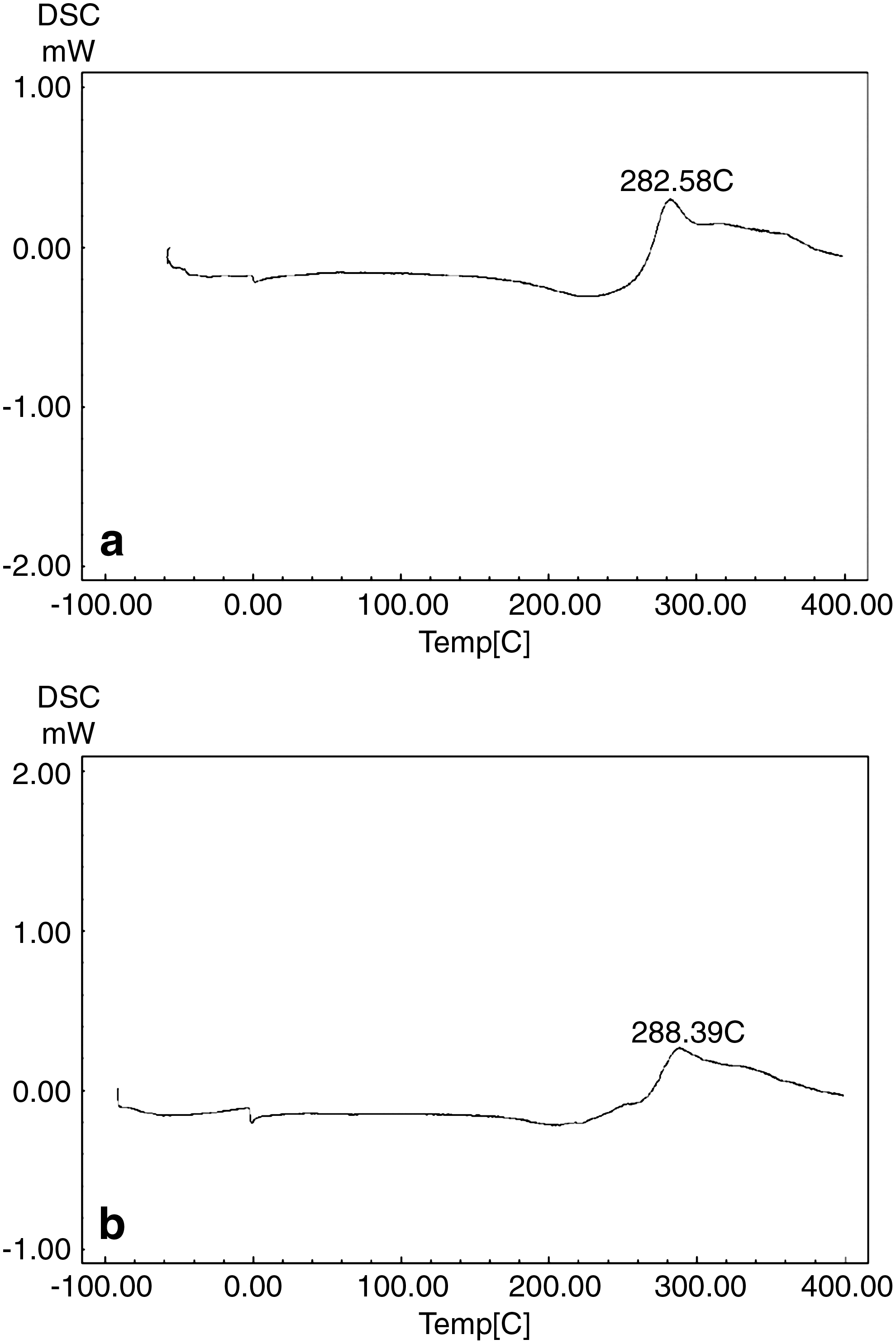
Differential scanning calorimetry thermoanalysis of
ATR-FTIR analysis
Attenuated total reflectance fourier transformed infrared spectroscopy spectral data were used to confirm the chemical stability of TM in chitosan film. Figure 2 shows the ATR-FTIR spectra of blank matrix chitosan film, TM, and drug-loaded film, respectively. Placebo chitosan film showed a broad band at 3,359 cm−1 attributed to O-H stretching vibrations of hydroxyl groups. The C-H stretching vibration of the polymer is manifested through peaks that appear at 2,872 cm−1. The characteristic split bands at 1,651 cm−1 are associated with amide and amine vibrations. In the case of TM, a broad band appearing at 3,351 cm−1 is due to the O-H/N-H stretching vibrations, while the bands at 2,945 cm−1 are due to aliphatic C-H stretching vibrations. Spectra of TM-loaded chitosan films preserved similar peaks (3,356 and 2,876 cm−1) of placebo chitosan, thus confirming that those booths are not characteristically different. TM band characteristics, such as 2,945 and 1,603 cm−1, appear in drug-loaded films, in turn indicating the chemical stability of TM in chitosan film. On the basis of these results, it can be concluded that TM is indeed present in the matrix film. However, it is important to confirm if the drug maintains its activity in the drug-loaded films through in vivo experimental assays.
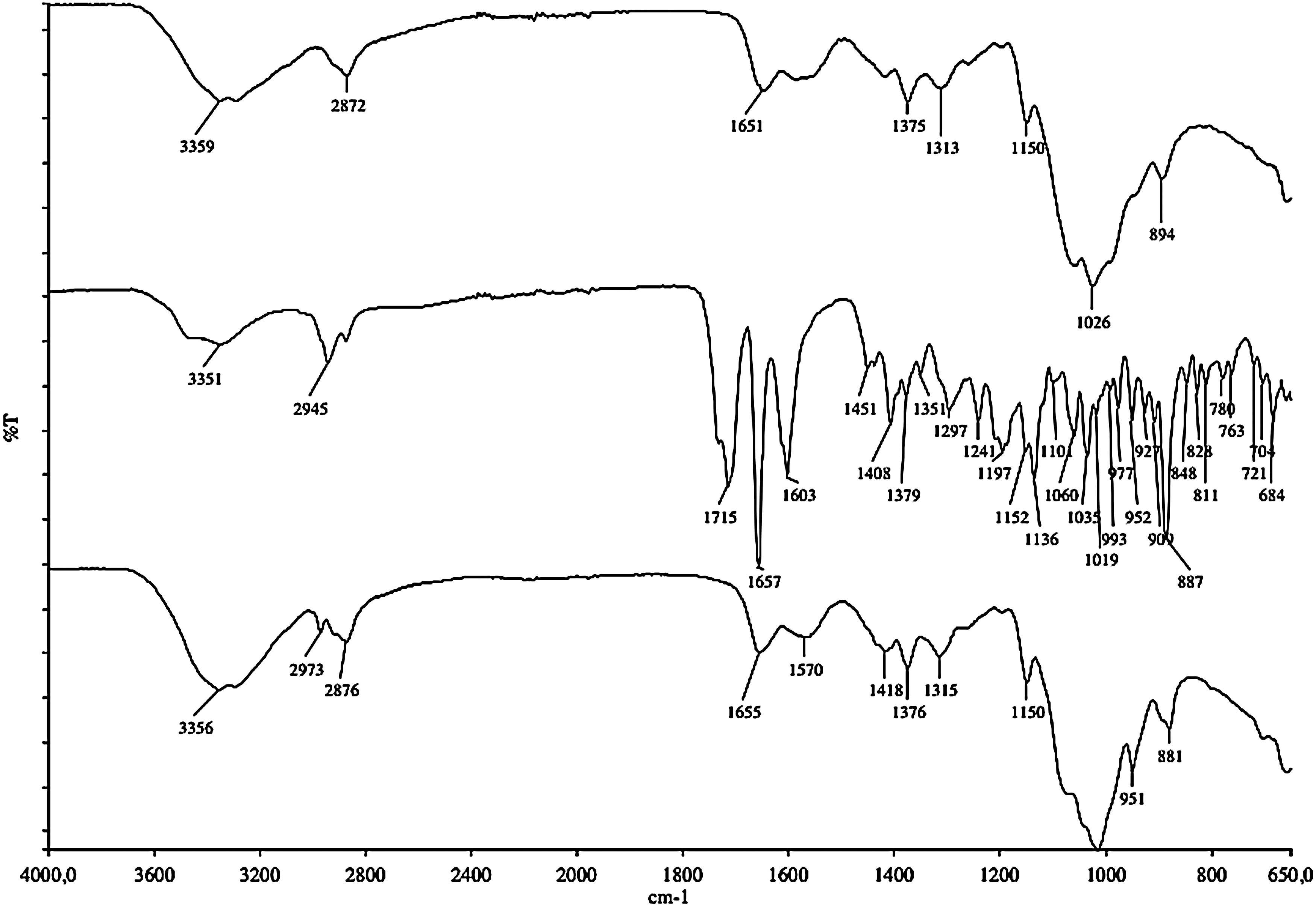
ATR-FTIR spectra. ATR-FTIR spectra of placebo chitosan films, TM, and TM-loaded chitosan films, respectively.
In vitro release and in vivo studies
The percentage of TM released from chitosan films was shown in detail in Fig. 3. In 2 weeks, 85% of the drug was released from the films, whose total in vitro content was released within 4 weeks. The possibility of eye irritation due to the film's administration and dislocation, or the swelling capacity of the mucoadhesion film on the bulbar or palpebral conjunctiva surface, was evaluated. Before administering the chitosan film, hydratation in saline is necessary to optimize the mucoadhesion process and avoid conjunctival lesions. Figure 4 shows a dry chitosan film before and after hydration. No symptoms of ocular lesions, such tearing, redness, edema, and inflammation, could be observed during the experimental assays (Fig. 5). Furthermore, fluorescein staining did not indicate corneal or conjunctival ulcerations. Ocular surface structures and intraocular tissues proved to be normal.
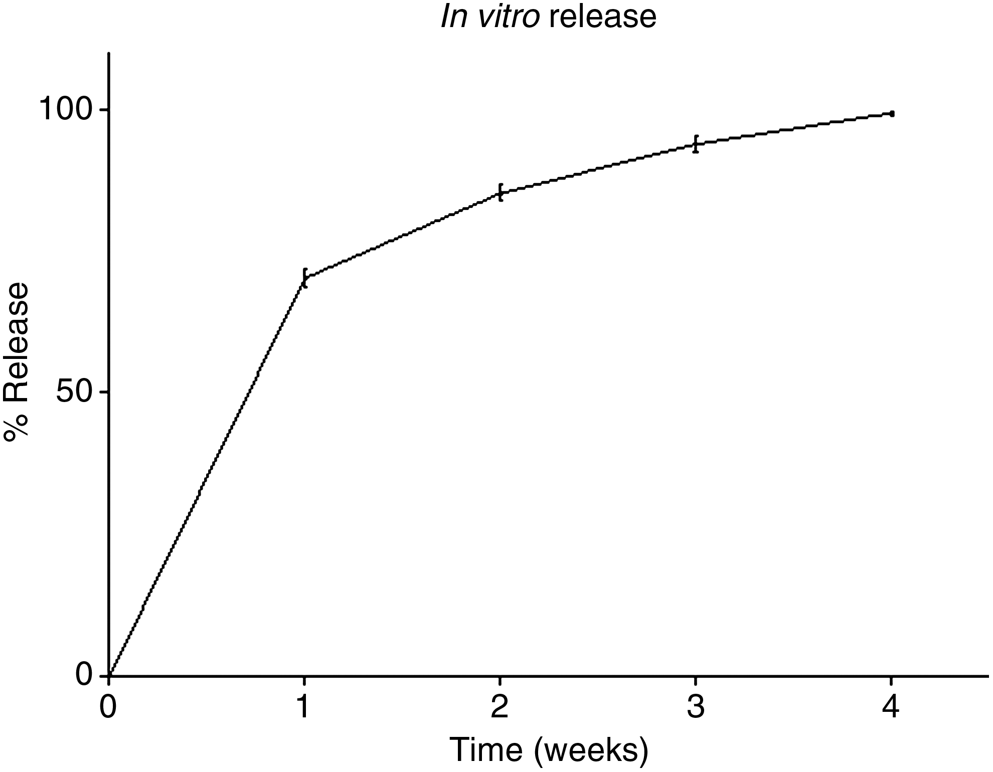
Cumulative TM-released profile, in vitro, from the developed chitosan films; 85% of the drug was released from the films over a 2-week, ending at 4 weeks. The values are shown as mean±standard deviation (n=6).
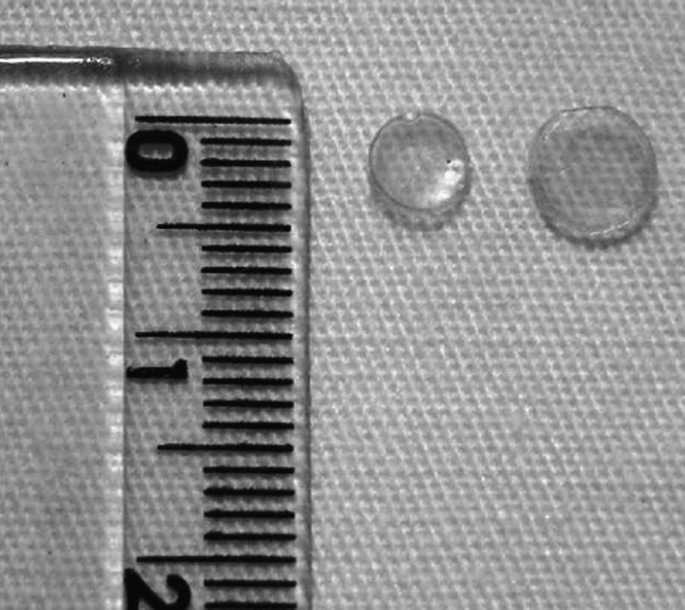
Photograph showing TM-loaded chitosan film used in this study. The films weighed 8.5 mg, were 130 μm thick, and were 4 and 6 mm in diameter before and after hydration, respectively. TM-loaded chitosan film already cut (left) and chitosan film after hydration in phosphate-buffered saline (right).
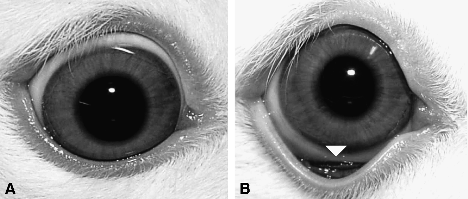
Rabbit eyes.
On the basis of microscopic evaluation, the structures that on direct contact with films, such as bulbar and palpebral conjunctiva from both the control (A) and treated eyes (B, Timoptol commercial collyrium; C, TM-load chitosan films) are shown in Fig. 6. The histological examination of the experimental eyes showed no structural changes. The bulbar and palpebral conjunctiva displayed normal cell layers and morphology from controls and treated eyes. These structures demonstrated no signs of toxicity that included vascular and cellular alterations or inflammatory cell infiltration.
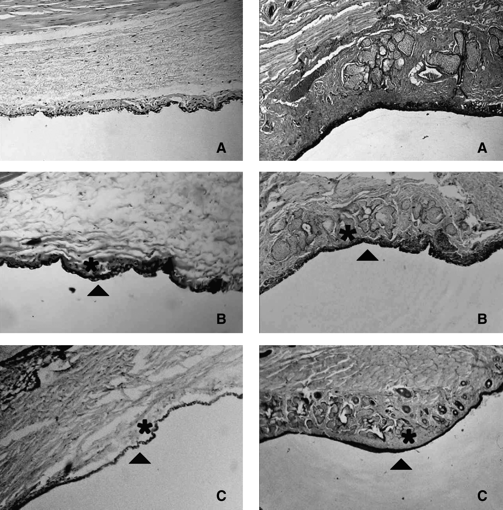
Histological sections of ocular surface structures of eyes from control group
Figure 7 shows the reduction of IOP in OD at the 5th to 12th measurements after the administration of collyrium and TM-loaded chitosan films. No statistical difference between the 2 treatments (P<0.05) could be observed. After the 12 measurements, the Timoptol commercial collyrium treatment was interrupted and the IOP levels of the rabbits returned to values that were similar to those of the control group. However, animals that received TM-loaded chitosan films maintained their IOP levels at lower than those of the control and treated groups during the 35th measurement over a 10-week period. The impact the IOP levels on OS proved to be a consequence of the treatment on OD. As expected, Fig. 8 demonstrated the reduction of IOP levels in OS from the 5th to 12th measurements in animals that had received Timoptol and TM-loaded chitosan films. No statistically significant difference could be observed when the 2 treatments were compared (P<0.05). After the 12th measurement, the Timoptol commercial collyrium treatment was interrupted and the IOP levels of the rabbits returned to values that were similar to those of the control group. However, animals that received the TM-loaded chitosan films maintained IOP levels of lower than the control and treated groups during the 35th measurement over a 10-week period.
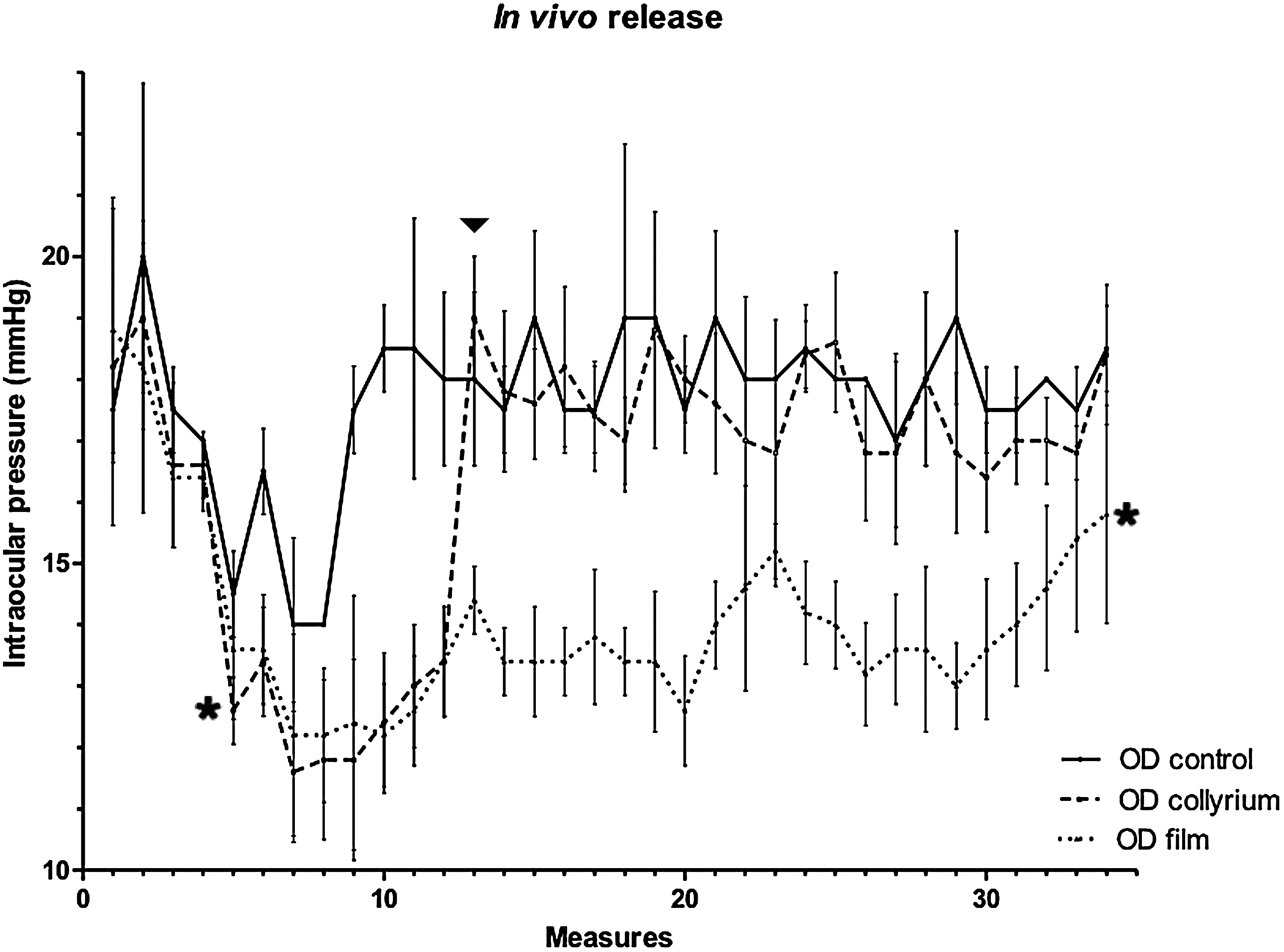
Decrease in IOP levels on the fifth measurement of right eye after beginning the treatment with Timoptol commercial collyrium and TM-loaded chitosan films (asterisk). Note that the IOP values of rabbits treated with Timoptol returns to normal levels (similar to those of the control group) after having interrupted the collyrium at the 12th measurement (arrowhead). TM-loaded chitosan films decreased the IOP levels over a 10-week period up to the 35th measurement (each measure lasting for 48 h) (asterisk). Representative values are defined as the means (±S.D.; n=5) of at least 3 measurements, and proved not to be significantly different when compared to collyrium and TM-loaded chitosan films (P<0.05). IOP, intraocular pressure.

Decrease in IOP levels on the fifth measurement of left eye after beginning the treatment with Timoptol commercial collyrium and TM-loaded chitosan films on the right eye (asterisk) (left eye received no treatment). Note that the IOP values of rabbits treated with Timoptol return to normal levels (similar to those of the control group) after having interrupted the collyrium at the 12th measurement (arrowhead). TM-loaded chitosan films decrease IOP levels over a 10-week period up to the 35th measuring (each measure for 48 h) (asterisk). Representative values are defined as the means (±S.D.; n=5) of at least 3 measurements, and proved not to be significantly different when compared to collyrium and TM-loaded chitosan films (P<0.05).
Discussion
Mucoadhesive films represent alternative methods aimed at improving conventional ocular therapy. The prolonged time released from the TM-loaded chitosan films in vitro observed in the present study are much higher than the 14 h recently reported by the same research group concerning chitosan dexamethasone-loaded films. 26 However, they do show a similar release profile, with ∼90% of the drug released in half the time necessary for a total release. These results, at least in part, were predicted, as both chitosan films underwent a similar manufacturing process. On the basis of the prolong release profile in vitro, those films raised expectations regarding the maximum time needed for an in vivo release.
The TM release was evaluated indirectly through the pharmacodynamic effects in normotensive eyes. Pharmacodynamics is a quantitative study of a functional relationship between the drug concentration and its effects. In antiglaucomatous drugs, the correlation between the effects and drug concentration in ocular tissues has been reported to optimize the ophthalmic formulation, in turn improving ocular drug delivery.28,29 The administration of timolol to one eye is associated with a decrease in IOP levels in both eyes.30,31 The reduction in contra-lateral eye is generally about one half of the IOP of a treated eye.30,32 The penetration of the drug into contralateral eye through the systemic route has been well documented in the literature in both animals and humans.31,33 Our results demonstrate that both implants and TM with 0.5% commercial collyrium are equally effective in reducing IOP levels in rabbits. However, the magnitude of IOP reduction proved not to be significantly different (P<0.05) in eyes treated with Timoptol compared to the TM-loaded chitosan implant during the 5th to 12th measurements.
Once the film had been administrated in the conjunctival sac after topical anesthesia, it did not move. This is important because, if the film dislocates, it can touch the cornea, causing epithelial damage, such as an ulceration, or can be lost completely, thus compromising the therapy. In addition to the safety, correct ocular therapy guarantees a prolonged delivery of the drug when the film remains in original administration site. After experimental assays, the ideal time at which to immerse the films in saline could be determined. Ten seconds of immersion the films in saline proved to be enough for water absorption. This is an important feature for its correct and safe implantation in ocular conjunctiva. Therefore, without immersion, films can cause lesion in ocular mucosa as a consequence of its capacity of absorption. This film is easily applied by pulling up the lower eyelid, exposing the lower conjunctival sac. In this study, no physical or pharmacologic contention (sedation or systemic anesthesia) it was used to perform this application.
The facility of administration on an ocular surface is a common feature of the hydrogels, nanoparticles, and TM-loaded chitosan films developed.15,16,20–22,25 However, preparation of nanoparticles is more complex and may require organic solvents in the manufacturing process. 16 This fact represents a disadvantage when compared to the simple technique of production of the TM-loaded chitosan presented in this study. The methodology for the production of chitosan film is similar to those that successfully carry dexamethasone, a finding recently reported by Rodrigues et al. 26 The authors described that the mono- and bilayer dexamethasone-chitosan films released ∼85% of the drug in vitro over 8 h and 4 weeks, respectively. The incorporation of a second layer of chitosan film significantly increases and prolongs the drug release profile. 26 Therefore, there is a real potential to develop bilayer TM-loaded chitosan films to increase the 10 weeks of IOP reduction in vivo. Future studies are warranted to develop effective bilayer timolol-loaded chitosan films as well as to maintain suitable, comfortable, and nontoxic characteristics. Bioadhesion is the state in which 2 materials, at least one of which is biological in nature, are held together for extended periods by interfacial forces. Mucoadhesion occurs when the attachment adheres to the mucus and/or the mucous membrane. 34 This adhesion has been of interest in the pharmaceutical sciences as a means through which to enhance localized drug delivery in locations such as the ocular surface. 35 The mucoadhesion properties of chitosan are determined by the formation of either secondary chemical bonds or ionic interactions between the positively charged amino groups of chitosan and the negatively charged sialic acid residues of mucus. 17
Prolonged used of TM may produce morphologic changes to the conjunctiva of rabbits, including an increase in the density of the collagen fibers. 36 These alterations were not observed in the present study, which is most likely associated with a short treatment period (2 and 10 weeks for collyrium and chitosan films, respectively). The absence of histologic alterations and abnormal inflammatory cells in the cornea, conjunctiva, and eyelids was consistent with the lack of clinical signs in the exposure to chitosan film. As the animals tend to lick or scratch their eyes and periocular tissues, even if only a minimal irritation exists, whether caused by foreign body or other ocular diseases, the use of an eye protector, such as Elizabethan collars, becomes necessary. Rabbits in this study did not use any form of protection against automutilation, as they did not demonstrate any behavioral signs of discomfort.
The beta-blockers work to reduce IOP levels so as to primarily have an effect on the β-adrenergic receptors located on the ciliary processes and ciliary body of the eye, thereby decreasing aqueous humor production and IOP levels. 37 One serious disadvantage of ophthalmic timolol therapy is the systemic beta-blockade produced by drug absorption from the eye into the systemic circulation. 38 Many reports of serious respiratory and cardiovascular events, and even death, have been attributed to the use of ophthalmic timolol in human beings.12–14 However, these events are rarely described in rabbits that undergo ocular treatment with TM. 39 Although an electrocardiogram was not performed, no adverse effects in rabbits could be observed in the present study. These systemic adverse effects result partly from the nonselective inhibition of β-adrenergic receptors.40,41
Although the major mechanism of action for timolol occurs in the anterior eye, it has been established in humans and animals that this drug more commonly affects the posterior segment, as well as retinal and optic nerve head circulation.42,43 Grunwald reported that timolol increases retinal blood flow in normotensive and hypertensive eyes.42,44 However, other authors have demonstrated a reduction in retinal and optic disc blood flow.43,45 Although an increased blood flow is important in the management of glaucoma, as it tends to represent potential therapeutic benefits, it has yet to be clearly demonstrated how timolol influences the posterior segment blood flow. In the present study, no alterations could be observed in the blood vessels in the choroid, retina, and optic disc in rabbit eyes treated with Timoptol, the chitosan implant, and the control group by direct and indirect opthalmoscopy and slit-lamp biomicroscopy. In the histologic evaluation, no difference could be identified in the vessel calibers of these groups. Further studies, by means of specific methodologies, such as laser Doppler flowmetry, are warranted to evaluate the effects of TM chitosan implants released in posterior segment blood flow in rabbit eyes.
Although rabbit and human ocular surfaces are not equal, a good correlation between rabbit and human eye irritation data does in fact exist. 46 On the basis of the effectiveness, safety, and absence of toxicity, this film appears to promising for future tests in humans. However, further studies are warranted to determine whether chitosan films can deliver drugs for the efficacious treatment of glaucoma in animals and humans beings.
Conclusion
Regarding ocular tolerance, efficacy, and safety, the present study's results demonstrated that mucoadhesive TM-loaded chitosan films are promising candidates for ocular drug delivery, as they present no signs of toxicity, are well tolerated in vivo, and have proven to be efficient in lowering IOP levels in normotensive eyes in a rabbit model.
Footnotes
Acknowledgments
The authors would like to thank the many collaborators from the School of Veterinary Sciences and the School of Pharmacy from the UFMG.
Author Disclosure Statement
No competing financial interests exist.