
Abstract
Abstract
Purpose:
To evaluate the safety of intracameral and subconjunctival injection of a novel mucoadhesive polysaccharide isolated from Bletilla striata in rabbit eye.
Methods:
One hundred microliters (100 μL) of Bletilla striata polysaccharide (BsP) at concentrations of 10, 40, and 80 mg/mL was intracamerally or subconjunctivally injected into rabbit eyes. Phosphate-buffered saline and 10 mg/mL hyaluronic acid solution were also injected as controls. BsP safety was evaluated via clinical follow-up and histological analysis. The dead corneal endothelial cells were observed by vital staining with alizarin red and trypan blue at 14 days after intracameral injection. Finally, in the intracameral injection study, scanning electron microscopy was performed for evaluation of the structure of the corneal endothelium and anterior lens capsules.
Results:
Subconjunctival injection of 10 mg/mL BsP does not cause pathological changes or an inflammatory response. Concentration greater than 10 mg/mL of BsP (40 or 80 mg/mL) leaded to a slight inflammatory response, but the rabbits recovered well in 3 days. The pathological observation further confirmed the safety of subconjunctival injection of BsP, and subconjunctival injection of 80 mg/mL BsP caused no lesion of the ocular tissues. Intracameral injection of 80 mg/mL of BsP did not cause a significant inflammatory reaction, and an even lower inflammatory response was observed in rabbits intracamerally injected with 40 or 10 mg/mL BsP. All rabbits intracamerally injected with BsP recovered within 7–14 days. BsP had little effect on the blood-aqueous barrier's integrity when the concentration was 10 mg/mL; at 40 or 80 mg/mL, a mild effect was observed, and the rabbits recovered in 1–3 days. Intracameral BsP injected at a concentration of 80 mg/mL had a negative impact on the corneal endothelium and lens, but concentrations of 40 or 10 mg/mL could be injected safely.
Conclusions:
BsP injection into the subconjunctival space and anterior chamber in rabbits at low concentrations (such as 10 mg/mL) did not have adverse effects.
Introduction
Bletilla striata has been used medicinally for over 1500 years as a traditional Chinese medicine. 1 Its root shows antibacterial, anti-inflammatory, and demulcent properties.1,5 There is no record of adverse effects caused by Bletilla striata during its long history of use as a medicinal herb. Today BsP is widely used in clinics and is administered through routes including external and internal application and arterial embolization, and there have been no reports of adverse reactions or toxicity. 6 Although the safety of BsP has not been explicitly tested in the past, our previous cytotoxicity and ocular irritation tests demonstrated that BsP is safe to use as a method of ophthalmic topical drug delivery.
Similarly to other polysaccharides, such as chitosan,7,8 hyaluronic acid (HA),9,10 and larch arabinogalactan,11,12 BsP may have a wide range of uses in ophthalmology, but its safety, especially for intraocular use, remains to be evaluated. The purpose of this study was to investigate the safety of BsP after conjunctival and intracameral injection and to provide basic safety data to guide the further use of BsP in ophthalmology.
Materials and Methods
Drugs and animals
Ketamine hydrochloride was obtained from Jiangsu Hengrui Medicine Co., Ltd. (Lianyungang). Chlorpromazine was purchased from Shanghai Harvest Pharmaceutical Co., Ltd. HA injections at a concentration of 10 mg/mL were obtained from LG Life Science. Proparacaine ophthalmic solution and balanced salt solution (BSS) were obtained from the Alcon Pharmaceuticals Company. Fluorescein solution (25%) was purchased from Guangzhou Baiyunshan Mingxing Pharmaceutical Co., Ltd., and diluted to 0.5% with BSS before its use.
Male and female New Zealand albino rabbits were obtained from Qingdao Kangda Foodstuffs Co., Ltd. [License No:SCXK (Lu) 20070023]. Animal care and procedures were conducted according to the Principles of Laboratory Animal Care. The experimental animals weighed between 2 and 2.5 kg and were housed individually in an air-conditioned and light-controlled room at 25°C±2°C and at 70%±5% relative humidity. They were given standard pellet feed and were provided with water ad libitum. All animals were healthy and free of clinically observable ocular abnormalities. The animal study was approved by Shandong Eye Institute Ethics Committee for Animal Experimentation, Qingdao, Shandong, China.
Preparation of BsP and BsP solution
BsP was purchased from the Nanjing Institute for Comprehensive Utilization of Wild Plants. The compound had the appearance of small white fibers after freeze drying. It was soluble in water but not in organic solvent. Neither proteins nor nucleic acids were detected in BsP. The BsP contained 98.6% (w/w) total sugar and no uronic acids, and it had a molecular weight of 99,658 Da as indicated by gel filtration chromatography. Glucose and mannose were the only two monosaccharides in BsP, and the molar ratio of mannose and glucose was found to be 1.6:1. 4 BsP was dissolved in 0.02 M phosphate-buffered saline (PBS) at pH 7.4 to form isotonic 80, 40, and 10 mg/mL solutions, which were subsequently sterilized by flowing steam at 121°C for 20 min.
Subconjunctival injection
A total of 40 rabbits were randomly distributed into five groups with 8 animals in each group, and the left eye was designated as the experimental eye. The groups received injections of PBS, 10 mg/mL HA solution, and BsP solutions containing 80, 40, and 10 mg/mL BsP. Anesthesia was induced with an intramuscular injection of 25 mg/kg ketamine hydrochloride and 25 mg/kg chlorpromazine. Topical anesthesia was achieved with administration of one drop of proparacaine ophthalmic solution to the eye. Subconjunctival injection of 100 μL of the solutions into rabbit eyes was performed with a 29-gauge needle under local anesthesia. A clinical examination was performed by slit-lamp on days 1, 3, 7, and 14 after injection. The hyperemia of the conjunctiva and episclera as well as the chemosis and lachrymation induced by the different injected solutions were graded from 0 (absence) to 3 (highest) according to the clinical evaluation scale described in Table 1. 13 Scores of 2 or 3 in any category were considered positive: total score=A+B+C; maximum=9. Two rabbits from each group were sacrificed for histology on days 1, 7, and 14 after injection. The tissues were fixed in 10% formol and embedded in paraffin. Hematoxylin-eosin (HE) or periodic acid-Schiff (PAS) staining was performed.
Intracameral injections
The groups of animals were assigned the same as for the “Subconjunctival Injections” with 10 rabbits each group. After anesthesia, one hundred microliters (100 μL) of aqueous humor was removed from each eye using a 29-gauge needle, and then 100 μL of experimental solution was subsequently injected.
Rabbits were examined at 1, 3, 7, and 14 days after injection. A slit-lamp experiment was performed to examine the interaction between the injected solution and the tissue in the anterior chamber. The ocular grading method used for biomicroscopic examinations is shown in Table 2. The corneal epithelium was examined under blue light after instillation of fluorescein solution to detect any epithelial damage. At each study point, the intraocular pressure (IOP) was measured using an Icare VET tonometer (TonoVet) and compared with the IOP measured in the noninjected contralateral eye. To standardize the measurements, all IOP procedures were performed at 2:00 P.M. 14 During clinical assessment, nine parameters were recorded from rabbit eyes and numerically graded on an increasing severity scale of 0–4.15,16 The means of the ocular scores at each follow-up time point were quantitatively calculated to be the sum of the scores for each group divided by the total number of eyes in that group.
At days 1, 7, and 14, two rabbits chosen randomly from each group were euthanized, and the injected eyes were harvested. After fixation in 10% formaldehyde solution for at least 24 h and dehydration with an alcohol gradient, the eyeballs were prepared for HE and PAS staining using routine methods and analyzed with light microscope to examine the conjunctiva, the cornea, the limbus, the chamber angles, and the sclera. The presence of neutrophils, lymphocytes, macrophages, fibroblasts, and giant cells was regarded as evidence of a tissue response. Every sample was treated simultaneously to reduce variations related to the fixation procedure.
At day 14, the remaining four rabbits were euthanized, and the eyes were enucleated, and then cornea and lens of each eye were separated. Two corneas were placed with the endothelial side up, and 0.25% trypan blue was applied dropwise to stain the dead cells. After 90 s, the stain was poured off, and the corneas were briefly rinsed twice in normal saline and replaced in the corneal cup. Then 0.20% alizarin red S was applied to cover the endothelium for 90 s. 17 After the staining procedure, the corneas were immersed in glutaraldehyde fixative solution for 10 min. They were then evaluated for endothelial damage using light microscope.
The remaining two corneas and the crystalline lens of each eye were fixed in 2.5% glutaraldehyde in 0.1 M sodium cacodylate buffer and prepared for scanning electron microscopy (SEM). The corneas were bisected and postfixed in 2% osmium tetroxide for 2 h. For SEM, samples were prepared using critical point drying. The tissue was glued to a stub, sputter coated with gold palladium, and viewed with a JSM-840 scanning electron microscope (JEOL).
Statistical analysis
Statistical analysis for the determination of differences in the measured properties between groups was accomplished using one-way analysis of variance. A P value<0.05 was considered significant.
Results
Subconjunctival injections
Direct observation of the rabbit eyes after the subconjunctival injection was performed. After the subconjunctival injection of 100 μL of BsP solution, there was a bubble over the conjunctiva (Fig. 1), but the bubble disappeared from the injection site of each sample within 1 day. The mild inflammation was related to the BsP concentration injected. The results of biomicroscopic evaluations are presented in Table 3. From the table it can be seen that during the first day after injection, BsP triggered slight hyperemia and chemosis, graded 1.25±0.71 and 1.00±0.35 in rabbits injected with 80 and 40 mg/mL BsP, respectively. However, no lachrymation and no discharge were observed, and the adjacent cornea was perfectly clear, indicating that there was a mild inflammatory reaction. This mild inflammatory reaction significantly decreased by day 3. One to 3 days after injection, the inflammation caused by subconjunctival injection of 80 and 40 mg/mL BsP was more severe than injection of 10 mg/mL HA or BsP (P<0.05), and the inflammatory response to subconjunctival injection of 10 mg/mL BsP was similar to that induced by injection of 10 mg/mL HA (P>0.05). By day 7, all of the eyes that had a mild inflammatory reaction recovered well, and there was no significant difference (P=0.144), compared with the control eyes. Figure 1 illustrates the presence of 80 mg/mL BsP solution after subconjunctival injection. Slight vasodilatation of conjunctival vessels over the injection site can be seen on day 3, but there is no obvious inflammatory reaction.

Representative time-course slit-lamp biomicroscopic images of rabbit eyes after subconjunctival injection of 80 mg/mL BsP. During the 14-day period of observation, no lachrymation and no discharge were observed, and the adjacent cornea was perfectly clear. BsP, Bletilla striata polysaccharide.
P<0.05 versus to PBS.
P<0.05 versus to HA.
BsP, Bletilla striata polysaccharide; PBS, phosphate-buffered saline; HA, hyaluronic acid.
Histopathologic results confirmed the biocompatibility of BsP. A transient inflammatory reaction was observed in the subconjunctival area with a few neutrophils and lymphocytes around the conjunctival cell at day 1. This inflammatory reaction disappeared within 7 days after the subconjunctival injection, and no inflammatory cells could be found at day 7. During the 14-day period of observation, no histological evidence of macrophages, fibroblasts, giant cells, or tissue exudates appeared after the subconjunctival injection, which was similar to our observation after injection with PBS. Moreover, the histological analysis of all sections of the corneas showed that there was a normal organization of the architecture after the injection of BsP throughout the experiment. Conjunctival goblet cells were abundant in the treated eyes, and they were filled with secretory product as determined by PAS staining (see in Fig. 2).

Representative time-course of histopathological images of rabbit eyes after subconjunctival injection of 80 mg/mL BsP. During the 14-day period of observation, no histological evidence of macrophages, fibroblasts, giant cells, or tissue exudates appeared after the subconjunctival injection.
Intracameral injections
On clinical observation, the injected BsP appeared as a transparent gel in the anterior chamber immediately after injection, but the BsP could not be easily identified under a slit-lamp because it dissolved into the aqueous humor within several minutes after injection. The results of biomicroscopic evaluations are presented in Table 4, and the detail descriptions were following.
P<0.05 versus to PBS.
P<0.05 versus to HA.
n=8 as in the two eyes; inflammatory reactions of the iris could not be observed because of exudative fiber in the anterior chamber.
Cornea
There was no corneal edema observed throughout the experiment period in any of the groups, and the corneas became increasingly clear throughout the experiment period in all groups. As for the keratic precipitate (KP), there was only one eye in 80 mg/mL BsP group scored 1 on day 1, but it recovered well and no KP was found in any eyes.
Anterior chamber
For our observation of aqueous flare in the anterior chamber, there were four eyes in the group injected with 80 mg/mL BsP that scored 1 and one eye in the group injected with 40 mg/mL BsP that scored 2 at day 1, but all of these eyes recovered and received a score of 0 in the following days of observation. With respect to aqueous cells in the anterior chamber, there were three eyes in the group injected with 80 mg/mL BsP that scored 1, and one eye in the group injected with 40 mg/mL BsP that scored 1 at day 1, but all of these eyes recovered and received a score of 0 in the following days of observation. The other three groups showed no aqueous flare or cells throughout the experiment (see in Fig. 3).
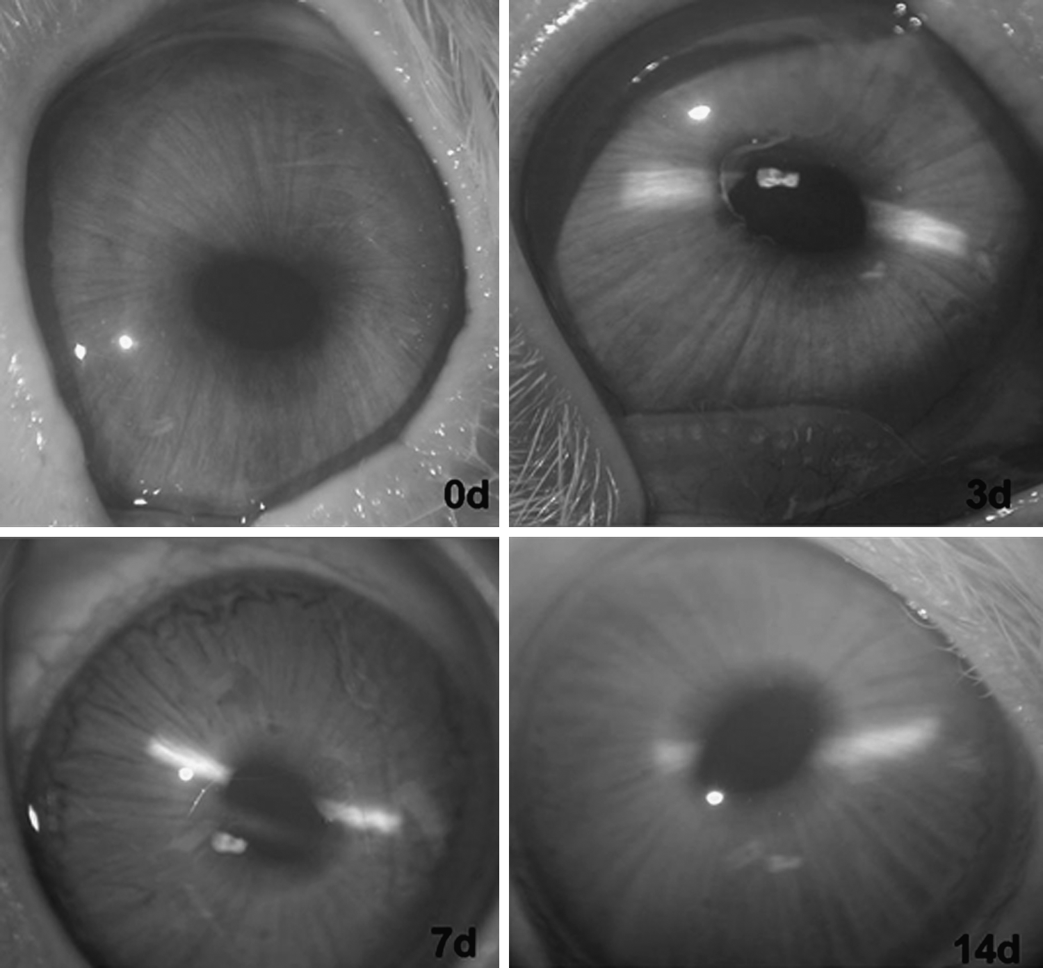
Representative time-course slit-lamp biomicroscopic images of rabbit eyes in the group that received intracameral injection of 80 mg/mL BsP. Fibrin exudates could be seen around the pupil at day 3 after injection, but all of the signs recovered well at day 14 after injection.
Fibrin exudate could be observed in the anterior chamber in eyes injected with 80, 40, and 10 mg/mL BsP at day 1 after intracameral injection. There was one eye that scored 3, two eyes that scored 2, and one eye that scored 1 in the 80 mg/mL BsP group. The mean scores of fibrin exudate of 80 mg/mL were significantly higher than that of PBS and HA groups (both P<0.05) at day 1 after injection. There was much less fibrin exudate in eyes injected with 40 and 10 mg/mL BsP. Among the eyes injected with 40 mg/mL BsP, only one eye scored 2, and four eyes scored 1 (both P<0.05, when compared with PBS and HA groups), and among the eyes injected with 10 mg/mL, only one eye scored 1 at day 1 after injection. After 3 days, the fibrin exudate became slighter in all of the eyes. Among the eyes injected with 80 mg/mL BsP, one eye scored 3, two eyes scored 2, and one eye scored 1, and four eyes scored 1 at day 3 after injection. In the group that was injected with 10 mg/mL BsP, there was no fibrin exudate at day 3 after injection. The fibrin exudate became much slighter in all eyes at day 7, though fibers were still observed in some eyes in the rabbits injected with 80 mg/mL BsP, there was no significant difference when compared with 10 mg/mL HA and PBS (P≥0.05); at this time point, only two eyes scored 2 in the 80 and 40 mg/mL BsP groups, respectively. At the end of the observation (day 14), all of the fibrin exudate had disappeared. The groups injected with 10 mg/mL HA and PBS had no fibrin exudate.
Iritis was observed at day 1 in the groups injected with 80, 40, and 10 mg/mL BsP. In the group that received 80 mg/mL BsP, four eyes scored 1, and two eyes could not be observed due to the fibrin exudate in the anterior chamber at day 3, but only one eye scored 1 at day 7. In the group injected with 40 mg/mL BsP, four eyes scored 1 at day 1, one eye scored 1 at day 3, and two eyes scored 2 at day 7. At day 14, none of the eyes in the groups injected with 80 and 40 mg/mL BsP had iritis. For the group injected with 10 mg/ml BsP, only three eyes scored 1 on day one, and one eye was observed with iritis at the later time points. The groups injected with 10 mg/ml HA and PBS had no iritis.
Lens
There were four eyes with tiny white deposits on anterior lens capsule in the group injected with 80 mg/mL BsP and one eye in the group injected with 40 mg/mL BsP, but no deposits were observed in the eyes of rabbits in the other groups. At day 3, there was one eye in the group injected with 80 mg/mL BsP and two eyes in the group injected with 40 mg/mL BsP with tiny white deposits on the anterior lens capsule, but by day 7, none of the eyes in these two groups had tiny white deposits. In the group injected with 10 mg/mL BsP as well as the groups injected with 10 mg/mL HA and PBS, no tiny white deposits were observed. At the end of experiment observation, no phacoscotasmus was observed in any eyes from any of the groups.
Intraocular pressure
The effect of BsP injected at different concentrations on the IOP in rabbit is depicted in Fig. 4. Mean IOP values ranged from 9 to 15 mmHg in all of the experimental rabbits before injection, and no significant difference (P>0.05) was observed between any of the groups. There was a decline in the IOP at days 1–3 after injection of 80, 40, and 10 mg/mL BsP and 10 mg/mL HA. Then, the IOP change reverted to about 8–14 mmHg in all of the rabbits at 7 and 14 days, while the PBS groups maintained a relatively stable IOP during the experiment.
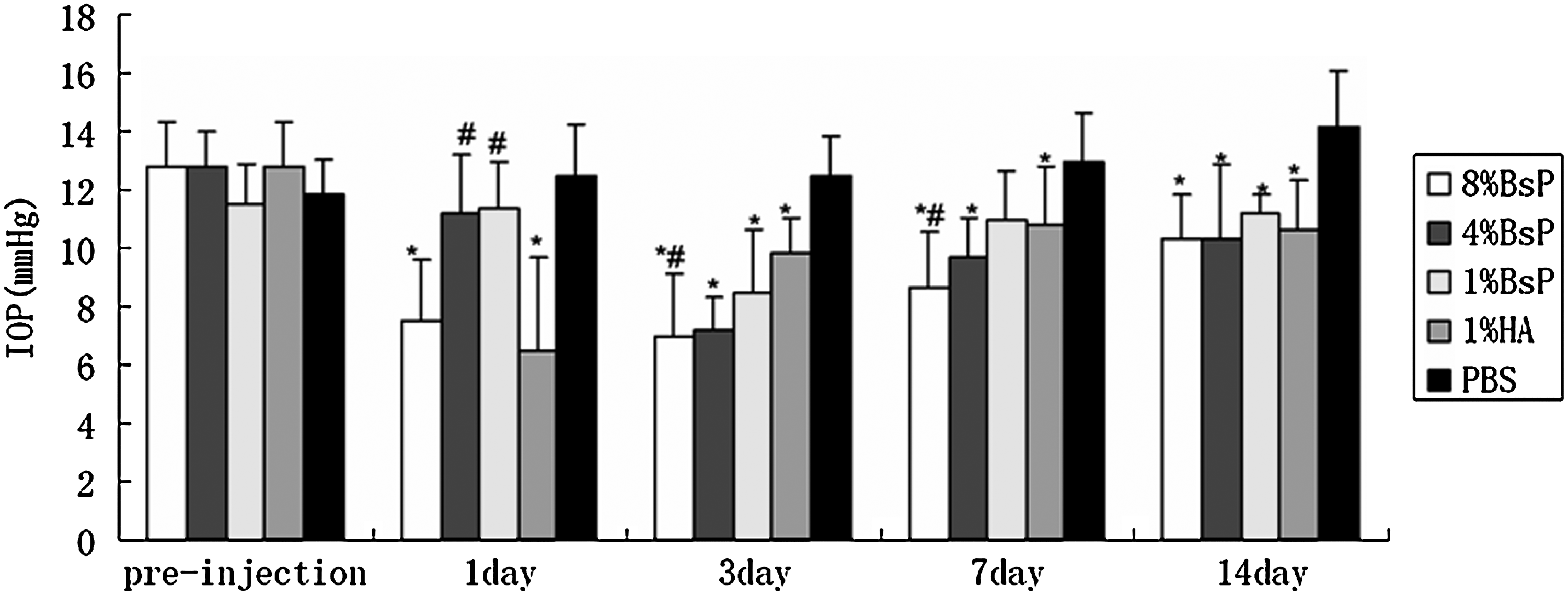
IOP before and after intracameral injection of BsP. (*P<0.05 versus to PBS, #P<0.05 versus to 1% HA). There was a decline in the IOP at days 1–3 after injection of 80, 40, and 10 mg/mL BsP and 10 mg/mL HA groups, but the IOP change reverted to about 8–14 mmHg in all of the rabbits at 7 and 14 days. IOP, intraocular pressure; PBS, phosphate-buffered saline; HA, hyaluronic acid.
Histological analysis
The histological analysis of the eyes is presented in Fig. 5, and the integrity of the cornea can be observed. Neither cell infiltration to cornea nor alteration of the endothelial cell layer was found. For intracameral injection, there was no sign of tissue necrosis. All eyes in all groups had a clean vitreous cavity, explicit retinal structure, and a compact sclera. The structure of the chamber angle was normal, but fibrin was observed and inflammatory cell infiltration in the chamber angle was detected. The structure of the iris was also normal but with vasodilation, and fibrin could be found around the iris. At day 7, the inflammatory cell infiltration in the chamber angle and vasodilation in iris were significantly reduced, and the fibrin had completely disappeared in the above tissues. At day 14, the inflammatory cell infiltration in the chamber angle and vasodilation in the iris had disappeared, indicating that the eye had recovered completely. All eyes in the groups injected with 40 or 10 mg/mL BsP and 10 mg/mL HA or PBS were normal throughout the study.

Representative time-course histopathologic images of rabbit eyes in the 80 mg/mL BsP groups after intracameral injection (×100). At day 1, there was no sign of tissue necrosis; the structure of the vitreous cavity, the retinal, and the sclera were normal; the structure of the chamber angle was normal with fibrin and inflammatory cell infiltration in it; and the structure of the iris was normal but with vasodilation, and fibrin could be found around the iris. At day 7, the inflammatory cell infiltration in the chamber angle and vasodilation in iris were significantly reduced, and the fibrin had completely disappeared in the above tissues. At day 14, the inflammatory cell infiltration in the chamber angle and vasodilation in the iris disappeared.
The endothelial cells of cornea were observed by light microscope (see Fig. 6) and electron microscope (see in Fig. 7) at the end of the observation period. The results from light and electron microscopy showed that all eyes in the 40 and 10 mg/mL BsP, 10 mg/mL HA, and PBS groups had well-preserved hexagonal endothelial cell mosaics and normal microvilli on their endothelial cell surfaces, and no edema or necrosis cells were found (see Fig. 6B–E). The 80 mg/mL BsP group exhibited increased nonhexagonal endothelial cells by electron microscopy (see Fig. 7A).
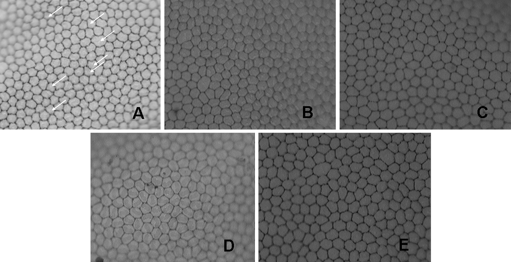
Light microscopic photographs of rabbit cornea endothelial cells stained with trypan blue and alizarin red at day 14 after intracameral injection (

Scanning electron micrographs of rabbit cornea endothelial cells at day 14 after intracameral injection (
The anterior lens capsule was observed with electron microscope under low magnification (×3500) and high magnification (×10000) as presented in Fig. 8. In the group that was injected with 80 mg/mL BsP, the morphology of the anterior lens capsule was normal under low magnification, whereas it was slightly rough under high magnification. The anterior lens capsule showed slight shrinkage, appearing as the fiber under the anterior capsule, but no further damage or cell deposition was found on the lens capsule surface. There was significantly less shrinkage of the anterior lens capsule in the group treated with 40 mg/mL BsP, and the structure was normal in rabbits injected with 10 mg/mL BsP and 10 mg/mL HA or PBS even under high magnification.
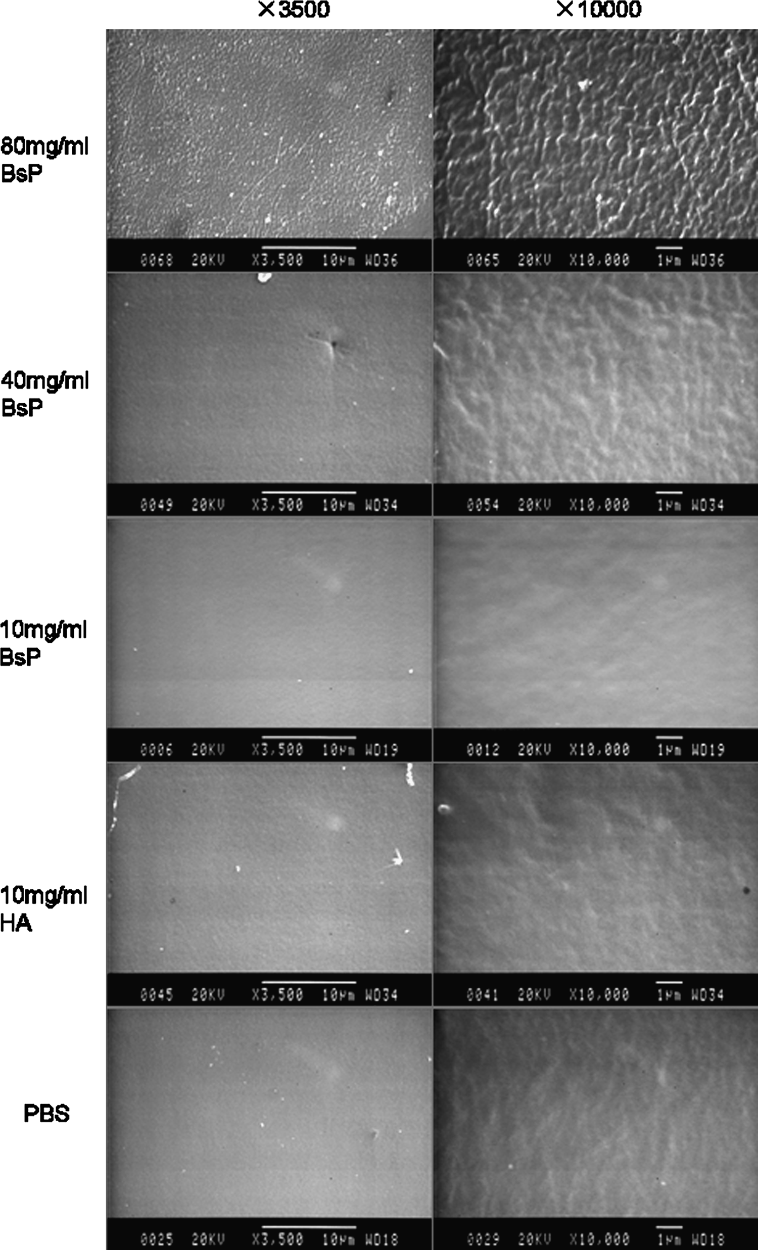
Scanning electron micrographs of rabbit anterior lens capsules at day 14 after intracameral injection. The anterior lens capsule was normal under 3,500 magnifications. To 80 mg/mL BsP group, the anterior lens capsule showed slight shrinkage, appearing as the fiber under the anterior capsule, but no further damage or cell deposition was found on the lens capsule surface under 10,000 magnifications. There was significantly less shrinkage of the anterior lens capsule in the group treated with 40 mg/mL BsP, and the structure was normal in rabbits injected with 10 mg/mL BsP, 10 mg/mL HA, or PBS under 10,000 magnifications.
Discussion
BsP is not an inert material, and its use has been widely investigated in recent years. In a previous study, it was found that BsP can significantly induce the proliferation of HCECs, suggesting that BsP may reduce the toxicity of some drugs toward the cornea. 4 BsP has good biocompatibility with lacrimal fluid, and it enhances intraocular absorption and corneal drug accumulation. All of these characteristics suggest that BsP may be useful for ophthalmic drug delivery through routes such as topical application and subconjunctival or intraocular injection.
A viscoelastic agent is an essential auxiliary drug that is injected into the eye tissue compartment during ophthalmic microsurgery. These agents can provide lubrication, serve as a barrier, or promote infiltration in order to improve surgical safety, increase the success rate, and reduce complications.18,19 BsP has the characteristics of a viscoelastic agent, and it is a sterile, transparent, uniform gel liquid that causes no stimulation, contains no pyrogen, and does not cause allergic reaction. Therefore, BsP may have a wide range of uses in ophthalmology, but the safety of its use must be investigated. As subconjunctival and intracameral injections are widely used in ophthalmic drug delivery systems and ophthalmic microsurgery, these two routes of application were investigated in this study.
After subconjunctival injection of BsP, signs of inflammation were observed, including conjunctival hyperemia and edema in the injection area. Higher concentrations of BsP resulted in slightly more severe inflammation. However, all of the inflammation observed was mild, and 3 days after injection, all of the eyes had recovered well. According to the classification criteria, 13 at day 1 after conjunctival injection, 80 mg/mL BsP caused moderate irritation, 40 mg/mL BsP caused mild irritation, and 10 mg/mL BsP or HA did not cause irritation. Additionally, the irritation was resolved in all of the groups at later time points. Therefore, we conclude that subconjunctival injection of 10 mg/mL BsP does not cause pathological changes or an inflammatory response; higher concentrations of BsP (such as 40 or 80 mg/mL) can lead to a slight inflammatory response, although all animals recovered well in 3 days. The pathological observation further confirmed the safety of subconjunctival injection of BsP; subconjunctival injection of 80 mg/mL BsP caused no lesion in the ocular tissues.
For intracameral injection, the presence of cells in the aqueous humor is a reliable indicator of inflammation, and the number of cells present reflects the severity of inflammation. 20 In this study, rabbits that received intracameral injection of 80 or 40 mg/mL BsP showed a weak inflammatory response (grade 1 aqueous cells) 1 day after injection. No aqueous cells were observed at later time points, suggesting that the animals recovered well. Therefore, the results suggest that intracameral injection of BsP does not cause a significant inflammatory reaction.
Aqueous flare is caused by increases of protein concentration in the anterior chamber. 21 Some factors, such as inflammation, trauma, and IOP, can damage the blood-aqueous barrier, allowing proteins in the plasma to penetrate into the anterior chamber, which causes aqueous flare. 22 Therefore, the presence of aqueous flare indicates that the integrity of the blood-aqueous barrier has been compromised. The results of aqueous flare observation suggest that BsP had little effect on the integrity of the blood-aqueous barrier at a concentration of 10 mg/mL. When the concentration was increased to 40 or 80 mg/mL, a mild effect was observed, but the rabbits recovered well in 1–3 days.
Exudative fiber observed in the anterior chamber was one of the major inflammatory responses after intracameral injection of BsP, and the severity of the fibrous exudate was proportional with the BsP concentration. In the group injected with 80 mg/mL BsP, on the first day after injection, exudative fibers observed were more severe than the reaction observed in rabbits injected with PBS or HA (P<0.05). The exudative fibers in the group injected with 40 mg/mL BsP were mild, but the effect was more severe 1 day after injection than it was in the groups injected with 10 mg/mL HA and PBS (P<0.05) on the first day after injection. After 1 day, no significant difference was found between rabbits injected with 40 and 10 mg/mL HA or PBS group (P≥0.05). The group injected with 10 mg/mL BsP was similar to the groups injected with 10 mg/mL HA or PBS (P≥0.05).
For intracameral injection, vascular engorgement of the iris was a major inflammatory response, and the severity of vascular engorgement was proportional with the concentration of BsP. From day 1 to day 3 after injection, there was more severe vascular engorgement in the group injected with 80 mg/mL BsP than the group injected with PBS (P<0.05), but at day 7 after injection, the vascular engorgement of the iris was significantly reduced (P≥0.05, when compared with the PBS group). In rabbits injected with 40 and 10 mg/mL BsP, a lower inflammatory response was observed, and the animals recovered in 7–14 days after injection.
It has been reported that cellular hexagonality is a sensitive indicator of endothelial damage or instability and may be valuable in qualitative observations of cell appearance.15,23 In this study, after intracameral injection of 80 mg/mL BsP, the number of nonhexagonal endothelial cells increased when compared with the group injected with PBS, but there was no difference observed between the groups injected with 40 and 10 mg/mL BsP, 10 mg/mL HA, and PBS. Therefore, 80 mg/mL BsP has a negative impact on the corneal endothelium, but when the concentration decreases to 40 or 10 mg/mL, there was no effect. We conclude that a BsP concentration of 40 mg/mL or lower is safe for intracameral injection with respect to the corneal endothelium.
Long-term use or exposure of the lens or lens capsule to toxic substances would cause impairment due to increasing permeability and loss of the barrier function, finally leading to lens opacity or the formation of drug-induced cataracts.24,25 The safety of the lens is essential for drug administration by intracameral injection. In this study, the impact of BsP on the lens was examined by slit-lamp observation and SEM. The results of slit-lamp observation showed that even in the group injected with 80 mg/mL BsP, no lens abnormalities occurred. When the lens was observed with a scanning electron microscope under ×3500 magnification, no abnormalities were detected. A slightly wrinkled appearance of the lens capsule could be detected at a magnification of ×10,000 in rabbits injected with 80 mg/mL BsP. The results suggest that 80 mg/mL BsP may have some effect on the lens, while 40 mg/mL or 10 mg/mL BsP and 10 mg/mL HA are safe for the lens.
IOP is considered a major risk factor for the development or progression of glaucoma. 16 In this study, the IOP was measured as an indicator of the effect of BsP on the balance between aqueous humor production and drainage. During the experiment, the IOP decreased after BsP or HA injection, and then a reversion was observed at subsequent time points. Although there was a significant difference (P>0.05) when compared with the PBS group, the IOP in all of the BsP and HA groups was normal; that is, there was no risk of development or progression of glaucoma.
In summary, the safety of BsP in rabbits after subconjunctival or intracameral injections was investigated. The results demonstrated that BsP exhibited a high level safety when injected to the subconjunctival space and the anterior chamber of rabbits at a concentration of 10 mg/mL. When the concentration was increased to 40 or 80 mg/mL, there were some signs of an inflammatory response, although the rabbits recovered within several days.
Footnotes
Acknowledgments
This research was supported by the Taishan Scholar Program, Shandong Province of China (project No. ts20081148); the National Natural Science Foundation of China (project No. 81000369); the Young and Middle-Aged Scientists Research Awards Fund of Shangdong Province, China (project No. BS2009SV053); and the Natural Science Foundation of Shangdong Province, China (project No. Q2008C02).
The authors would like to thank Professor Weiyun Shi, Shandong Eye Institute, Qingdao, China, for reviewing the article.
Author Disclosure Statement
We have no proprietary or commercial interest in any of the mentioned drugs or companies.