
Abstract
Abstract
Purpose:
To investigate Pseudomonas aeruginosa biofilm formation on silicone hydrogel contact lens in various artificial tears.
Methods:
P. aeruginosa was cocultured with contact lenses (Senofilcon A, Acuvue Oasys Hydraclear plus®; Johnson & Johnson, Jacksonville, FL) in artificial tears containing hyaluronic acid (HA) (0.3%, 0.1%, or 0.1%+benzalkonium chloride) or carboxymethylcellulose (0.5% or 0.3%+dextran). P. aeruginosa biofilm was stained with crystal violet (0.1%) after 48 h of incubation. The optical density of the dissolved biofilm was measured by a spectrophotometer at 570 nm and was compared.
Results:
P. aeruginosa biofilm formation in 3 solutions (0.3% HA, 0.1% HA+benzalkonium chloride and 0.3% carboxymethylcellulose+dextran) was significantly decreased compared to the other 2 solutions (0.1% HA and 0.5% carboxymethylcellulose) (P<0.01).
Conclusions:
P. aeruginosa biofilm formation on silicone hydrogel contact lens was affected by the composition of artificial tears. A higher concentration of HA and addition of benzalkonium chloride or dextran in artificial tears were helpful to prevent P. aeruginosa biofilm formation.
Introduction
Contact lens wearers are more likely to report dry eye symptoms than spectacle wearers or clinical emmetropes. 9 Contact lens wear increases tear film evaporation, 10 and it is considered an important risk factor for dry eye syndrome.11,12 Many contact lens users use artificial tear solutions to relieve the symptoms of dry eye. Various compositions of commercially available tear solutions are now available. However, some compositions such as hyaluronic acid (HA) or carboxymethylcellulose (CMC) can be used as an energy source by some bacteria.13,14 To our knowledge, there is no published report assessing biofilm formation in artificial tears of various compositions. In this study, we evaluated the effect of various commercial artificial teardrops on P. aeruginosa biofilm formation on silicone hydrogel lenses.
Methods
Bacterial strains and culture conditions
P. aeruginosa (ATCC 27853) was obtained from American Type Culture Collection (Manassas, VA). Bacteria were grown in a trypticase soy broth (TSB) at 37°C.
In vitro biofilm formation by P. aeruginosa on silicone hydrogel contact lens
To assess the adhesion of P. aeruginosa on hydrogel contact lens in different artificial tear solutions, we employed a static microplate assay using a modification of previously reported protocols.15,16 Briefly, a fresh colony of P. aeruginosa on a blood agar plate was inoculated into 50 mL of TSB and was grown for 18 h at 37°C to obtain a mid-exponential growth culture (1×10⁹ colony-forming units/mL). One milliliter of cell suspension was centrifuged at 5,000 g for 15 min, and the cell pellets were washed 2 times with sterile phosphate buffer solution (PBS). Then, the cell pellets were dissolved in 1 mL of each artificial tear solution and placed onto a microplate. The composition of each artificial teardrop used in the experiment is described in Table 1. The contact lenses (Senofilcon A, Acuvue Oasys Hydraclear plus®; Johnson & Johnson, Jacksonville, FL) were washed to remove preservatives, placed in the microplate with convex surface up, and incubated stationary at 37°C for 48 h. The microplate wells were supported by a sterile silicon base to provide an interface between air and liquid for the inoculated lens. After incubation, the medium was discarded, and the lens was gently washed 3 times with 1 mL of PBS. Thereafter, lenses were air-dried, and stained with 0.1% crystal violet for 15 min. Excess stain was decanted off, and the plates were washed 3 times with PBS. The biofilms were dissolved in 400 μL of 95% ethanol, and the optical density at 570 nm (OD570) was measured in an automatic spectrophotometer (SpectraMax plus 384 microplate reader; Molecular Devices, Sunnyvale, CA). Each experiment used 2 contact lenses per each tear solution, and the experiments were performed in triplicate and averages calculated.
Statistical analyses
SPSS version 13.0 software (SPSS, Chicago, IL) was used for statistical analyses. Data normality was assessed with the Shapiro–Wilk test. An analysis of variance with the Bonferroni correction was used to compare the means of different groups. P values<0.05 were considered significant.
Results
Significant differences were found in P. aeruginosa biofilm formation in different artificial tear solutions. The quantification of biofilm cultured in 3 solutions [0.3% HA, 0.1% HA+benzalkonium chloride (BAK) and 0.3% CMC+dextran] showed the lowest amounts of biofilm formation (P<0.01 vs. 0.1% HA or 0.5% CMC) (Fig. 1). There were no statistical differences among these artificial tears. The comparison with 2 negative controls also showed no significant differences in biofilm formation. In contrast, relatively higher biofilm formation occurred in the presence of the other 2 compounds (0.1% HA and 0.5% CMC). Although no statistical difference was found between these 2 solutions in biofilm formation, 0.5% CMC showed biofilm formation equivalent to the positive control (P=0.070). Overall analysis revealed higher biofilm formation in 0.1% HA and 0.5% CMC compared to the other solutions (Supplementary Fig. S1; Supplementary Data are available online at www.liebertpub.com/jop).
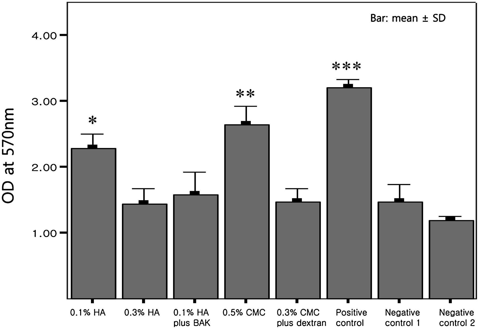
Pseudomonas aeruginosa biofilm quantification. Three drugs [0.3% HA, 0.1% HA plus BAK and 0.3% CMC plus dextran] showed the least amount of biofilm formation. No statistical differences were found among these drugs, and comparison to negative controls also showed no significant differences. Two drugs (0.1% HA and 0.5% CMC) showed higher amounts of biofilm formation. No statistical differences were found between these 2 solutions; however, 0.5% CMC showed biofilm formation equivocal to positive control. Optical density (OD) at 570 nm was measured by crystal violet methods. Positive control was grown in the Luria-Bertani medium, and negative controls were grown in a phosphate buffered solution (control 1) or contact lens without adding bacteria (control 2). HA, hyaluronic acid; CMC, carboxymethylcellulose; BAK, benzalkonium chloride. *P=0.001 versus 0.3% HA, 0.009 versus 0.1% HA plus BAK, 0.002 versus 0.3% CMC plus dextran, 0.001 versus positive control, 0.002 versus negative control 1, <0.001 versus negative control 2, **P<0.001 versus 0.3% HA, <0.001 versus 0.1% HA plus BAK, <0.001 versus 0.3% CMC plus dextran, <0.001 versus negative control 1, <0.001 versus negative control 2, ***P<0.001 versus all samples, except 0.5% CMC (P=0.070). Analysis-of-variance test was used.
Discussion
In this study, we compared the effect of various commercial artificial tear solutions on P. aeruginosa biofilm formation on silicone hydrogel lenses. We found that P. aeruginosa can produce biofilm on silicone hydrogel contact lenses when cultured with some artificial tear solutions. We also found that a higher concentration (0.3%) of HA or addition of BAK or dextran is helpful in prevention of P. aeruginosa biofilm formation.
Some components of artificial tears can be used by bacteria as nutritional sources. High-molecular-weight components such as HA and CMC are popular ingredients in formulating artificial tears due to their ability to hold water and maintain wet ocular surfaces. CMC is a cellulose derivative with carboxymethyl groups bound to the hydroxyl groups of the glucopyranose monomer that make up the cellulose backbone. CMC is used as an active ingredient in artificial tears, because it forms an occlusive film and protects eyes from drying. 14 However, the catalytic product of CMC is glucose. P. aeruginosa can produce a catalytic enzyme, carboxymethylcellulase, which can hydrolyze CMC and use the degradation products as carbon and energy sources.17–19 It has been reported that CMC in Refresh Plus® (Allergan, Irvine, CA) supports the growth of the planktonic form of P aeruginosa. 14 Our results support this previous finding, even with the knowledge that the biology of the planktonic and biofilm forms of bacteria is not identical.
HA is a glycosaminoglycan made up of glucuronic acid and N-acetyl glucosamine disaccharide units. It is a main component of extracellular tissues abundant in skin, connective tissue, synovial fluid, and vitreous humor. 13 HA polymers organize into a reticular structure and produce a molecular framework. 20 This characteristic enables HA to be used as an ingredient in several artificial tears. In contrast to CMC, we found antibiofilm effects of HA in a higher concentration (0.3%). Some bacteria produce hyaluronidase and use HA as a source of energy; however, it is generally acknowledged that P. aeruginosa does not produce hyaluronidase. 21 Instead, P. aeruginosa is known to synthesize its own HA (about 5% of the slime produced by the bacteria), and HA is the main component of Pseudomonas-induced biofilm. 22 Dose-dependent antibacterial effects of HA were reported in some gram-positive (Staphylococcus epidermidis) and gram-negative (Escherichia coli) bacteria, and the addition of HA in the culture medium can invoke relatively inhibitory effects on growth of planktonic P. aeruginosa, although the exact mechanism remains unclear.13,23 Further research regarding the antimicrobial effects of HA is necessary, but is beyond the scope of this article.
Antimicrobial components may also be added into artificial tears. Dextran and BAK are examples. Neutral polysaccharides, including dextran, glycogen, and mannan, are known to inhibit the adhesion of P. aeruginosa to buccal epithelial cells.24,25 We also found that the addition of dextran to 0.3% CMC significantly decreased P. aeruginosa biofilm formation in this study. BAK is a commonly used preservative in many topical eye drugs, although there is growing body of evidence describing its corneal toxicity.26,27 BAK has bactericidal, fungicidal, and virucidal activities. 28 The addition of BAK to 0.1% HA significantly decreased P. aeruginosa biofilm formation in our study, as expected. However, bacterial tolerance to antimicrobial agents is a general concern, and a recent report described that chronic and continuous exposure of P. aeruginosa to BAK can lead to more virulent and tolerant biofilm formation through bacterial adaptation. 29
Biofilm produced by P. aeruginosa is one of the major risk factors for contact lens-induced infectious keratitis.7,30 The ocular surface of patients with dry eye already has a disruption of the normal defensive mechanisms, making them more prone to microbial adhesion and invasion. In addition, the mechanical and chemical alteration of the normal epithelial barrier in contact lens users predisposes the abnormal dry ocular surface to microbial infection.11,12 Therefore, a careful strategy to reduce biofilm formation on contact lenses in patients with dry eye is very important to prevent microbial keratitis.
In summary, we found that different artificial tear solutions have different effects on P. aeruginosa biofilm formation on silicone hydrogel contact lenses. Because contact lens-induced microbial keratitis can be caused by many different microorganisms, including P. aeruginosa, it is difficult to conclude which artificial tear formulation is superior from our study. However, the concept that the most commonly prescribed eye drops, artificial tears, can sometimes promote bacterial biofilm formation is important to recognize.
Footnotes
Acknowledgments
This work was supported in part by the Basic Science Research Program through the National Research Foundation of Korea (NRF) funded by the Ministry of Education, Science and Technology (NRF 2010-0002532) (CYP), and an unrestricted core grant from Research to Prevent Blindness (RSC).
Author Disclosure Statement
No author has a financial or proprietary interest in any material or method mentioned.
References
Supplementary Material
Please find the following supplemental material available below.
For Open Access articles published under a Creative Commons License, all supplemental material carries the same license as the article it is associated with.
For non-Open Access articles published, all supplemental material carries a non-exclusive license, and permission requests for re-use of supplemental material or any part of supplemental material shall be sent directly to the copyright owner as specified in the copyright notice associated with the article.