
Abstract
Abstract
Purpose:
The study aimed to prepare a new type intra ocular lens (IOL), in which 5-Fu, as an antimetabolite drug, could be sustained released to prevent posterior capsule opacification (PCO), and evaluate its efficacy and safety in vitro and in vivo.
Methods:
5-fluorouracil chitosan nanoparticles (5-Fu-CSNP) were prepared by template polymerization and selected by a cell counting kit (CCK-8). The 5-Fu-CSNPs that inhibit Human Lens Epithelial Cells (HLECs) proliferation most efficiently were chosen for further analysis. We then investigated cell death type in vitro by flow cytometry and fluorescent probe. IOLs were surface modified by 5-Fu-CSNP after being activated by a low energy fluorine ion beam, and then were implanted into rabbits' eyes after transparent lens enucleation to evaluate the efficacy of preventing PCO and safety in anterior chamber. Transparency and haze of the 5-Fu-CSNP-surface-modified intraocular lens (Nano-5-Fu-IOL) were detected by a haze meter. Drug release of the Nano-5-Fu-IOL was measured by UV spectrophotometry.
Results:
The CCK results showed that the half inhibition dose (ID50) of the HLECs in 5-Fu-CSNP group and in 5-Fu solution group was 0.2 μg/mL and 1 μg/mL, respectively. The transparency of the 5-Fu-CSNP modified IOL was observed to have no significant difference with the blank control. The IOLs can continuously release 5-Fu in 4 days in vitro. In vivo, 5-Fu-CSNP IOL group had little aqueous flare 3 days after the surgery, and lighter PCO 4 weeks after the surgery than the blank control group. The results of the light microscope and electron microscope further confirmed the above results in vivo.
Conclusions:
The new IOL modified by 5-Fu-CSNP could be prepared and have a sustained release of 5-Fu to prevent PCO by promoting HLEC apoptosis. The drug-loaded CSNP could reduce the anterior chamber toxicity of 5-Fu markedly, which was probably due to the occurrence of endoplasmic reticulum stress and induction of apoptosis.
Introduction
Lens epithelial cells (LECs), left behind in the capsular bag after any type of extra capsular cataract surgery, are mainly responsible for PCO development. 2 Proliferation, migration, epithelial-to-mesenchymal transition, collagen deposition, and lens fiber regeneration of LECs are the primary causes of the PCO. At present, the occurrence of PCO can be reduced by completely clearing the LECs and cortex in the operation or by implanting the IOL with a sharp optic edge. 3 However, the complete clearance of the LECs and cortex through surgical technique is inconceivable. In addition, the IOL with a sharp optic edge is very expensive. Although the Nd: YAG laser capsulotomy is effective for PCO treatment, it carries vision-related complications like cystoids macular edema and puts a significant financial burden on the health care system. 4 Therefore, an effective method to prevent the PCO in surgery is urgently needed, which would be helpful to maintain postoperative visual quality for patients, reduce the operation cost and risk.
Theoretically, the drugs which could prevent LECs from proliferation and migration can be used to prevent PCO. 5 However, due to the toxicity and side effects, none of them could be used directly in clinic. The surface modifications of the IOLs would be an effective method to inhibit the LECs proliferation and migration. As reported, many bioactive molecules have been widely used for the surface modification of the IOLs, 6 including heparin, 7 alpha-allyl glycoside, 8 and poly (ethylene glycol). 9 However, all of them cannot eliminate PCO. 5-Fu, as an antimetabolite drug, carried by chitosan nanoparticles (CSNP) was found to inhibit the LECs in an extremely low concentration by promoting their apoptosis, 10 further decreasing the local inflammation caused by necrosis and extremely low toxicity on the anterior chamber.
The controlled-release drug delivery systems are effective to regulate the release of bioactive molecules with defined kinetics and control the specific therapeutic concentrations, which could further reduce the toxicity of the drugs. With the goal of reducing the incidence of PCO, we have developed a sustained and controlled release system for 5-Fu, which was further used to modify the IOL. The safety and efficacy of the surface modified IOLs in vitro and in vivo was evaluated. 5-Fu was passively transported through being surgically implanted and enabled to be released slowly in the lens capsule to inhibit the proliferation of LECs and finally prevent the occurrence of PCO.
Methods
Materials and animals
Chitosan (deacetylate degree 90%, average molecular weight 40–80 kDa) was purchased from Shanghai Ruji Biology Technology Co., Ltd. Poly acrylic acid (molecular weight 100 kDa, 35%), 5-fluorouracil (5-Fu) standards and Human Lens Epithelial Cells (HLECs) were obtained from Sigma Aldrich Co., Ltd. The IOLs used in this study were poly (methyl methacrylate) (PMMA) IOLs which were produced by Rafi Systems, Inc. (model: CJ55). Propidiumiodide (PI), AnnexinV, and CCK8 were purchased from Sigma Corporation.
New Zealand white rabbits were offered by Chedun experimental animals farm in Songjiang district, Shanghai, whose license number was shxk20020013. All the animals were treated according to the ARVO Statement for the Use of Animals in Ophthalmic and Vision Research.
Preparation of 5-Fu-CSNP-modified IOLs
In this work, CSNP was prepared by adding chitosan solution into polyacrylic acid, 11 and the 5-Fu-CSNP drug delivery system was prepared by adding 5-Fu and chitosan solution separately into polyacrylic acid. PMMA lens were surface modified with F ions and 5-Fu-CSNP as described in our previous study. 12 Briefly, the lens surface was first treated with carrier energy F ion beam, which was 5×10¹³ ions/cm2∼5×1015 ions/cm2, and then they were soaked in 5-Fu-CSNP suspension at 25°C for 24 h. The morphology of the nanoparticles on the surface of the IOL was detected by scanning electron microscopy.
The haze of the IOL modified or not was detected by a haze meter (NDH5000, NIPPON DENSHOKU) with 0.05% accuracy. The ASTM method for optical characteristics of plastics was selected. Four parallel samples per measurement were performed and the obtained values were presented as means±standard deviation.
For knowing the drug content of each IOL, an acid dissolution method was used and the drug content was determined by UV spectrophotometry. Specifically, the standard 5-Fu aqueous solution with concentration of 100 μg/mL was prepared at first. Then the solution was diluted to the final concentrations of 4, 6, 8, 12 and 16 μg/mL. The absorption intensity at 265.5 nm was detected using UV-Vis spectrophotometer (SHIMADZU, UV mini-1240). Using these solutions of known concentrations, a calibration was performed to obtain the best-fit equation that will determine the concentration at any given absorbance. The equation was then obtained, A=0.0399c+0.0314, where r2=0.9981, (1), A represents the absorption, and c represents the concentration of 5-Fu (μg/mL).
After that, one 5-Fu-CSNP modified IOL was immersed in dilute hydrochloric acid solution (0.1 mol/L). Then the absorbance values of the solution at 265.5 nm were measured by UV spectrophotometry. The concentration of 5-Fu in the modified IOL was calculated according to the regression equation.
For the drug release studies, 5 pieces of IOL modified by 5-Fu-CSNP were immersed in 10 mL phosphate buffer solution (PBS) (pH=7.2) and were left in a thermostatic cultivation wave bed with shaking speed of 100±0.5 rpm at 37°C±1°C. At specified time points (0, 0.25, 0.5, 1, 2, 4, 8, 12, 24, 48, 72 and 96 h), 5 mL solutions were withdrawn, and the release of 5-Fu was estimated by detecting the absorption intensity at 265.5 nm using UV-Vis spectrophotometer (SHIMADZU, UV mini-1240). For each sampling, the solution was added with fresh PBS at 37°C±1°C, maintaining the total volume constant. The absorbance was used to calculate the concentration, and knowing the exact volume, the mass of the drug in solution can be quantified. The cumulative release was calculated by the equation. The release rates of 5-Fu from the IOLs modified by 5-Fu-CSNP were calculated using Eq. (2).
Where, C is the concentration of 5-Fu at different time points, V1 is the volume of PBS that were withdrawn each time (mL), V2 is the total constant volume of PBS, m is the total weight of 5-Fu (μg/mL) of the 5 IOL modified by 5-Fu-CSNP. The cumulative release of surface-modified IOLs is plotted as a function of time.
Efficacy of 5-Fu-CSNP suspension on inhibiting LECs proliferation in vitro
Groups: Artificial LEC lines were cultured and passaged in vitro. According to different treatments, the experiments were divided into 4 groups. Group A: PBS blank control; Group B: 250 μg/mL CSNP group; Group C: 5 μg/mL 5-Fu solution group; Group D: 5-Fu-CSNP group. In terms of concentration gradients, the above drugs were added into cells and the following tests were performed. Group B and Group D shared the same content of chitosan, while Group C and Group D were of the same 5-Fu concentration.
Detection of the impact of 5-Fu-CSNP on the proliferation of LECs
The impact of the 4 groups on the proliferation of LECs after 24 h was detected with CCK8 method. The proliferation efficacy was calculated using Eq. (3).
Where, OD1 is the OD value per pore, OD2 is the OD value of the blank control pore.
Fluorescence staining and flow cytometry: The apoptosis and necrosis rates of LECs after 24 h treated by the 4 groups were detected with flow cytometry and annexin V/PI fluorescence staining.
Safety and efficacy of 5-Fu-CSNP-modified IOLs in vivo
Researchers in vivo randomly distributed twenty female New Zealand white rabbits, weighting between 2.8 and 3.5 kg, into 4 groups with 5 per group (Table 1). Only the right eye of each animal underwent surgery. Preoperatively, oxybuprocaine 0.4%, tropicamide 1% eye drops were used for local anesthesia and pupil dilation. The eyelids were disinfected with 5% povidon iodine solution and a drop of 0.3% povidon iodine was instilled into the eye. In the first postoperative week, the eyes were treated with tobramycin dexamethasone (15 mg tobramycin, 5 mg dexamethasone/5 mL) eye drops 4 times daily.
IOL, intra ocular lens; 5-Fu-CSNP, 5-fluorouracil chitosan nanoparticles.
Surgery started with an intravenous injection of nembutal (30 mg/kg) in the ear vein for initial sedation and keeping animals at surgical depth anesthesia. Then, the rabbit was positioned on its left side under a binocular microscope with a fiber optic, coaxial illumination with the right eye facing upward. A wire eyelid speculum was used to retract the eyelids. A corneal incision was made with a 3 mm diamond keratome and the anterior chamber filled with a highly viscous Amvisc solution (1.7% sodium hyaluronate solution; Bausch Lomb). A second incision (paracentesis) was created with a 15° knife. The anterior capsule was teared circularly and continuously with a capsulorhexis forceps and 6 mm capsulorhexis was created. The anterior chamber was kept under pressure of an infusion system with PBS, while the natural lens substance was aspirated. After removal of the lens the anterior chamber infusion was removed and the chamber and the capsular bag were filled with Amvisc solution to protect the cornea from the following treatment. For groups a, b, and c, the unmodified IOL, the IOL modified with CSNP and 5-Fu-CSNP were implanted directly and separately. For groups d, e, and f, before the implantation, flush type posterior capsular polishing device was used to inject the solutions, including 5-Fu-CSNP suspension, CSNP suspension, and 5-Fu injection separately as shown in Table 1. Then, they were implanted with nonsurface-modified IOL.
Inflammation in the anterior chamber and cloudiness in the back peplos in vivo
The inflammation in the anterior chamber and cloudiness in the back peplos was observed by slit lamp biomicroscope in vivo at designated time points, including 3 days, 1 week, 2 weeks, and 4 weeks, respectively, after surgeries. Aqueous flare in the anterior chamber is graded as 13 : 0: no aqueous flare; 1: slight aqueous flare; 2: medium aqueous flare with iris and crystalline lens identified; 3: obvious aqueous flare with iris and crystalline lens difficult to identify; 4: severe aqueous flare with aqueous humor coagulated and massive cellulose exudation. The grading criteria of cloudiness of the central optic zone in the posterior lens capsule 14 : 0: no cloudiness; 1: little cloudiness; 2: slight cloudiness; 3: medium cloudiness; 4: severe cloudiness.
Histopathology
Four weeks after the surgeries, the right eyes of the rabbits were removed and then were observed with light microscope focusing on the parts of cornea, corneoscleral limbus, iris, and ciliary body. The posterior lens capsule was also isolated and then observed with transmission electron microscope focusing on the central part.
Statistical data analysis
All the data was calculated by SPSS12.0. Aqueous flare and the cloudiness in the central optic zone of the posterior lens capsule were analyzed by Kruskal–Wallis rank sum test with P<0.05 as the minimal level of significance.
Results
Characteristics of 5-Fu-CSNP in vitro
Spheroid particles of different size are distributed in the surface of 5-Fu-CSNP surface-modified IOL (Fig. 1). Most of the particles have diameters below 100 nm. Several large particles have diameters between 100 and 400 nm. The haze of the IOL modified or not, was measured. The result indicated that the IOL has a haze of 2.8±0.1, while the IOL modified by 5-Fu-CSNP has a haze of 3.1±0.1. The photograph of the IOL modified or not was also taken by Canon EOS 500D. As shown in Fig. 2, both the IOL and IOL modified by 5-Fu-CSNP were almost completely transparent. It is worth noting that the IOL modified by 5-Fu-CSNP was transparent with a light yellow color. Although the haze of the modified IOL was a little higher than the IOL, it does not create any vision problems. The5-Fu-CSNP surface-modified IOLs of different concentrations can be produced with different fluorion energies. In our research, the average 5-Fu content was 19.55±1.31 μg per IOL. The release of 5-Fu from IOLs modified by 5-Fu-CSNP was characterized over a 96 h (4-day period), and the cumulative release percent of 5-Fu from these samples are shown in Fig. 3. As expected, the surface modified IOLs exhibit a burst release of 5-Fu in 2 h, with the amount of 5-Fu released decreasing rapidly with time. It was initially hypothesized that the surface absorbed 5-Fu on the IOLs was rapidly released and then the entrapped 5-Fu was released with the degradation of chitosan. During the formation of the CSNP, the carboxylic groups of PAA are dissociated into COO- groups which complex with protonated amino groups of CS through electrostatic interaction to form the polyelectrolyte complex. In the meantime, 5-Fu was entrapped in the CSNP and could not be released easily. 11 The electrostatic interaction acts as a rate-limiting barrier to the drug, enabling better control of drug release and allowing a steady, sustained release. 15

Photograph of scanning electron microscope of 5-fluorouracil chitosan nanoparticles (5-Fu-CSNP) surface-modified intra ocular lens (IOL).
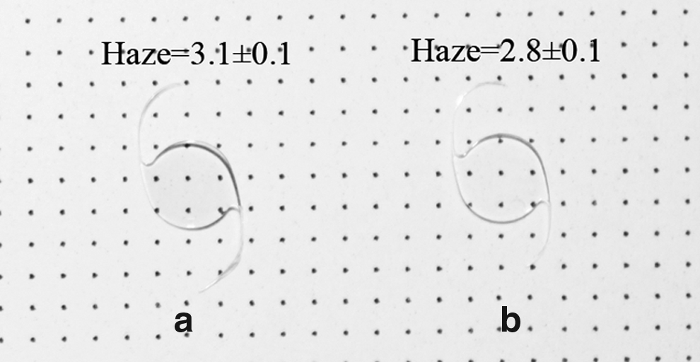
Photograph of the IOL modified by 5-Fu-CSNP
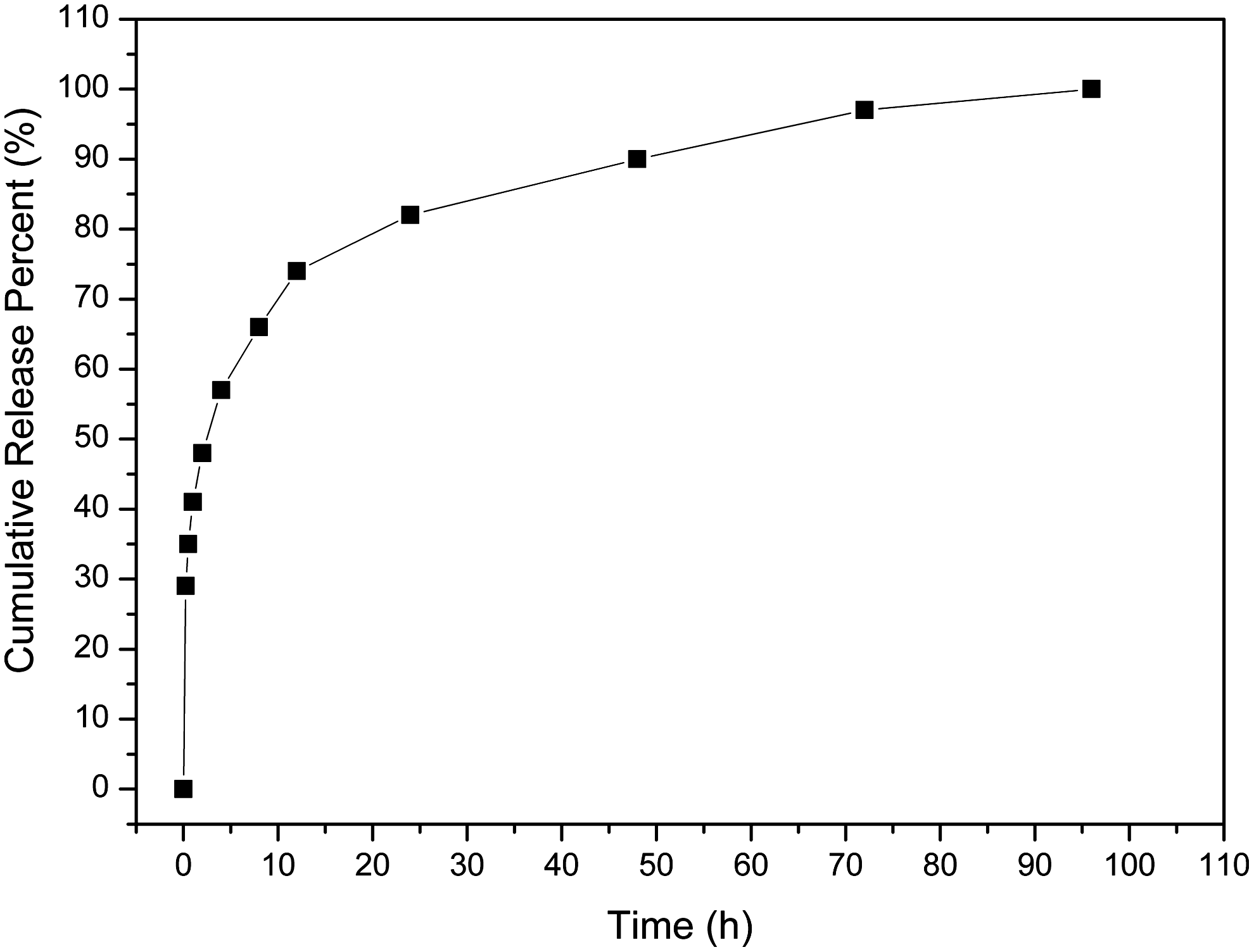
Release characteristics of 5-Fu from IOLs modified by 5-Fu-CSNP.
Efficacy in vitro
The impact of 5-Fu-CSNP on LECs was detected by CCK8 method
Different groups were treated with drugs of different concentrations and the cell proliferation efficacy was measured as well as the median lethal dose (ID50) (Fig. 4). The ID50 was about 1 μg/mL for the 5-Fu solution group and 0.2 μg/mL for the 5-Fu-CSNP group.
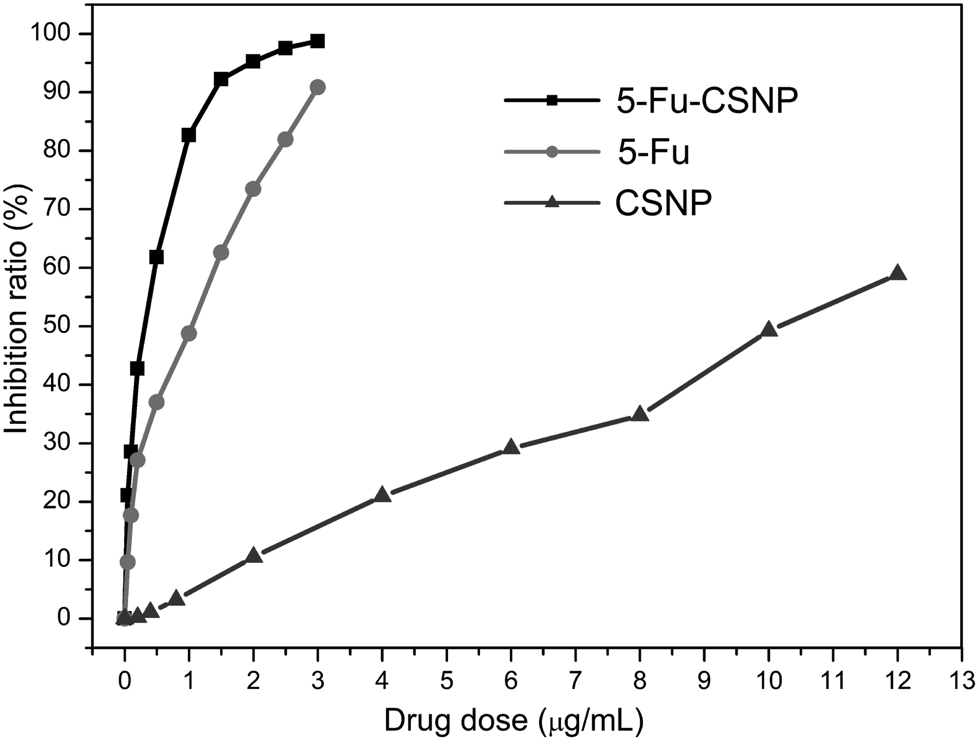
Human lens epithelial cells growth depression curve.
Cell apoptotic rate with fluorescence staining and flow cytometry
Multiflow cytometry tests showed that the 5-Fu-CSNP, which promoted the apoptosis and lowered the necrotic rate, could inhibit the LECs proliferation rate more effectively compared with the 5-Fu solution (Fig. 5).

Flow cytometry of each group. The picture in the upper left is flow cytometry of the control group. Apoptotic rate and necrosis rate of this group is 1.59% and 0.21%, respectively. The upper right picture is flow cytomentry of the 250 μg/mL CSNP group. Apoptotic rate and necrosis rate of this group is 13.94% and 4.57%, respectively. The picture in the bottom left is flow cytomentry of the 50 μg/mL 5-Fu solution group. Apoptotic rate and necrosis rate of this group is 27.75% and 11.04%, respectively. The bottom right picture is flow cytomentry of the 50 μg/mL 5-Fu-CSNP group. Apoptotic rate and necrosis rate of this group is 37.22% and 6.86%, respectively.
Safety and efficacy in vivo
Anterior chamber and the posterior capsule observed with slit lamp
Inflammation in the anterior chamber: 3 days after the surgeries, slit lamp examinations were performed with mydriasis. Aqueous flare in Groups b and c was lighter compared with the other 4 groups with obvious significance (χ2=11.949, P=0.008) (Table 2). The anterior chamber inflammations in the 6 groups were all alleviated 3 days and 1 week after the surgeries. Cloudiness in the posterior lens capsule: 4 weeks after surgery, cloudiness in Group b, d, and f was lighter compared with Group a, c, and e with statistically significance (χ2=9.244, P=0.026). Cloudiness among Group f, d, and b plus a, c, and e had no significant deviation (χ2=1.280, P=0.527) (Table 3). In the examinations of fundus among these groups, detachment of retina, cystoids macular edema, and other complications were not observed.
Cornea, corneoscleral limbus, iris and ciliary body
In Groups a and b, inflammation of anterior chamber was not detected, which could be found in other groups. Group f was of the most serious inflammation: bleeding in the angle of anterior chamber, infiltration of inflammatory cells, vacuolization of ciliary processes epithelial cells, and vasodilatation of the ciliary processes (Fig. 6).
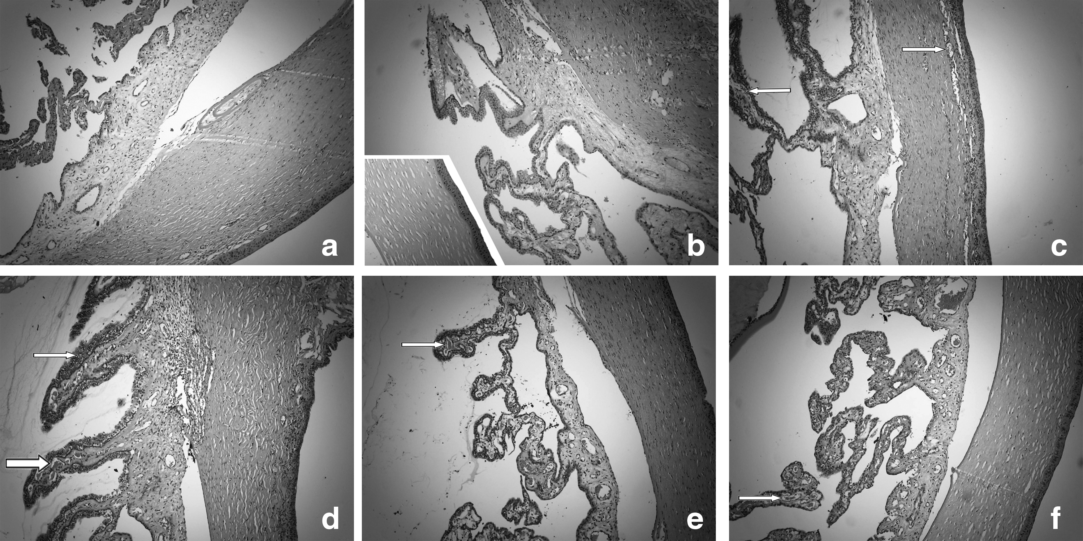
Light microscope photos of corneoscleral junction of each group.
Cloudiness in the posterior capsule with electron microscope
Proliferation of LECs was not observed in the other groups except in Group a. The local posterior capsule of LECs was abscised in Group b and degeneration of posterior capsule of lens was observed in Group f (Fig. 7).
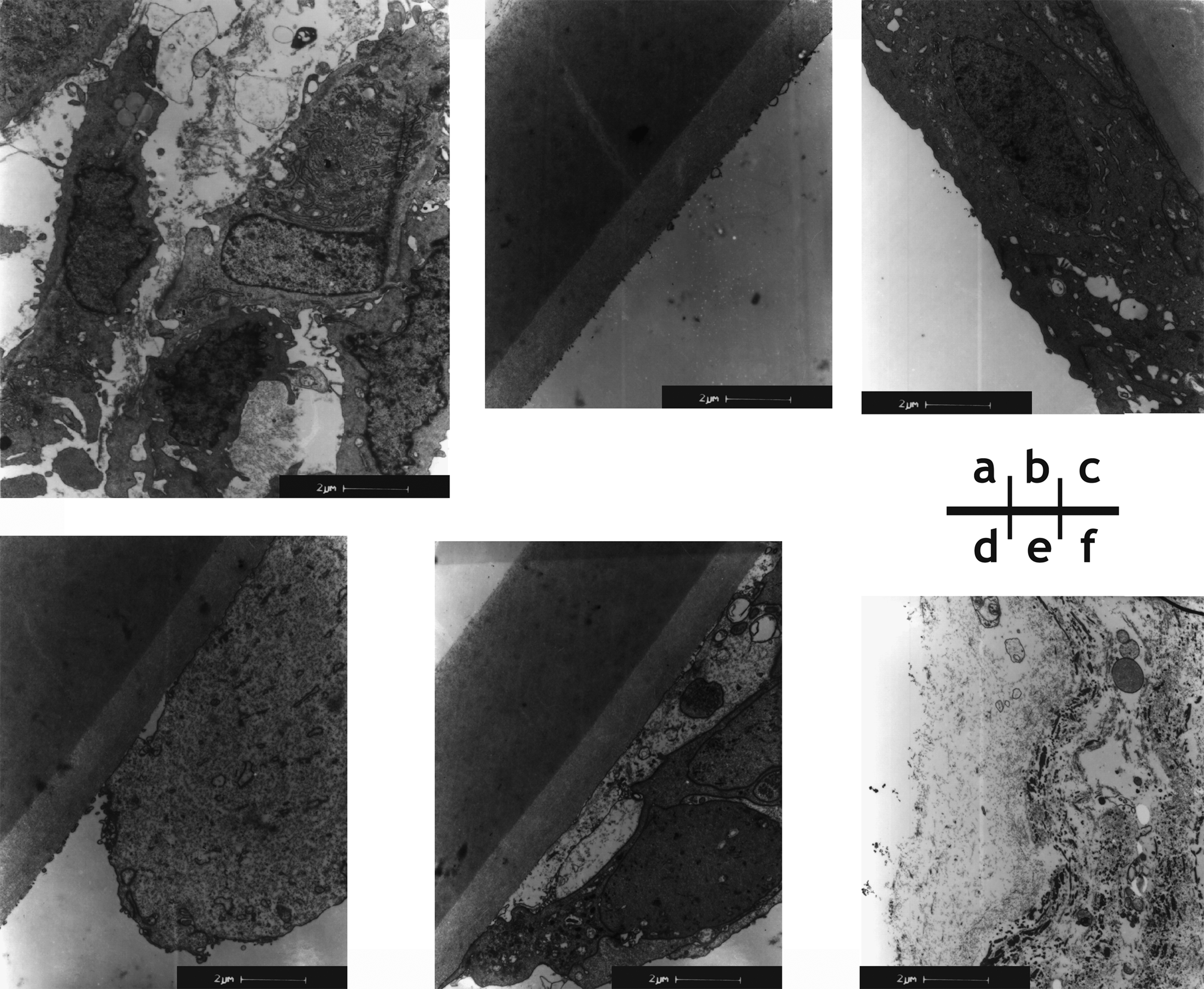
Posterior capsular electron micrograph of each group. Lens epithelial cell hyperplasia was obvious in
Discussion
Our experiments indicate that the toxicity of the 5-Fu-CSNP was decreased due to the slow-releasing effect. When the 5-Fu-CSNP modified IOL was implanted into the capsule, the drug acted through passive transportation and finally prevented the PCO. In addition, the toxicity of 5-Fu after modification was lowered and affected the normal intraocular tissue less.
We found that the role of the 5-Fu-CSNP is more significant in promoting the apoptosis of LECs and inhibiting cells' subculturing. In our study, after exposure in vitro for 24 h, the 5-Fu CSNP group had a ID50 of 0.2 μg/mL. While in other studies, ID50 of the5-Fu solution was respectively, 0.6 μg/mL and 30 μg/mL after exposure for 2 weeks 16 and 24 h. 17
To minimize the toxicity of 5-Fu on the normal tissues in the anterior chamber, some researchers have tried to inject a mixture of sodium hyaluronate into the empty capsular bag. 18 Some researchers implanted a ring with sustained release of 5-Fu to the equatorial LECs in the capsular bag. 19 While some researchers prepared the human capsular bags from donor eyes and sealed with the Perfect Capsule device. 20 Then 5-Fu was introduced for 2 min. The bags were then perfused with Eagle's minimum Essential medium and an IOL was inserted before the bags were dissected and pinned to the base of plastic culture dishes. The toxicity of the 5-Fu to the anterior chamber was not obvious in the mentioned 3 studies. However, in the first 2 studies, they did not show the inhibition of PCO, respectively with 33 mg/mL of transient effect of 5-Fu and 1 μg/mL lasting slow-release. In the third study, it was ineffective in the concentration of 250 μg/mL but apparent in the concentration of 25 mg/mL. Nevertheless, the third study utilized the additional device and the surgical skill was much more challenging. Once the capsule was not completely closed, only 25 mg/mL 5-Fu solution could induce the devastating inflammation in the anterior chamber.
Moreover, the surgeons did not need the intended training for implanting 5-Fu-CSNP modified IOLs but followed the similar procedure of common lens implantation. We are expecting a wide clinical use of our modified IOLs.
Conclusion
This study utilizes the technology of lower-energy F ion beam to produce 5-Fu-CSNP modified IOLs, which is convenient and economic with a mild and nontoxic process. Through the regulation of intensity of the beam, 5-Fu-CSNP modified IOLs with various drug concentrations can be obtained. As 5-Fu can be continuously and slowly released from the surface-adhered nanoparticle, the slow-releasing effect was definite.
Concerning the function of 5-Fu, experiments in vitro of 5-Fu nanoparticle suspension suggested that the new dosage of 5-Fu-CSNP could promote the apoptosis of LECs, while inhibit cellular necrosis with an overall enhancement of cell proliferation. In contrast, CSNP itself had a certain function of promoting apoptosis which was occasionally related to cell death.
Studies on the safety and effectiveness in vitro showed that 5-Fu-CSNP modified IOLs could obviously inhibit the occurrence of PCO without apparent inflammation, while incidence rate of PCO was high in the CSNP-modified IOL group, CSNP in-bag injection group and the blank control group. In the 5-Fu solution group and the 5-Fu nanoparticle in-bag injection group, although the incidence rate was low, the ocular inflammation was apparent, especially in the 5-Fu solution group with posterior capsule degeneration in the electron microscope.
Footnotes
Acknowledgments
This study was supported by nano special fund from Shanghai Science and Technology Commission (0952nm02900), surface project fund from Shanghai Health Bureau (2010245) and young startup fund from Shanghai Changzheng hospital (200905).
Author Disclosure Statement
No competing financial interests exist for any of the authors.