
Abstract
Abstract
Eye is a unique organ of perfection and complexity, and is a microcosm of the body in many ways. It represents a great opportunity for nanomedicine, since it is readily accessible—allowing for direct drug/gene delivery to maximize the therapeutic effect and minimize side effects. The development of appropriate delivery systems that can sustain and deliver therapeutics to the target tissues is a key challenge that can be addressed by nanotechnology. Dendrimers are tree-like, nanostructured polymers that have received significant attention as ocular drug delivery systems, due to their well-defined size, tailorable structure, and potentially favorable ocular biodistribution. In this review, we highlight recent developments in dendrimer-based ocular therapies for both anterior and posterior segment diseases.
Introduction

A schematic representation of critical ocular barriers that offers challenge to ocular drug delivery.
The above-mentioned anatomical and physiological barriers are a challenge to drugs, xenobiotics, and drug delivery vehicles. 5 Topical applications are mostly used for treating anterior segment-related diseases. 6 Typically, drugs, such as anti-inflammatory drugs, steroids, anti-fungal (imidizoles, polyenes), antibiotics, and miotics, are used as liquid formulations via the topical route.6–8 These drugs have poor permeability through an intact corneal epithelium,9,10 and are susceptible to be washed out by a tear (30 s) or an occurrence of drug–protein binding in the tear fluid. Approximately 90% of these drugs are diluted and washed by the lachrymal gland secretion and its nasolachrimal drainage. Some part of the drugs is lost by tear dilution and tear turnover, and typically less than 5% of the administered drug reaches the aqueous humor by crossing the precorneal and corneal barriers (the epithelium, stroma, and endothelial tight junctions).11,12 To maintain and deliver a therapeutic concentration to the target site, repeated dosage is adopted, which results in poor patient compliance. Due to low ocular bioavailability, invasive methods, such as peribulbar, subtendon, subconjunctival injections, are used. Systemic drug delivery to posterior segments has been attempted, but only a small quantity of the injected drug reaches the vitreous cavity due to the BRB selective permeability and drug metabolization in the systemic route. Therefore, frequent administration of high amount of drugs is required, leading to systemic side effects. 12 Intraocular injections, such as intravitreal, suprachoroidal, and retrobulbar methods, have been attempted, which do result in higher drug concentrations in the vitreous cavity and retina leading to accumulation and side effects such as drug toxicity, retinal detachment, endophthalmitis, and intravitreal hemorrhages. 13
Implantable drug delivery systems made up of nonbiodegradable and biodegradable polymers are popular choices as they offer an adjustable drug release profile for a sustained period of time, but these implantable devices are not the only choice available owing to the rapid advancement in polymer synthesis and nanotechnology.14–16 The ultimate goal is to develop suitable drug delivery systems that can overcome the problems of conventional therapies and circumvent the protective barriers with improved bioavailability of drugs, increased retention time, better cellular targeting, lowered side effects, providing therapeutic effects for extended periods of time with improved patient compliance.
Ocular Nanomedicine: Clinical and Pharmacological Opportunities
Recent years have witnessed an unprecedented growth of various drug/gene delivery nanoparticles, which provide tremendous versatility in the structure and function for tailored formulations. 17 Nanoparticles are being used in a variety of ophthalmological applications and can be fabricated with various simple techniques, such as solvent evaporation, emulsification encapsulation, ionotropic gelation, surface conjugation, which require less purification procedures, leading to high encapsulation efficacy, drug loading, and improved scale-up.14,18–20 Variety of materials, such as organic biopolymers (chitosan, albumin, collagen, hyaluronic acid, dextran, and gelatin), organic biocompatible synthetic polymers (polycaprolactones, polyacrylates, polyalkylcynoacrylates, polylactides (PLA), polylactide-co-glycolide (PLGA) etc.), inorganic materials (cerium oxide and calcium phosphate), metallic and magnetic materials, are being investigated. Various particulate and vesicular nanoformulations, such as nanoparticles, nanoemulsions, nanospheres, niosomes, liposomes, dendrimers, cyclodextrins, have been reported in literature with improved pharmacokinetic and pharmacodynamic properties for various drugs. These provide significant benefits, such as increased efficacy at lower concentrations for prolonged period of time, and lower side effects. 5 Nanomaterials can be formulated with desired physicochemical characteristics and ligand attachment to enhance cellular delivery of poorly permeable drugs. 3 Hydrophobic drugs, such as dexamethasone, triamcinolone, budesonide, ganciclovir, are unable to cross the epithelial barriers due to their poor permeability. However, colloidal nanosuspensions and nanoparticle formulations of the above drugs enhanced solubility and permeability through cell layers both in vitro and animal models.20–24 In this focused review, we highlight recent developments in nanoparticle-based ocular therapies, with a special emphasis on dendrimers. The structures of the different dendritic architectures highlighted in this review are provided in Fig. 2. A summary of the various dendrimer-based, preclinical studies focusing on ocular applications is provided in Table 1, and provides an overview of the potential of dendrimers for ocular applications.
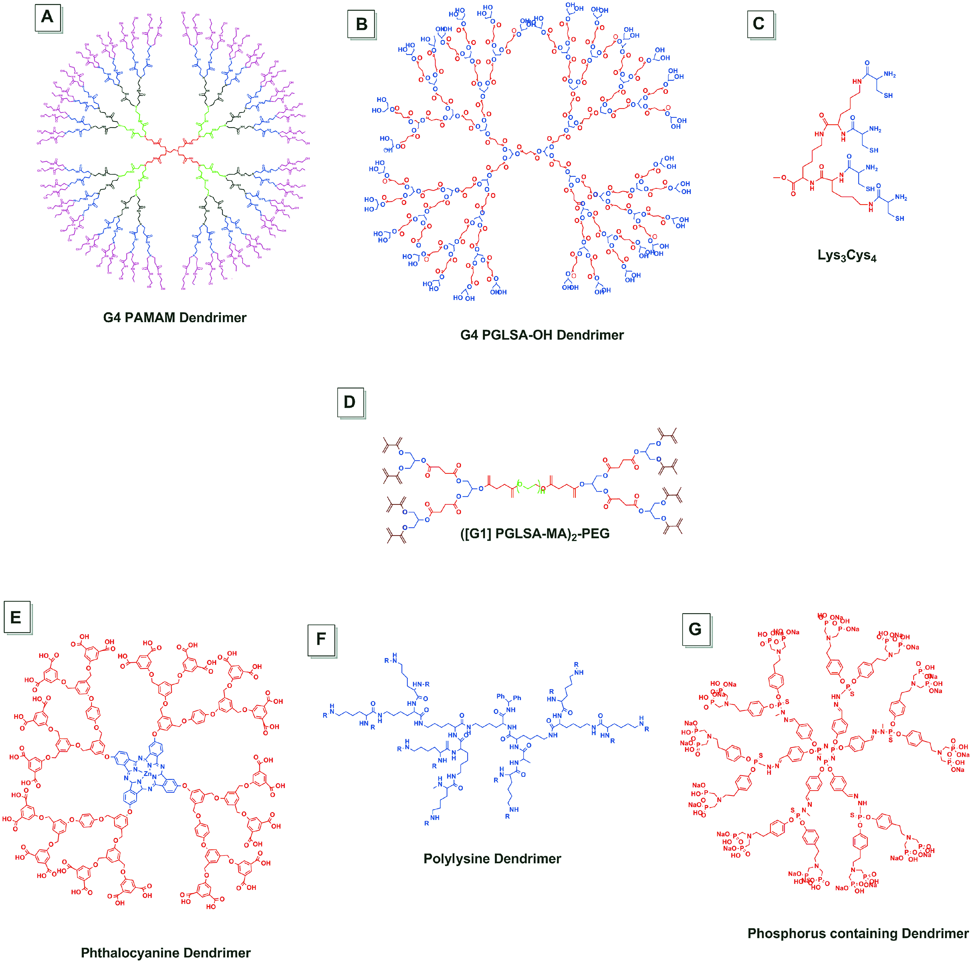
A schematic representation of the structure of some of the dendrimers featured in this review.
AMD, age-related macular degeneration; CNV, choroidal neovascularization; IOP, intraocular pressure; PDT, photodynamic therapy; VEGF, vascular endothelial growth factor; ODN-1, anti-VEGF oligonucleotide.
Improvement in permeation and corneal translocation
Surface functionalization of nanoparticles with suitable ligands, size, surface groups, and surface charge plays a key role on the cellular internalization by endocytosis, phagocytosis, micropinocytosis, or fluid-phase endocytosis.25,26 Dendrimers have a well-defined shape, size (∼2–20 nm), and narrow polydispersity. They are readily transported into and out of cells. Their surface functional groups enable multiple drug molecules to be attached to the surface, enabling a high drug payload, compared to typical linear polymers. 27 The surface functional groups are highly tailorable enabling attachment of multiple imaging moieties, drugs, and ligands (Fig. 3). Polyamidoamine (PAMAM) dendrimers can have different cell entry pathways, depending on the surface functionality. Anionic PAMAM dendrimers are endocytosed primarily through a caveolin-mediated process, whereas neutral and cationic dendrimers were reported to follow clathrin-mediated internalization in cells. 28 These cellular internalization processes can be highly beneficial in the case of crossing the epithelial and retinal barriers in the cornea and retina. Moreover, surface modifications of dendrimers have been reported to enhance multivalent interactions of dendrimers with biological membranes, thereby improving the cellular internalization process and cell entry kinetics.29,30 Even though studies on the role of the physicochemical properties of the dendrimers on overcoming many of the ocular barriers outlined in Fig. 1 are in the early stages, some insights are emerging on their effect on corneal transport. Extensive studies have been carried out to identify the effect of size and surface charge PAMAM dendrimers on permeability into Caco-2 epithelial cells, which have continuous tight junctions similar to corneal epithelial cells. Anionic (the COOH surface group) dendrimers showed enhanced permeability (EPR) at higher generations. In contrast, cationic (the NH2 surface group) and neutral (the OH surface group) dendrimers showed higher permeability at lower generations. 31 Such size- and surface-dependent transport can be attributed to the high density of surface amine (PAMAM-NH2) and internal amine groups (all PAMAM) in dendrimers that actively interact with the mucin layers above the cells. Similar results have been reported for the permeability of PAMAM dendrimers through intact rabbit corneas. Topically instilled PAMAM dendrimer–Puerarin complexes (with amine and carboxyl dendrimer surface groups) showed longer residence time than puerarin itself, potentially improving drug availability. 32 The release of puerarin from dendrimer complexes were considerably slow both in deionized water, phosphate-buffered saline (pH 6.8) indicating strong interactions between the puerarin molecules and dendrimers, which could lead to increased corneal residence and overall improved permeation. 33 Pharmacokinetic studies of concentrations of topically instilled puerarin in the form of PAMAM–puerarin complexes were carried out using microdialysis and mass spectroscopy, which suggested that up to a 3.5-fold increase in the drug concentration could be achieved in groups treated with PAMAM dendrimer–drug complexes, compared to the free drug. 34 The above studies indicated that dendrimers act as transporters of drug molecules across the corneal membrane without damaging the epithelial and endothelial structures, avoiding precorneal washing of drug molecules by tear fluids. Both lipophilic and hydrophilic drugs can be conjugated to, or encapsulated in PAMAM dendrimers with appropriate surface functionality. PAMAM- NH2 dendrimers have been reported to decrease the transepithelial electrical resistance, thereby increasing the permeability through corneal layers. 31 Similarly, Holden et al. showed that dendrimer hydrogel (DH) formulations enhanced the bioavailability of antiglaucoma drugs in corneal layers. The enhancement was attributed to the PAMAM dendrimer opening the tight junctions of the corneal epithelium and a temporary disruption of corneal barriers.31,67
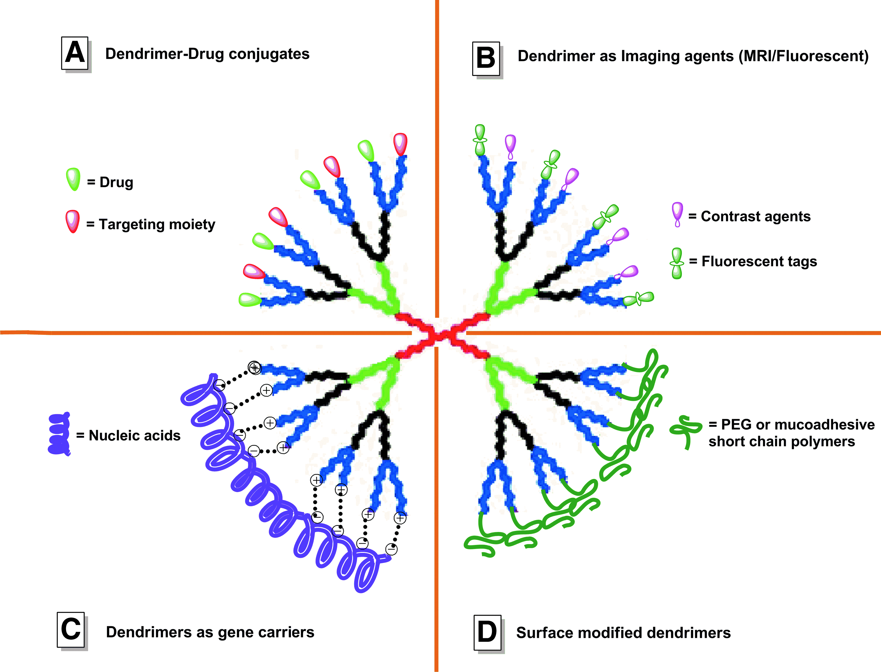
A diagrammatic representation of the versatility of the dendrimer structure that enables tailorability.
Various polymeric nanoparticles, such as chitosan, PLGA, alginate, collagen, albumin, polycaprolactones, and gelatin, have been reported to have good bioadhesive and permeation properties through corneal epithelial layers and could overcome the hurdles presented by lacrimal drainage and tight junctions of corneal epithelial cells.35,36 Among the above class of polymers, chitosan has been extensively studied for its superior mucoadhesiveness. The electrostatic interactions between the positive amine groups of chitosan and negative sialic acid groups of mucins present in the tear film, which is intact with the corneal epithelium, enabled an increase in corneal residence time, and increased penetration of drug-loaded nanoparticles into the intact corneal epithelium.35–37
Engineering desired biodistribution in ocular tissues
Biodistribution and bioavailability of the administered nanoparticle drug delivery systems in ocular tissues are key factors for achieving an effective drug concentration to the specific tissue of interest, especially in treating various vitreoretinal diseases. This is somewhat poorly understood due to the relatively complex anatomy of the eye (Fig. 1). Delivery of drugs to retinal layers is a difficult task as the retina itself is isolated from blood circulation and vitreous fluid similar to the brain by the inner and outer BRB to maintain its homeostasis and clear vision. These regulatory barriers are perturbed only at later stages of retinal disease where treatment becomes complicated and requires higher and frequent dosages. Various parameters, such as the chemical structure, composition, particle size, surface charge, and mode of administration, play a key role in the effective distribution of nanoparticles in ocular tissues.12,38,39 Ganciclovir-loaded albumin nanoparticles, when administered by the intravitreous route, diffused through vitreous space. Two weeks after administration, they were broadly distributed in the vitreous cavity, a thin layer over the retina, and in the region near the blood–aqueous barrier. These particles were well-tolerated over a prolonged time and caused no alterations in the tissue architecture. 21 Rhodamine-conjugated liposomes, loaded with the vasoactive intestinal peptide (VIP-Rh-Lip), were injected intravitreally into the eyes of Lewis rats and after 24 h of injection, these particles were distributed mainly in the vitreous and inner layer of the retina; some particles were detected in the iris root and ciliary body. Liposomes were found internalized by retinal Müller cells and ocular resident macrophages, free rhodamine was found in the internal layers of the retina suggesting the release of drugs from the liposomes in the retina.40,41 Intravitreal injection of 2.2 μg of PLA nanoparticles loaded with Rh-6G or Nile red was well-tolerated. Six hours after injection, these nanoparticles were found in deep retinal layers such as the ganglion cell layer and the photoreceptor outer segments layer; within 24 h, nanoparticles were found accumulated in RPE cells. These nanoparticles were found to be stable in RPE after 4 months of injection, releasing loaded dye in a sustained fashion. 42 Such formulations may provide opportunities for sustained delivery of drug molecules into the retina.
The influence of the size of nanoparticles on biodistribution in intraocular tissues and BRB transport has been investigated. Polystyrene nanospheres of different sizes 2 μm, 200 nm, and 50 nm were injected intravitreally, and their subsequent retinal biodistribution was studied. Larger nanospheres (2 μm) were found accumulated near the trabecular meshwork and discharged out from the eye, whereas the smaller diameter nanospheres were found accumulated in the retina and retinal boundaries, even 2 months after injection. 43 Similar effects were observed in the case of metallic gold nanoparticles of different sizes of 20 nm and 100 nm administered intravenously. The 20-nm gold particles were found to cross the BRB via systemic circulation and well distributed in retinal layers and retinal cells, such as endothelial cells, retinal neurons, and periendothelial glial cells, within 24 h of injection and showed no structural or cellular damage. About 100 nm particles were not found in retinal layers suggesting the influence of size on transport across the BRB. 44 Surface charges also played an important role in the movement of human serum albumin nanoparticles (HSA-NPs) in the retinal layers and crossing the vitreous barrier. Anionic HAS-NPs more easily penetrated the vitreal barrier and were found inside the retina than cationic HAS-NPs within 5 h of intravitreal injection. 45 They were found in deep retinal layers and most of these nanoparticles were colocalized within the RPE layer and Müller cells in the ILM, ganglion cell layer, inner nuclear layer, outer plexiform layer, and the outer nuclear layer (ONL) both in the normal and laser photocoagulated retina. In the laser photocoagulated retina, more HSA-NPs were detected in the choroidal space, compared to the normal retina. 45 These albumin nanoparticles carry different reactive groups, such as thiol, amino, and carboxylic groups, on their surfaces that can be used for attaching ligands or surface modification for various purposes.
Pathology-dependent biodistribution of dendrimers has been utilized for targeted, sustained intracellular delivery. 46 Upon intravitreal administration, hydroxyl-functionalized PAMAM dendrimer (G4-OH), labeled with FITC or Cy5.5, was found to selectively localize into activated microglial cells in the outer retina and photoreceptor outer segments, in rat models of retinal degeneration. Such cellular biodistribution was not observed in the case of healthy rat eyes. The above specific localization was observed for a period of 30 days, and the dendrimer–drug conjugate (PAMAM dendrimer-fluocinolone acetonide) was shown to provide sustained attenuation of neuroinflammation, long-term neuroprotection, along with maintaining photoreceptor health. 46 Such selective targeting to cells associated with neuroinflammation can improve drug efficacy and minimize side effects, with implications in indications associated with retinal neuroinflammation.
Polylysine biodendrimers, carrying antivascular endothelial growth factor (anti-VEGF) oligonucleotides (ODN-1) targeting the retinal pigment layers (RPE) of the outer retina to suppress neovascularization, have been investigated. These poly-cationic polylysine dendrimers, built on a lipid core, were shown to be biocompatible, exhibiting no toxic effects on retinal structures. Delivering biomolecules and peptides across the retinal layers is a challenge, as they are often subjected to be metabolized by cellular lysosomal enzymes and nucleases and other biological intracellular barriers and secretions before reaching the target.47,48 AntiVEGF oligonucleotides, complexed to polylysine dendrimers and injected intravitreally, were found in the ONL and RPE layers of the outer retina 2 weeks postinjection. At later times, these dendrimers migrated from the ONL layers and were concentrated in the RPE layers and further in the nucleus rather than cytoplasmic structures of RPE cells, indicating the effective translocation of ODN-1, and suppressing neovascularization. 49 These results demonstrate the potential of dendrimers in targeting specific ocular cells to treat neovascularization in age-related retinal diseases.
Ligand-targeted PLGA nanoparticles have been explored for systemic delivery to the eye. AntiVEGF intraceptor plasmids were delivered, using PLGA nanoparticles surface functionalized with transferrin and the RGD peptide through intravenous administration. Delivery of these biomolecules to the retina via systemic circulation, overcoming various biological barriers encountered in the intravenous route through ligand targeting. 50 The extent of targeting and distribution patterns within the retina were found to be depended on functionalized moiety on the nanoparticle surface. Transferrin, RGD, or dual targeted (both transferrin and RGD) functionalized nanoparticles accumulated in photoreceptor layers and RPE within 24 h of injection. They also localized in retinal blood vessels around the laser-induced lesions. Such localization patterns were not observed in nonfunctionalized nanoparticles and healthy eyes. 50 Surface conjugation of polystyrene nanoparticles with deslorelin and transferrin showed increased uptake in the corneal epithelium, compared to nonfunctionalized nanoparticles. 51 Surface immobilization of RGD peptides on poly(lactide-co-2-methyl-2-carboxytrimethylene carbonate) nanoparticles can be used to target corneal epithelial cells for drug delivery to treat corneal neovascularization, dry eye syndrome, and neurotropic keratopathy. 52 Protein nanoparticles can be biodegradable, noncytotoxic, nonantigenic, and amenable for surface modifications so that drugs and ligands can be covalently attached to the surface easily. 53 These nanoparticles have improved tolerability, increased intraocular half-life, and adhere to inflamed ocular tissue, significantly better than normal tissue.54,55
Dendrimers for Topical and Anterior Segment Diseases
Conventional eye drops have various limitations such as short precorneal residence time, poor absorption and penetration of drugs into the cornea, and nonspecific inactivation of drugs by enzymes. Since more than 95% of the instilled eye drop is washed away by tear secretions and nasolacrimal drainage, there is a need for modified formulations that can overcome the above problems, and lead to enhanced drug availability and sustained release.
Topical administration for corneal diseases
As discussed earlier, specific interactions between the nanoparticulate surface and tear film is one of the key factors for corneal permeation and increased residence time. Ocular mucins and mucin-like o-glycan layers on the glycocalyx structures of the surface epithelium have a vital biological role of protection, preventing bacterial adhesion, maintaining structural integrity, and promoting boundary lubrication of the corneal and conjunctival epithelium. 56 Investigating the type of interactions between the nanoparticle surface and glycocalyx secretions is highly useful for gaining insights into factors that may contribute for the development of new strategies for improving the adhesiveness. PAMAM dendrimers showed remarkable interactions with membrane-associated mucin layers. Surface plasmon resonance was used to probe the interfacial interactions between dendrimer surfaces and ocular mucins at the molecular levels by Bravo-Osuna et al. 57 Nonionic interactions via hydrogen bonding between the PAMAM dendrimer surface moieties and mucins were observed. 57 Both cationic and anionic PAMAM dendrimers show similar mucoadhesive multiple surface interactions, which were much stronger compared to linear polymers. Therefore, it may be possible to use dendrimers in eye-drop formulations with drug molecules covalently conjugated to the dendrimer surface groups. pH-dependent interactions of PAMAM dendrimers with ocular mucins were also investigated, suggesting stronger interactions at pathological conditions (pH 5.5 for tear fluid) compared to physiological conditions. At lower pH, a larger fraction of the primary amine surface groups of PAMAM-NH2 dendrimers are protonated, and the tertiary amine groups are partially protonated, resulting in increment of net-positive charge leading to an increased association with mucins. On the other hand, the mucin interactions with the PAMAM-OH dendrimer remained the same at both neutral and acidic pH. 57 The above results are in agreement with the previous research findings by Yao et al., who reported increased permeation and retention time of cationic PAMAM dendrimer–puerarin complexes with increased bioavailability of puerarin in aqueous humor indicating the transport of drug molecules across the corneal layers.33,34 This provides a promising strategy for treating various diseases such as cataracta gluca, ocular hypertension, and also the ability of puerarin in decreasing the intraocular pressure (IOP) and improving the ocular blood flow.32–34,58 Anionic PAMAM dendrimers (PAMAM-COOH) were expected to have decreased adhesion to the mucin surface due to their electrostatic repulsion between the negatively charged mucins and carboxylate groups of the dendrimer surface. Interestingly, these dendrimers also showed adhesive interactions on the corneal surfaces. 57 These interactions were also observed by Griffiths et al. in gastric mucins, where the anionic dendrimers establish hydrogen bonds with the sugar residues of the mucin side chains. 59 Nonglycosylated domains of the mucins possess positively charged amino acid residues. Therefore, electrostatic interactions can occur with the negatively charged carboxylate groups. The above findings provide ample evidence that PAMAM dendrimers, may have potential to compete with other mucoadhesive polymers, such as chitosan, and may enable development of new hydrophilic dendrimer eye-drop formulations for treating a myriad of anterior segment diseases such as corneal infections, inflammations, wound healing, glaucoma, and corneal neovascular abnormalities.
Applications in corneal scarring, glaucoma infiltration, and cataract surgeries
Glaucoma infiltration surgery is often performed to reduce IOP buildup and to drain the excess aqueous humor. The surgery can sometimes result in scar tissue formation, corneal neovascularization, and inflammation. 60 Carboxylated PAMAM dendrimers were explored for their role in enhancing the efficacy and availability of amino-saccharides in preventing scar tissue formation, thereby increasing the long-term success of the surgery. 61 Shaunak et al. synthesized PAMAM generation 3.5 dendrimer conjugates with glucosamine and glucosamine-6-sulfate, and showed improved antiangiogenic and immune modulatory properties. 61 The conjugates actively inhibited lipopoly-saccharide (LPS)-induced proinflammatory chemokines and cytokines in human dendritic cells and macrophages. Dendrimer–glucosamine conjugates effectively prevented the activation and maturation of dendritic cells by suppressing and inhibiting the TLR-4 pathway activated by LPS. On the other hand, dendrimer glucosamine-6-sulfate conjugates inhibited the endothelial cell proliferation by blocking the fibroblast growth factor-2, thereby preventing microtubule formation and neoangiogenesis. Combination therapy with both the conjugates by subconjunctival administration inhibited scar tissue formation and increased the long-term success of the surgeries from 30% to 80% in rabbit models of glaucoma infiltration surgery. Moreover, these conjugates did not exhibit any signs of tissue inflammation, clinical, hematological, and biochemical toxicity, or systemic microbial infections throughout the experimental period. 61 Inhibition of the TLR pathway, which plays a major pathogenic role in a variety of inflammatory and neovascular diseases such as blunt traumatic corneal ruptures and wounds, by these dendrimer–drug conjugates, is highly beneficial and could pave the way for new treatment options using PAMAM dendrimer formulations for various surgical wounds often associated with inflammation in the cornea.
Open-angle glaucoma
Delivery of pilocarpine for treating open-angle glaucoma is a major challenge because of its poor permeability and low stability, resulting in rapid drug clearance by lachrymal secretions with 1%–3% bioavailability of the total instilled drug. 62 To overcome this problem, the drug is administered frequently, which can lead to patient noncompliance. Hence, a delivery system that penetrates through corneal layers delivering the parasympathomimetic drugs, such as timolol, epinephrine, tropicamide, pilocarpine, to reduce IOP in a sustained manner would be desirable. Nanoparticulate formulations were shown to have better bioavailability of pilocarpine with effective reduction of IOP in albino rabbit glaucoma models, when compared to commercially available conventional eye drop formulations. 55 Vandamme et al. investigated PAMAM dendrimers as drug delivery vehicles for delivering pilocarpine and tropicamide. 63 A systematic study was conducted to determine the effect of size, surface charge, and molecular weight of PAMAM dendrimers influencing the corneal residence time, permeability, and efficacy in prolonging the miotic and mydriatic activity in the pupil. 63 Comparison of various physical, chemical, and biological parameters suggested that dendrimer formulations were more efficient compared with commercially available Carbopol® eye drop formulations. 63 The physical properties such as surface tension, refractive index for all different dendrimer formulations were comparable with Carbopol® eye drops and mimicked the same that of tear fluid, showing no disturbance in the visual acuity when instilled on the corneal surface. Hydroxyl-terminated PAMAM dendrimers did not create any ocular irritation, and other dendrimers PAMAM-NH2 and PAMAM-COOH had a consistently significant lower ocular irritation index compared with Carbopol® solutions. The increased bioavailability was attributed to decreased washing out during the initial lachrymal drainage phase, and improved mucoadhesive properties of PAMAM dendrimers. 63 This improved biological response, along with other studies relating to long-term release of entrapped and surface-conjugated drug molecules from dendrimers, are providing positive evidences that the dendrimer formulations can be candidates for eye drop formulations in the form of emulsions or suspensions.
Phosphorous dendrimers possess antiprion properties, and can be effective transfection agents for delivering genetic materials safely into the nucleus. They promote neuronal cell growth and maturation, and offer excellent antimicrobial property avoiding the mal-conformational changes of various proteins and anti-inflammatory. 64 These dendrimers are designed to have suitable electrostatic interactions between the drug and the surface groups and to have chemical entities that can act as a substitute preservative for drug molecules. Spataro et al. have built a dendrimer with quaternary ammonium salt as a core, which serves as a stabilizer for carteolol (an antihypertensive drug used to treat glaucoma), as the traditional preservative benzalkonium chloride is toxic and damages the protective barriers in the cornea. 65 Topically instilled dendrimer–carteolol conjugate solutions produced no irritation even after several hours in rabbit corneas and showed increased precorneal residence time as compared to PAMAM–COOH dendrimers. Moreover, the quantity of carteolol in aqueous humor was ∼2.5-fold higher when compared to concentrations in animals treated with carteolol solutions, suggesting improved bioavailability. 65
As seen in many synthetic antiglaucomatic drugs, timolol and brimonidine are hydrophobic and have very limited solubility in aqueous ophthalmic solutions and dramatically influence their cellular uptake, corneal transport, and bioavailability. Further, their salt forms have rapid tear clearance due to the permi-selective nature of the cornea, thereby requiring frequent dosage leading to patient noncompliance.66,67 Holden et al. have developed DH consisting PAMAM (G3-NH2) dendrimers with poly ethylene glycol (PEG) acrylate chains (crosslinkers) conjugated to their surface and brimonidine or timolol maleate encapsulated into their hydrophobic cores, aimed to increase the bioavailability of drugs after topical instillations. 67 Upon photoinitiation, they crosslink to form a water soluble viscous solution that, when topically administered on to the cornea, offers continuous availability of drug molecules.66,67 DH formulations possess better mucoadhesive properties due to numerous surface anionic groups, sustained release with no cytotoxic effects, and able to enhance corneal transport of encapsulated drugs. Moreover, it is noteworthy to mention that the DH formulation acts as cargos, significantly enhancing (77.6% increase) the solubility of brimonidine and can be attributed to its superior cellular uptake and crossing of corneal barriers to aqueous humor facilitating a therapeutic concentration for long-term efficacy.66,67
Corneal infections: keratitis and conjunctivitis
Corneal infections caused by a variety of microbial organisms such as bacteria, fungi, and virus often subjected to take various forms keratitis, which are very difficult to differentiate. They project as a major health problem if untreated, causing corneal reddening, opacification, rupture, irritation, and inflammation, leading to obscure vision. 68 Under adverse conditions, these microbes even penetrate into deep layers reaching the posterior segment and affecting the retina. Various antimicrobial drugs used for systemic infections are being reinvestigated for corneal infections, but the major problems projected are their poor corneal permeability, solubility, and stability, and hence, poor bioavailability. 69 Repeated or frequent dosage with high concentrations cause physical damage to the microbes, but at the same time also causes secondary damage to corneal cells. Nanoparticle drug formulations as potential agents to treat corneal infections are being investigated.
Dendrimers can be both topical antimicrobial agents, and delivery vehicles for antimicrobial drugs. Their core, surface charge and functionality, size and three-dimensional conformation affects the antimicrobial activity. PAMAM dendrimers were reported to show an excellent bactericidal activity in the case of gram-negative E. Coli-induced chorioamnionitis, an intra-amniotic infection, which can result in congenital neuroinflammatory diseases such as cerebral palsy and preterm birth. 70 Different surface groups on dendrimers exhibited different bacterial cell destruction mechanisms. PAMAM G4-NH2 dendrimers showed a greater entry dynamics into eukaryotic cells with good antibacterial properties by destabilizing both the inner and outer membrane of the bacterial cell wall, thereby exposing the bacterial contents for denaturation. The probable mechanism proposed is that at physiological pH, primary amines at the dendrimer surface and more than half of the tertiary amines in the core become protonated and, thereby, act as polycations and actively interact with polyanions of the cell wall surface causing major structural changes resulting in bacterial cell death. 70 Interestingly, PAMAM G4-OH and G4-COOH also showed an appreciable antibacterial activity by altering the cell wall structure. At physiological pH, carboxylic acid-terminated dendrimers can chelate the positive calcium and magnesium ions of the lipid heads, destabilizing the membrane of the cell wall of the E. coli. 70
Amine-terminated PAMAM dendrimers have reported to be cytotoxic to cells at moderate to high concentrations. Therefore, a PEGylation stealth strategy for terminal amine groups was adopted to show the antibacterial activity against gram-negative and -positive bacteria, such as Pseudomonas aeruginosa and Staphylococcus aureus, respectively. These bacteria often cause various ocular infections such as conjunctivitis and keratitis. Partially PEGylated dendrimers were found to be nontoxic to corneal epithelial cells at the concentrations that are toxic to P. aeruginosa, but were less effective against S. aures. Therefore, conjugating antibacterial drugs to these dendrimers can have a dual effect of desired antibacterial and reduced cytotoxic effects.71,72 Quinolones are exploited for their potential antibacterial activity against both gram-positive and -negative bacteria, but often they have issues such as low aqueous insolubility, poor penetration, destruction of mucin layer leading to dry eye, and toxic to corneal epithelial cells. PAMAM dendrimers were investigated for their potential in enhancing the solubility of hydrophobic quinolones such as nadifloxacin and prulifloxacin. 73 The dendrimers, due to their hydrophobic and open cavities in their internal structure, offer a greater surface area and the primary and tertiary amine groups on the dendrimer surface and the core interacts with the carboxylic groups of nadifloxacin and prulifloxacin, thereby forming dendrimer–drug complexes with greater aqueous solubility. 71 These complexes showed a 2-fold increase in the antibacterial activity than free nadifloxacin and prulifloxacin. 73 Dendrimeric polyguanidilyated translocators (DPTs) are a class of dendrimers with triolyl branches and surface guanidine groups. 74 DPTs were investigated as potential ophthalmic carriers for gatifloxacin (GFX), a fourth generation fluoroquinolone approved for the treatment of conjunctivitis. DPT-GFX formulations were found to increase the solubility of GFX by 4-fold and increased transport and entry across human corneal epithelial layers. GFX were found to complex with DPTs via ionic, hydrophobic, and hydrogen bond interactions and were found to kill microbes at a 2 to 4 times faster rate than GFX indicating the active transport of drug molecules into the eukaryotic cells. 74 A different class of dendrimers, such as glycodendrimers, polypropylenimine, anionic amphiphilic dendrimers, polylysine dendrimers, and oligosaccharide dendrimers, were reported to have antibacterial properties and can be used as potential antibacterial vehicles for antimicrobial drugs for treating a variety of corneal, conjunctival, and intraocular infections.
Ocular sealants: accelerated corneal wound healing
Dendrimer formulations are also used as potential ocular sealants for corneal wound repair. The cornea is a transparent tissue devoid of blood vessels and possesses a highly ordered arrangement of stromal collagen fibrils and critical for clear vision. Wounds caused due to trauma, cataract and LASIK surgeries, and corneal transplantation require sutures to fasten the corneal flaps, sometimes resulting in infection, corneal leaks, tissue scarring, inflammation, and neovascularization leading to alterations in the transparency and curvature of the cornea, thereby perturbed vision. 75 Grinstaff et al. have synthesized a new class of biodendrimers with biodegradable and biocompatible entities, such as glycerol, succinic acid or lactic acid, and PEG, as building blocks for photocurable corneal adhesives. 75 These DHs rapidly seal the corneal lacerations bridging the 2 edges of the corneal tear, forming transparent layers. These bioadhesives not only act as sealants, but also as temporary scaffolds for corneal regeneration. 76 An In vivo evaluation of the biodendrimer hydrogel in a leghorn chicken model showed no inflammation or scar tissue formation, whereas the sutured cornea showed irregular healing, inflammation, and scar tissue formation. 77
Dendrimers for the Intraocular Posterior Segment Diseases
In recent years, posterior eye segment diseases have become important therapeutic targets for nanotechnology. However, topical application is a challenge, since drugs have to travel a great distance overcoming the barriers in the cornea, before they reach the retina and choroid. Diseases, such as age-related macular degeneration (AMD), diabetic retinopathy, posterior uveitis, and retinal vascular diseases, are major causes for visual impairment and blindness for millions of patients across the world. Currently, intravitreal injection of drugs is widely used, which can lead to side effects such as ocular infections, endophthalmitis due to repeated injections, increased risk of cataract development, vitreous hemorrhage, retinal detachment, patient noncompliance, and high cost. Hence, an ideal drug delivery system is required that can provide sustained drug delivery, reducing the frequency of administration and the associated side effects.
Intraocular Infections: uveitis, cytomegalovirus retinitis, and endophthalmitis
Intraocular infections, often resulting from cataract surgeries and frequent intraocular injections or surgeries, may manifest in different diseases such as cytomegalovirus (CMV) retinitis, uveitis or microbial endophthalmitis leading to structural damage in the retina, retinal detachment or inflammation, resulting in a permanent vision loss. CMV retinitis is one of the leading causes of blindness in immunosuppressed patients who receive bone marrow, organ transplants, and in AIDS patients. Antiviral drugs such as ganciclovir, fomivirsen, maribavir are used, but associated with serious side effects such as hemolysis, kidney dysfunction, and systemic toxicity. 78 Luganini et al. have developed a novel class of peptide dendrimers containing SB105 and SB105 A10 peptides as building blocks built on a biocompatible lysine core. 79 These peptides show significant virucidal effects by inhibition of several viral strains by inhibiting the attachment of virons to the cell surface. CMV strains enter the living cells by attaching themselves to the heparin sulfate polysaccharides, which are linked to the proteins of the cell membrane. Once virons are attached, they induce conformational changes paving their entry in to the cells and replicate. Peptide dendrimers were found to block the heparin sulfate surface by attaching to them and inhibiting the viral entry. 79 These dendrimers were found to completely inhibit human CMV (HCMV) replication in both primary fibroblasts and endothelial cells. 79 A comparative study of the antiviral activity of polylysine dendrimers and PAMAM dendrimers against the herpes simplex virus (HSV) showed beneficial results. Polylysine dendrimers were found to be more potent antiviral agents than PAMAM dendrimers. These dendrimers showed inhibition of infection and destruction of viral cells via the cytopathic effect. These results suggest that these nanoparticles are potential agents for effective delivery of drugs for treating CMV retinitis in immunosuppressed patients. Antiendophthalmitis drugs, such as ofloxacin and fluconazole, available in commercial formulations show retinal toxicity when given intravitreally, which could be reduced by liposomal encapsulations. This offers new possibilities for treating various vision-threatening intraocular infections. 5 Anti-inflammatory drugs such as methylprednisolone loaded into copolymer nanoparticles, and piroxicam formulated into nanosuspensions inhibited endotoxin-induced uveitis in rabbit eyes, significantly better than microsuspensions of free drug. More importantly, these formulations manifested controlled release and a lower crystal formation of drug molecules.80,81
Intraocular tumors: retinoblastoma and uveal melanomas
Intraocular tumors such as retinoblastoma and uveal melanoma developing from critical structures of the retina are less common, but present a high risk of complications with high metastatic potential, leading to retinal detachment and a permanent loss of vision. 82 Limited number of studies have been done to develop nanodevices for delivery of drugs and detection of intraocular tumors. Carboplatin, a chemotherapeutic drug used to treat intraocular tumors is associated with complications such as local toxicity, orbital fat necrosis, and atrophy of the optic nerve. Kang et al. have explored the use of PAMAM dendrimers with carboxyl end groups (G3.5-COOH) for extended half-life and sustained delivery of carboplatin with lowered therapeutic toxicity. 83 Carboplatin-loaded PAMAM dendrimer complexes were explored in a transgenic murine retinoblastoma model, delivered via a subconjunctival route. The complexes were more effective in reducing the tumor mass than aqueous carboplatin formulations. These carboplatin-loaded dendrimers not only crossed the sclera to reach the contralateral eye via the local vasculature, but were also retained for a longer period of time in the tumor vasculature, thereby providing a sustained treatment effect. 83 Tumor cells overexpress mannose and lectin-like receptors on their surface, which can be utilized for tumor targeting and delivery of antitumor drugs into ocular tissues. Glycopolymers were found to enable multivalent interactions with surface lectins, and are being used to target and treat tumors. 84 Based on this strategy, Makky et al. have developed prophyrin-based glycodendrimers with the mannose-specific ligand protein Concanavalin A conjugated on to their surface, to specifically target the tumor cells in the retina. 84 These hybrid dendrimers are designed as photosensitizers for preferential accumulation in malignant ocular tissue, for enhancing the effectiveness of photodynamic therapy (PDT). The mannosylated dendrimers demonstrated specific interactions with the receptors in the lipid bilayer inducing protein channel rearrangement favoring the entry of the dendrimers into the cell. 84 Recently, Wang et al. synthesized α-mannosyl dendrimeric porphyrins, which exhibited good photoefficiency, superior cellular uptake, and significant phototoxicity in retinoblastoma cells. 85 These dendritic nanodevices were found to be highly sensitive to PDT treatment for killing tumor cells. This combined with specific targeting, and improved cellular uptake may lead to new photosensitizers that can also carry drugs to specific cells allowing a localized effect restricted to the tumor tissue.
Neurodegenerative and neovascular retinal diseases
Promising recent findings regarding the uptake of various nanoparticles by RPE and other retinal cells, suggest new ways for using them to treat retinal disorders associated with age and photoreceptor dystrophies. The purpose is to (1) reduce the frequency of intravitreal injections, (2) target the drug molecules to specific cells with specific targeting moieties, and (3) achieve a sustained efficacy. The vitreous cavity could be a reservoir for the nanoparticle systems for attaining therapeutic drug levels in the retina. The commonly used drugs to treat retinal diseases are steroidal or nonsteroidal anti-inflammatory drugs, which have poor solubility, and hence are excluded from intraocular tissues, or have high accumulation causing local toxicity. Another possible side effect of steroidal drugs is that they can elevate IOP, causing retinal ganglionic cells and optic nerve dystrophies. Therefore, targeted therapies can be highly beneficial. Neurodegenerative diseases, such as retinitis pigmentosa, AMD, are associated with neuroinflammation in the retina. Recently, Kannan, Iezzi and coworkers have developed PAMAM fluocinolone acetonide (FA) conjugates for targeting attenuation of activated microglia.46,100 These dendrimer-drug conjugates exhibited sustained release of FA for 3 months, thereby offering the potential to enhance the bioavailability of the drug to the target cells in the retina for an extended period of time.46,100 Upon intravitreal administration, the conjugates were found to preferentially accumulate into activated microglia and photoreceptors, and were present in the microglial cells even after 30 days postinjection. Interestingly, a single intravitreal injection showed significant attenuation of activated microglial cells at 30-fold lower dosage compared to free drug, with no cytotoxic effect. The electroretinography experiments showed favorable preservation of the photoreceptor function, indicating the neuroprotection activity in RCS rat models. 46 PAMAM dendrimers can also be used as effective gene carrier agents for delivering neuroprotective genes to the retina. Choi et al. have developed PAMAM–dexamethasone conjugates as efficient nuclear translocating agents for genes, where the drug molecules can have a dual role of therapeutic and gene translocation in to the nucleus. 86 It has been shown that glucocorticoid drugs dilate the nuclear pore up to 60 nm, allowing the genes into the nucleus. Nanoparticles can be effective gene delivery vectors for the retina. In a murine model of retinitis pigmentosa, nanoparticles compacted with opsin promoter genes, efficiently and safely delivered the DNA to the photoreceptors. The nanotherapy slowed degeneration and attenuated inflammation by inhibiting the cytokines, whereas the naked DNA did not show any signs of photoreceptor protection. 87 This study offers an effective gene delivery paradigm for treating photoreceptor degeneration. Betamethasone-encapsulated PLA nanoparticles were effective in controlling inflammation in autoimmune uveo-retinitis rat models. The therapeutic effect was achieved at a much lower concentration than at higher concentration dosages of betamethasone alone. 24
Retinal neo-vascular diseases such as wet AMD, diabetic retinopathy, macular edema, and choroidal neovascularization (CNV) cause irreversible changes, and are leading causes of blindness in the elderly population. Dendrimers have been explored as drug carriers and photosensitizers for the treatment of exudative AMD and CNV. Photodynamic therapy involves targeting photosensitizer agents to the respective retinal cells, followed by photoillumination, which causes destruction of the localized tissue. 88 However, the risk factor is the collateral destruction of other major tissues resulting in vision problems and retinal scars. Over the last few years, novel photosensitizers with improved targeting and efficacy have been developed. Porphyrin-based dendrimers developed by Nishiyama et al. were investigated as promising therapeutic modalities for solid tumors. 89 These porphyrin dendrimers are being investigated for their efficacy in treating retinal tumors and exudative AMD associated with CNV. 90 Dendrimer nanodevices are loaded into micelle structures to form aggregates of ∼50 nm. When injected into a CNV rat model, the nanodevices showed selective accumulation within 24 h in the CNV lesions. Since these CNV lesions share many common properties similar to that of a solid tumor such as hyperpermeability and leaky blood vessels,88–90 this approach may have applications in ocular tumors. Tamaki et al. developed polyion complex micelles containing the poly (aspartic acid) core and fluorescein isothiocyanate-labeled poly(L-lysine) branches and encapsulated it into PEG shell, forming dendritic micellar structures with a size of ∼50 nm. 91 These were investigated as photosensitizers for treating CNV lesions in PDT. The nanostructures, when injected intravenously, exhibited prolonged retention for as long as 168 h in CNV lesions in experimental CNV rat models. 92 This prolonged retention could be explained presumably by the EPR effect.
Nanoparticles hold promise for delivery of oligonucleotides for gene replacement, or siRNA for gene suppression, to knockout VEGF and VEGF receptors. 93 Marano and coworkers have explored the use of poly (lipid-lysine) dendrimers to deliver a sense oligonucleotide (ODN-1) to the retina, causing reduction in VEGF expression. 48 These dendrimers were found to be nontoxic to the retinal cells and offer a biocompatible treatment for delivering ODN-1 to the retina without causing toxicity and damage to critical retinal structures. 94 In vivo experiments also indicated significant reduction of VEGF expression for a prolonged period of more than 2 months indicating the sustained bioavailability of ODN-1 in the retina, whereas the free ODN-1 was subjected to enzymatic degredation. 49 PLGA nanoparticles with targeting ligands, such as transferrin and RGD peptides, efficiently delivered oligonucleotides targeted to the retina via intravenous injections, offering the eventual possibility of avoiding intravitreal injections. 50 Small interfering RNAs (siRNAs) are being investigated as potential agents for treating wet macular degeneration, and are in Phase I & II clinical trials.95,97 Dendrimers may enable superior delivery of the siRNAs. Polycationic dendrimers can complex the siRNAs, compacting them considerably and thereby avoiding enzymatic damage, apart from facilitating efficient cell transfection. 96 PAMAM dendrimers can be conjugated with various cell penetrating peptides, along with complexed siRNA, providing efficient translocation to targeted cells. For example, Kang et al. have developed cell permeating PAMAM dendrimers by conjugating Tat (cell penetrating peptide) with antisense oligonucleotides, which showed efficient nuclear transfection. 97 These dendrimers are designed to escape from the phagocytes and lysosomes to avoid premature degradation of the genes before reaching the target. Nishiyama et al. have developed phthalocyanine core-based dendrimer photosensitizers, which can be used to compact and deliver therapeutic genes with a targeting approach mediated by irradiation. 98 Upon subconjunctival injection of the above dendrimer formulation, followed by laser irradiation, transgene expression was observed only in the irradiated areas. Such strategies may be useful in the case of macular degeneration where neuroprotective genes can be administered intravitreally, and directed only to the macular area via photoillumination.
Ocular Nanomedicine–Potential for Clinical Translation
The eye is a unique and sensitive organ, yet ideally suited for the development of novel nanomedicine platforms. It has multiple tissue types confined to a small structure that are easily accessible. Increased incidences of vision-threatening diseases both in young and older generations have enhanced the need for development of new drug delivery platforms to achieve sustained release, facilitating improved drug bioavailability, and targeting. Zabrin et al. have highlighted the potential of nanoparticles in various ophthalmic clinical applications, such as imaging, diagnostics, regenerative medicine, and surgical technology, suggesting that a well-defined control of the composition, size, and shape are critical to translation. 101 Dendrimers may offer such well-defined control in the 3–30-nm-length scale. Dendrimers have exhibited potential as permeation and solubility enhancers for transdermal delivery of hydrophobic drugs leading to numerous patented studies that potentiate the utility of dendrimers in women's health, cosmetics, and personal care products.27,102 Poly(L-lysine) dendrimers were the first to undergo human clinical trials for HIV and bacterial vaginosis, showing significant efficacy against spreading sexually transmitted diseases and are being commercialized as Vivagel® (Starpharma).27,103 A biodendrimer-based corneal sealant has successfully passed clinical trials in Europe and is currently being commercialized under the trade name Ocuseal ™ liquid ocular bandage (HyperBranch Medical Technology, Inc. & BD medical) (search Ocuseal at www.bd.com). These formulations did not exhibit any side effects in humans. These hybrid formulations can reduce the dosing frequency, offer long-term patient compliance, and reduce costs.
Dendrimers have high synthetic control on critical nanoscale design parameters such as size, shape, surface functionality, and controlled architecture. 104 These parameters influence the biodistribution, receptor-mediated targeting, cellular internalization, and dosage frequency. Dendrimers can be tuned to release drugs in a different fashion based on the dosage, disease, and patient requirements. For treating ocular diseases, more localized and intraocular drug delivery may be desirable, since this can improve drug levels in the eye, while minimizing systemic toxicity. Blindness and irreversible sight impairment cost ∼$50 billion in the United States. 2 The commercial ophthalmic market is estimated to be $14 billion in 2009 and it is expected to reach $19 billion by end of 2014. 105 Nanomedicine approaches tailored to specific ocular therapeutics and indications, backed by appropriate regulatory evaluations that carefully balance both the risk and reward can lead to faster translation.
Conclusions
Dendrimers are a relatively new class of nanomaterials that hold significant promise for many ocular diseases. We have highlighted many recent preclinical developments that utilize the many unique aspects of dendrimers, where they have been shown to, for example, (1) enhance the corneal residence time of drugs administered as eye drops; (2) be effective as corneal glues to potentially replace sutures following corneal surgeries; (3) target retinal neuroinflammation and provide targeted, sustained neuroprotection in retinal degeneration; (4) increase the bioavailability of glaucoma drugs; (5) deliver drugs to the retina upon systemic administration. Even though dendrimers are not yet approved for clinical use in the eye, their promising preclinical results can provide significant opportunities (Table 1), when combined with careful planning relating to scale-up synthesis, safety, and a clear focus tailored to specific ocular indications.
A recent ARVO study outlined the important aspects of translating basic research to clinical translation for ocular diseases and identified the 5 barriers that need to be overcome. 2 These included (1) development of an effective and safe product; (2) identifying the best mode of delivery and the right delivery system; (3) assessment of the product in appropriate animal models, recognizing the differences in the human equivalence in different small and large animal models for the specific indication; (4) appropriate design of the clinical trials to attain a satisfactory endpoint; (5) commercialization through spin-offs or finding a commercial partner. Many of these aspects have to be considered for successful clinical translation of nanomaterial-based delivery technologies.
Author Disclosure Statement
No competing financial interests exist.