
Abstract
Abstract
Rho-kinase inhibitors affect actomyosin cytoskeletal networks and have been shown to significantly increase outflow facility and lower intraocular pressure in various animal models and human eyes. This article summarizes common morphological changes in the trabecular meshwork induced by Rho-kinase inhibitors and specifically compares the morphological and hydrodynamic correlations with increased outflow facility by Rho-kinase inhibitor, Y-27632, in bovine, monkey, and human eyes under similar experimental conditions. Interspecies comparison has shown that morphological changes in the juxtacanalicular connective tissue (JCT) of these 3 species were different. However, these different morphological changes in the JCT, no matter if it's separation between the JCT and inner wall in bovine eyes, or separation between the JCT cells or between the JCT cells and their matrix in monkey eyes, or even no separation between the inner wall and the JCT but a more subtle expansion of the JCT in human eyes, appear to correlate with the increased percent change of outflow facility. More importantly, these different morphological changes all resulted in an increase in effective filtration area, which was positively correlated with increased outflow facility in all 3 species. These results suggest a link among changes in outflow facility, tissue architecture, and aqueous outflow pattern. Y-27632 increases outflow facility by redistributing aqueous outflow through a looser and larger area in the JCT.
Introduction
P
The Rho and Rho-associated coiled coil-forming protein kinase (ROCK) pathway has been studied extensively for the past decade as a potential target for the treatment of glaucoma. More recently, several glaucoma drug candidates that target the Rho/ROCK pathway are undergoing phase I and phase II clinical trials,15–18 which underscores the importance on understanding the underlying mechanism behind Rho-kinase inhibitors that lower IOP. In the past several years, Y-27632, a Rho-kinase inhibitor, has been studied extensively in both animal and human models in an attempt to understand its mechanisms of increasing outflow facility. The purpose of this review was to summarize common morphological changes in the TM, induced by Rho-kinase inhibitors, and specifically compare the morphological and hydrodynamic correlations with increased outflow facility by Rho-kinase inhibitor, Y-27632, in bovine, monkey, and human eyes under similar experimental conditions.
Effect on Aqueous Outflow Facility and IOP
An overview of the Rho/ROCK pathway reveals that the activation of the Rho/ROCK pathways results in increased outflow resistance, thereby decreasing outflow facility and elevating IOP. Agonists of the Rho/ROCK pathway, such as endothelin-1, 19 transforming growth factor-beta, 20 lysophospholipids (lysophosphatidic acid and sphingosine-1-phosphate), 21 and expression of RhoAV14, 22 have been shown to decrease aqueous outflow and/or increase IOP. In contrast, inhibition of the Rho/ROCK pathways results in decreased outflow resistance, thereby increasing outflow facility and lowering IOP. Antagonists of the Rho/ROCK pathway, such as ROCK inhibitors (Y-27632, Y-39983, HA-1077, H-1152),23–37 myosin light-chain kinase inhibitor (ML-9), 38 and Lim kinase-2 inhibitor, 39 and silencing RhoA expression, 40 have all shown to increase aqueous outflow and/or decrease IOP in various animal models as well as in human eyes. A summary of the effect of the Rho-kinase inhibitors on aqueous outflow facility and IOP is shown in Table 1.
All Rho-kinase inhibitors increase outflow facility (C) and decrease intraocular pressure (IOP).
Clinical trial.
A popular Rho-kinase inhibitor used in studies of the trabecular outflow pathway has been Y-27632. Other groups have demonstrated that perfusion with 50 μM of Y-27632 for a minimum of 60 and 170 min in enucleated porcine 41 and monkey 42 eyes significantly increased the outflow facility, respectively. However, their results are difficult to compare because of the different perfusion pressures as well as possible different perfusion methods. On the other hand, Y-27632 in different species under similar experimental conditions (same concentration and perfusion time), studied by our group, has shown to have a greater increase in outflow facility in bovine and monkey eyes compared to human eyes.23–25 Human eyes required a longer perfusion time to achieve the similar percent increase in outflow facility as observed in nonhuman eyes (Fig. 1).
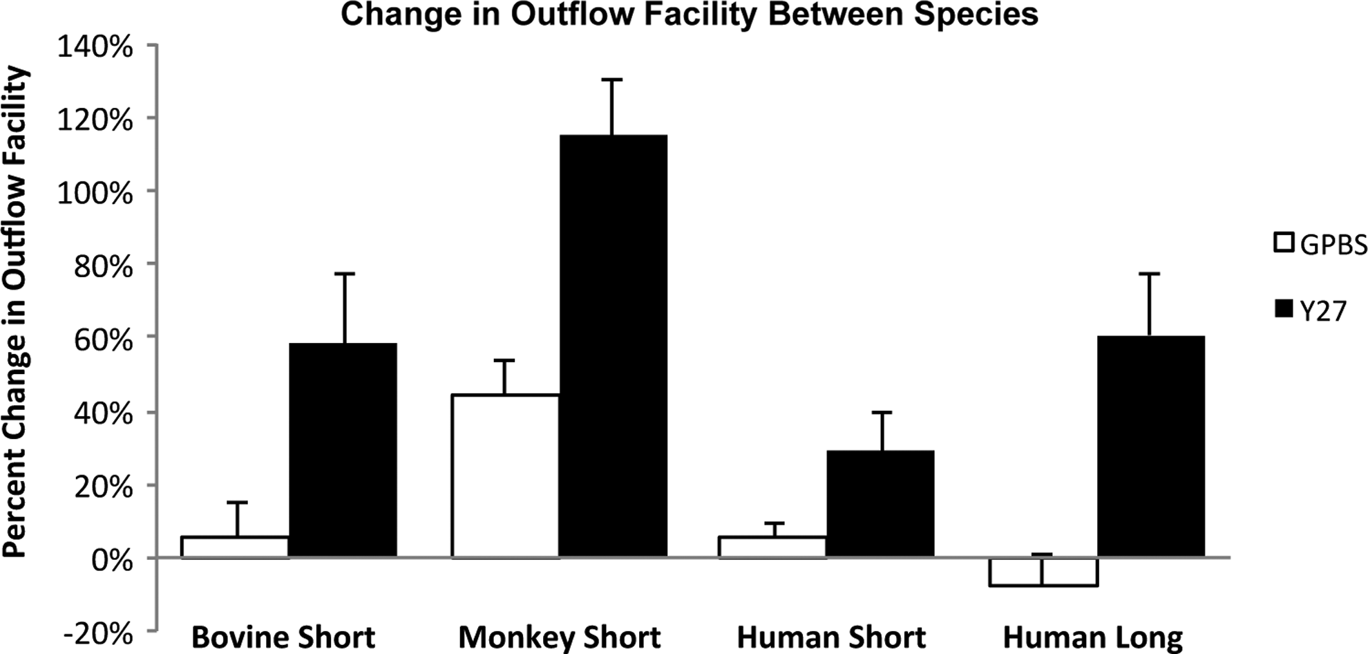
Comparison of outflow facility in bovine, monkey, and human eyes. Percent change in outflow facility in bovine, monkey, and human eyes after Y-27632 treatment (50 μM). Human eyes required a longer perfusion time (3 h) with Y-27632 to achieve a similar percent increase in outflow facility as found in bovine eyes when perfused for a short duration (30 min), whereas this increase was still less than the level achieved in monkey eyes when perfused for the same duration (30 min) with Y-27632 (from Yang et al. 25 ).
Morphological Changes Following Y-27632 Treatment
Morphological changes at the cellular level under cell culture conditions
TM cells
Studies have shown that TM cells possess smooth muscle-like properties, which can enable them to relax and contract and may play a role in regulating the aqueous outflow.41,43,44 In particular, one study has shown that Y-27632 can cause TM cell relaxation, disassembly of actin stress fibers, and focal adhesions, 41 which could potentially lower the aqueous outflow resistance by increasing paracellular fluid flow or alteration of the pathway through the JCT.
Inner wall endothelial cells of SC
In addition to TM cells, studies have shown that SC cells treated with Rho-kinase inhibitors or other cytoskeletal altering agents have a significant influence on the cell actin cytoskeleton, 41 cortex, 45 stiffness, and contractility. 46 Specifically, Y-27632 resulted in a decrease in junctional resistance of inner wall endothelial cells of SC from both human 41 and monkey 42 eyes, presumably due to the loss of actin stress fibers and a decrease in cell stiffness/contractility. Although the mechanism of how cell stiffness and contractility leads to an increase in outflow facility is still unclear, it has been hypothesized that decreasing cell stiffness and contractility can decrease outflow resistance by increasing the ability of SC cells to form giant vacuoles (GVs) and pores. 47 However, further studies are warranted to prove this hypothesis.
Morphological changes in ex vivo whole globe perfusion model
General morphological changes
Aqueous outflow tissue that has been treated with ROCK inhibitors often share common morphological features associated with increased outflow facility. Such features include the expansion of the JCT (human 25 ), SC inner wall endothelium and JCT separation (bovine 23 and monkey 24 ), and distension of the SC inner wall endothelium (porcine 41 ). These morphological observations are likely due to cell relaxation and disassembly of both actin stress fibers and focal adhesions. 41
Comparison of the morphological changes associated with increased outflow facility with Y-27632 treatment in bovine, monkey and human eyes
Our group has studied the morphological changes associated with increased outflow facility after Y-27632 treatment in bovine, monkey, and human eyes under similar experimental conditions.23–25 We found that, in both bovine and monkey eyes, light microscopy reveals a similar separation between the inner wall and JCT, and this separation showed a positive correlation with increased outflow facility23,24 (Fig. 2). However, upon closer inspection at the electron microscopic level, bovine eyes displayed a separation between the basal lamina of the inner wall endothelial cells and the extracellular matrix of the JCT (matrix–matrix separation), whereas monkey eyes revealed a separation between the JCT cells (cell–cell separation) or between the JCT cells and their matrix (cell–matrix separation) (Fig. 3). These similar morphological differences between bovine (matrix–matrix separation)48,49 and monkey (cell–cell and cell–matrix separations) 24 eyes were also reported following the washout effect (a perfusion volume-dependent phenomenon whereby outflow facility progressively increases during prolonged perfusion in nonhuman eyes 12 ), and similar cell–cell separation in the JCT was also observed in monkey eyes after treatment with latrunculin-B, which disrupts microfilament organization of the cells. 50 These results suggest that monkey eyes may have a stronger connection between the basal lamina of the inner wall cells and the JCT than that between the JCT cells and between the JCT cells and their matrix.

Light microscopic analysis in bovine and monkey eyes.
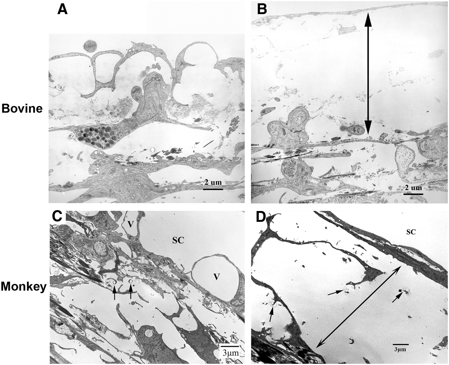
Electron microscopic analysis in bovine and monkey eyes.
Unlike the findings in bovine and monkey eyes, perfusion of human eyes with Y-27632 for the same time (30 min) or longer (3 h) showed no obvious separation between the inner wall and the JCT under light microscopy (Fig. 4). Interestingly, using a detailed imaging analysis, a more subtle expansion of the JCT region (2 μm increase in JCT thickness) was observed in human eyes 25 (Figs. 5 and 6). Despite this difference in morphological alterations, a positive correlation was found between the JCT thickness and increased outflow facility in human eyes, which was similar to the positive correlation between percent separation length and increased outflow facility in bovine and monkey eyes (Fig. 7). These results suggested that anatomical differences among species may affect the extent of changes in morphology and outflow facility, which is consistent with previous reports that human eyes do not exhibit the washout effect49,51 or inner wall/JCT separation following prolonged perfusion. 49 The morphological findings associated with an increase in outflow facility after Y-27632 treatment support the “funneling” model. 11 This hypothesis states that aqueous humor flowing through the JCT must cross through the pores of the inner wall endothelium of SC, where aqueous outflow resistance decreases with an increase in available area for flow and that the bulk of resistance is generated within the inner wall endothelium of the SC and JCT region. Therefore, Y-27632 could greatly attenuate the outflow resistance in the inner wall and JCT region by affecting the JCT/SC connectivity and increasing the thickness of the JCT through relaxing the inner wall and JCT cells as discussed above. However, the mechanism behind the changes in the outflow resistance as a result of Rho-kinase inhibitor treatment is likely not only due to JCT/SC connectivity and JCT geometrical changes, but the inner wall pore density/size may also play a role according to the funneling model, which is discussed further in the next section.

Light microscopic analysis in human eyes. JCT expansion and compact regions were observed in both long-duration Y-27632-treated eyes and its control groups. SC, Schlemm's canal (from Yang et al. 25 ).
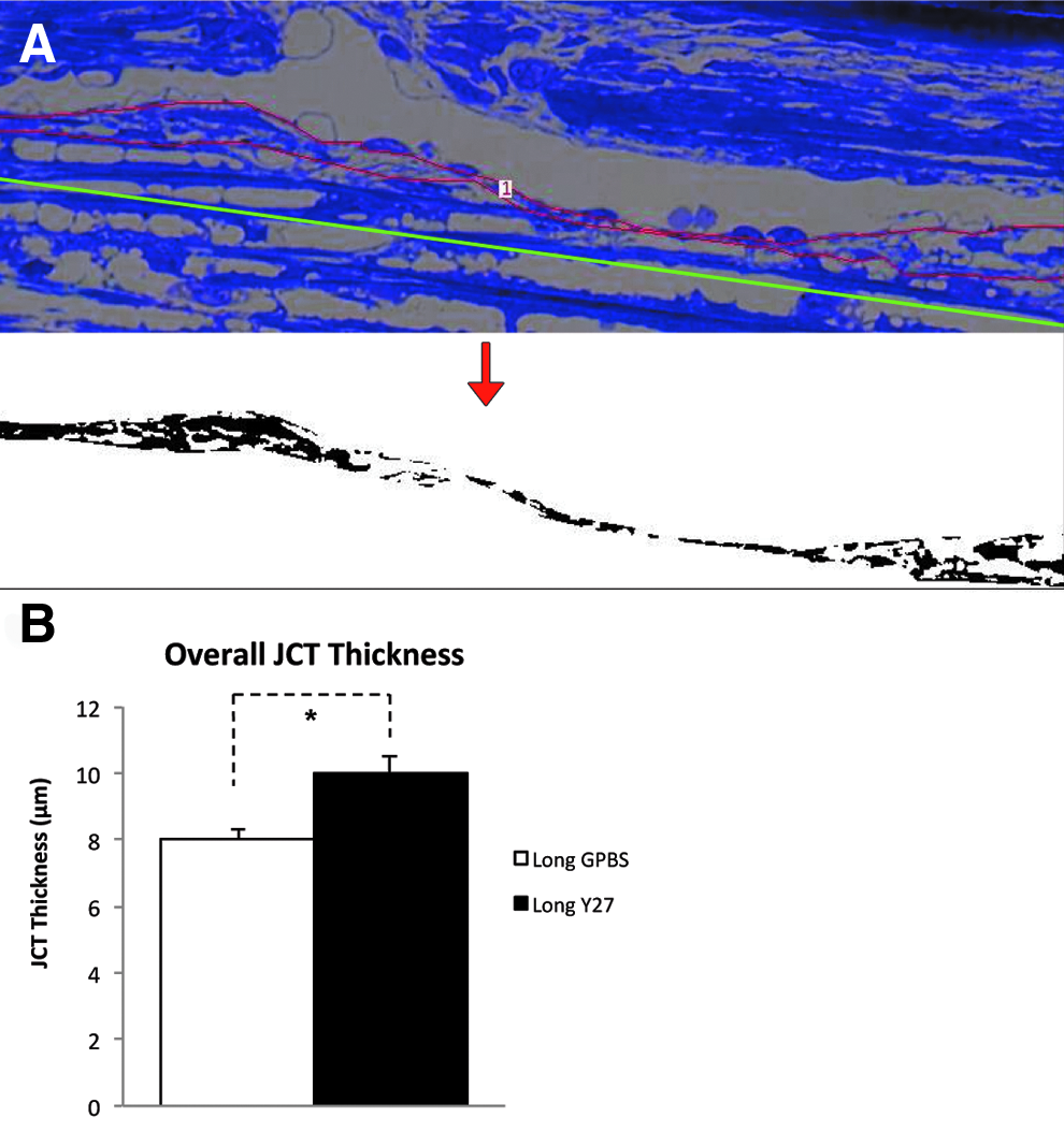
Morphological analysis of JCT thickness in human eyes.
JCT thickness in human eyes.

Correlation between changes in the JCT and outflow facility. In both bovine and monkey eyes, a significant positive correlation was found between percent separation length
Effect on pores and GVs
After traversing through the TM, the aqueous humor encounters the inner wall endothelium of SC, a confluent layer of cells lying upon a discontinuous basement membrane. The endothelium of SC forms 2 types of pores: intracellular pores and paracellular pores. 52 Aqueous humor is presumed to enter SC through these pores. To date, only one study has demonstrated an increase in number and size of paracellular pores after perfusing enucleated human eyes with a cytoskeletal disrupting agent, latrunculin-B. 53 Based on this study, one could infer that an increase in number and size of paracellular pores may also play a role in increased outflow facility by Rho-kinase inhibitors. On the other hand, the same group did not observe differences in paracellular pore density after perfusion with sphingosine-1-phosphate (S1P), an agonist of the Rho/ROCK pathway. 54 Therefore, whether drugs that target the Rho/ROCK pathway affect pore formation remains unclear.
Another feature of the endothelial lining cells of SC is the formation of GVs. GVs are pressure-sensitive structures caused by the pressure drop across the inner wall endothelium of SC. 55 When cells are attenuated and the cytoplasm becomes thin, intracellular pores are more likely to form and are often associated with GVs. An increase in GV density was reported near the CC ostia, where preferential flow exists. 56 Conflicting reports regarding increased GV formations after Rho-kinase inhibitors have made it difficult to associate these changes with higher aqueous outflow facility. There has been evidence that there is an increase in GV number compared to controls in Y-27632-treated enucleated porcine, 41 bovine, 23 and monkey24,42 eyes; however, there is also evidence that no increase in GV numbers exist in human eyes after Y-27632 treatment. 25 Conversely, agonists of Rho/ROCK pathway, lysophosphatidic acid or S1P, perfused in enucleated porcine eyes showed an increase in GV numbers, 21 whereas human eyes perfused with S1P showed no difference compared to its control. 54
Two potential mechanisms can be used to explain the conflicting findings regarding GVs: (1) modulation of the aqueous outflow resistance at the JCT/SC region affects the pressure drop across SC; (2) direct attenuation of SC endothelial cell stiffness and contractility. The first mechanism pertains to the modulation of morphology of the TM by expanding the JCT/TM, causing a more uniform flow and lower outflow resistance, according to the “funneling” model,11,48 and resulting in more GV collapse and therefore more difficulty detecting them. 53 The second possible mechanism relates to the direct attenuation of the SC cell stiffness and contractility and potentially increase GV formation. 47 However, whether Y-27632 has effects on the inner wall GV formation and pore density/size remains to be determined.
Effect on Effective Filtration Area
Aqueous outflow is segmental in normal eyes
Aqueous humor outflow through the trabecular outflow pathway has been reported to be segmental or circumferentially nonuniform through studies observing the distribution of pigment in the TM
57
and tracer perfused into the anterior chamber.23,24,57–65 These results suggest that at any given time, only a fraction of the outflow pathways are actively involved in aqueous humor drainage. This active area is termed the effective filtration area (EFA), which has been quantified by our group through the percent effective filtration length [PEFL=(length of the inner wall exhibiting tracer labeling
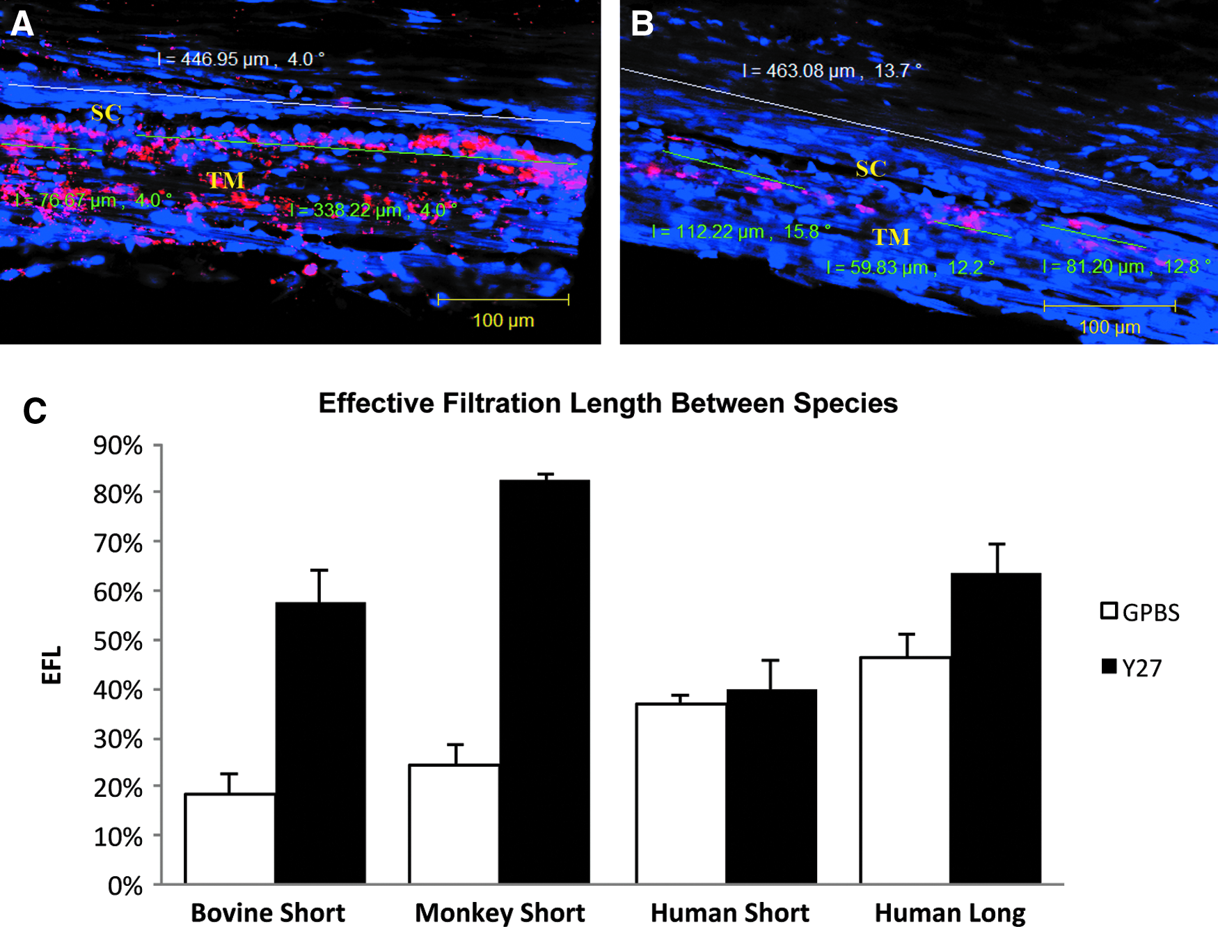
Confocal microscopy and percent effective filtration length (PEFL).
EFA decreases with increasing IOP and in POAG
EFA was found to reduce with acute elevation of IOP in bovine eyes. Outflow patterns in the JCT and inner wall transitioned from less segmental (more uniform) patterns at normal IOP (7 mmHg in enucleated eyes) to an increased segmental pattern at higher IOP (15–45 mmHg). 59 This decrease in EFA is associated with decreased outflow facility and is reversible when pressure is reduced from a high level to a normal level. 69 A decrease in EFA was also reported in chronic elevation of IOP in the laser-induced monkey glaucoma model, 70 with a lesser amount or no tracer was found in the areas of the TM where they had received laser injury, including the CC ostia region. Moreover, active outflow was found to be shifted away from areas with laser injury to areas without it. Significant reduction of EFA was also reported in POAG eyes compared to normal eyes in a tracer study (Cha ED, et al: Annual meeting of Association for Research in Vision and Ophthalmology (ARVO), Invest. Ophthalmol. Vis. Sci., 2013; 54:E-Abstract 2291). In addition, an inverse correlation between EFA and IOP was recently documented in an ocular hypotensive mouse model. 66 Collectively, these results suggest that the EFA is a valuable method of measuring outflow resistance and the effects of changes in IOP in humans and a number of different species.
EFA increases with Y-27632 treatment in bovine, monkey, and human eyes
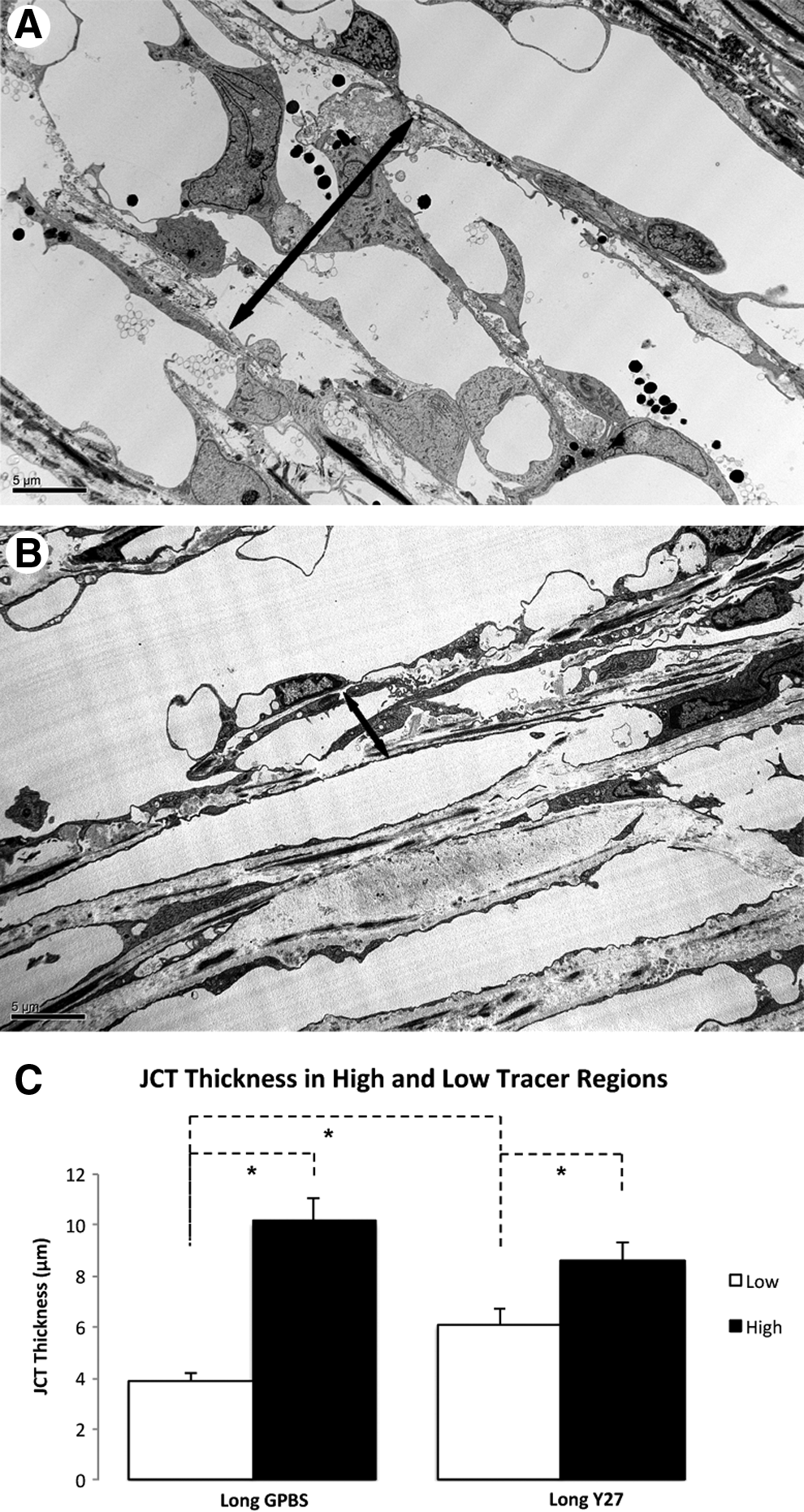
In addition to the different morphological changes that were found associated with increased outflow facility in bovine,23,24 monkey, 24 and human eyes 25 as described above, an increase in EFA was also found in all these 3 species following the Y-27632 treatment compared to their normal controls (Fig. 8). A positive correlation was found between outflow facility and EFA in all 3 species (Fig. 9). Similar to outflow facility, the magnitude of the increase in EFA varies among the species, where human eyes take a significantly longer time to observe a similar magnitude of change in outflow facility and EFA compared to bovine and monkey eyes (Fig. 8), presumably due to anatomical differences between species. It is also important to note that in these studies, an increase in outflow facility was shown to strongly correlate with an increase in EFA, regardless of the species or drug perfusion time (Fig. 9). Additionally, high tracer regions were also found to associate with an increase in TM thickness (innermost uveoscleral beam to the inner wall endothelium) in human eyes 25 (Fig. 10). These results suggest that active flow area can be used as a guiding tool to accurately assess morphological changes in the TM and their correlation with the changes in outflow facility and may also be used to study the effect of other drugs on the aqueous outflow pathway. One caveat of the studies involving the Rho/ROCK pathway is that none of its activators have studied whether EFA decreases with increased outflow resistance, presenting a void that needs to be filled in the future.
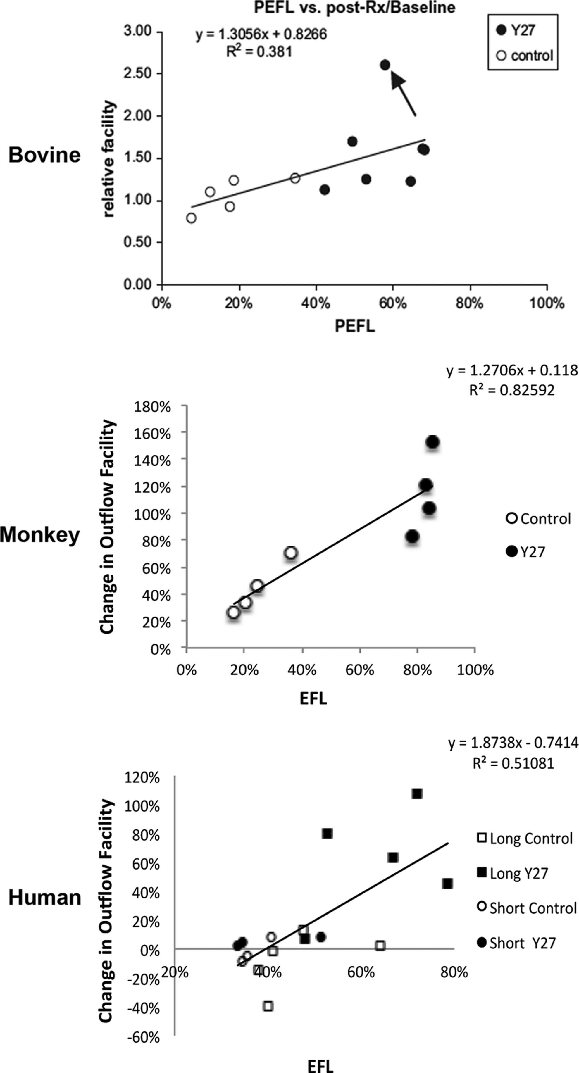
Correlation between the PEFL and outflow facility. A significant positive correlation was found between PEFL and change in outflow facility (with respect to baseline) in bovine, monkey, and human eyes with and without Y-27632 treatment. (Bovine data from Lu et al. 23 ; monkey data from Lu et al. 24 ; and human data from Yang et al. 25 )

TM thickness in high- and low-tracer regions of human eyes.
Summary
In summary, Rho-kinase inhibitors significantly increase outflow facility and IOP in various animal models and human eyes. The morphological changes in the TM associated with increased outflow facility include separation between the JCT and inner wall found in bovine eyes, separation between the JCT cells and between the JCT cells and their matrix found in monkey eyes, and an increased expansion in the TM and JCT in human eyes. Despite the different morphological changes in the JCT across these species, they all appear to correlate with percent changes of increased outflow facility. More importantly, these different morphological changes all resulted in an increase in EFA, which was positively correlated with an increase in outflow facility in all 3 species. These results suggest a link among changes in outflow facility, tissue architecture, and aqueous outflow pattern. With all these aqueous outflow-related changes considered, it is likely that Rho-kinase inhibitor Y-27632 increases outflow facility and thus lowers the IOP by redistributing aqueous outflow through a looser and larger area in the JCT. Whether the changes in the pore density/size of the inner wall also play a role in the increased outflow facility and EFA by Y-27632 remains to be determined. To date, all studies of the effect of Y-27632 on outflow facility and IOP have been performed in normal animal models and human eyes, and how its effect on altered structure of the trabecular outflow pathway in POAG eyes is warranted to be explored.
Footnotes
Acknowledgments
Original work was supported by BrightFocus Foundation (formerly American Health Assistance Foundation), NIH Grant EY018712, EY022634, the Boston University School of Medicine Wing Tat Lee Fund, and the Massachusetts Lions Eye Research Fund.
Author Disclosure Statement
The authors have no commercial relationships.