
Abstract
Abstract
Purpose:
The aim of the study was to determine the antimycotic effect of selected substances, povidone iodine at various concentrations and fluconazole, on the growth and development of Colletotrichum spp., which is one of the ocular pathogens.
Methods:
The materials used for the study consisted of 1-spore cultures of 4 fungal species of the genus Colletotrichum: C. dematium, C. gloeosporioides, C. acutatum, and C. coccodes. The method of poisoning culture media and the method of stippling the substance onto fungal colonies were used in the study. Different concentrations of fluconazole (1%) and povidone iodine (1%, 2% and 5%) were evaluated.
Results:
The growth of the studied fungal species was inhibited in 100% on the medium containing povidone iodine at the concentration of 1%, 2%, and 5%. After 24 h from the application of povidone iodine, a local disappearance of aerial mycelium was observed. In the case of C. coccodes, the colonies were not damaged. After 24 h from the application of fluconazole on C. dematium, C. gloeosporioides and C. acutatum colonies, slight disappearance of aerial mycelium was observed at these points. Despite dispensing the substance during the next few days, the inhibitory effect did not increase. After the application fluconazole on the C. coccodes colonies, the inhibitory effect of the preparation was not observed.
Conclusions:
The method of stippling of a preparation onto fungal colonies is a quick and reliable method to test many pharmacological substances. One percent, 2%, and 5% povidone iodine in culture medium is fungicidal for Colletotrichum spp. One percent fluconazole in culture medium is fungistatic for Colletotrichum spp. C. coccodes reveals the highest degree of insusceptibility to antimycotic treatment.
Introduction
M
Because of diagnostic problems and typical clinical symptoms, empiric antimycotic treatment is often required. A serious problem is the lack of available antimycotic substances in the form of eye drops. Great difficulties with such substances are observed because of their expensiveness. It is an economical problem for public hospitals. We observe not only infections caused by Candida, Aspergillus, and Fusarium, but also nowadays more often we notice Colletotrichum-induced ocular infections.2–4 Colletotrichum spp. can cause severe health problems for a human. Such infections spread especially among patients after ocular injuries caused by organic material, 3 and also among diabetic or immunosufficient ones. However, quite often they, as well, touch healthy people without any ocular problems.2,5 Colletotrichum dematium, Colletotrichum gloeosporioides, Colletotrichum coccodes, Colletotrichum acutatum, and Colletotrichum graminicola are regarded as dangerous and pathogenic for humans.6–8
Colletotrichum spp. are anamorphic stages of fungi of the family Glomerellaceae, class Sordariomycetes and type Ascomycota. 9 Conidia spreading these fungi are formed in acervuli under the cuticule or epidermis of above-ground parts of plants and in the animal tissues, including homeothermic animals.7,8,10–12
Colletotrichum spp. occur in different geographic regions of the world, both in hot and moderate climate, as polyphagic or monophagic species.10,12–14 Naturally, they may occur as saprotrophic species and cause diseases called anthracnose of various crop and wild species. Moreover, they can be harmful to homeothermic organisms, including humans.8,10,15,16 An eye infection may occur after mechanical introduction of spores into the eye during gardening, when tending to plants heavily infected by Colletotrichum spp. 8 These fungi can get into the eye tissues with dust particles carried by the wind.2,5 Because of the growing importance of Colletotrichum spp. in terms of crop production and human health, it is necessary to search the substances limiting the growth, development, and occurrence of these fungi. The sensitivity of these fungi to antifungal substances used in medicine has not been clearly defined yet.
Iodine preparations have been used for skin and mucous membrane disinfection for a long time. They have activity against bacteria, viruses, and fungi. Moreover, the ability to destroy fungal spores has been also described.17,18 In many cases, positive effects of treatment of giant conjunctival papillae and keratoconjunctivitis epidemica with povidone iodine have been observed.19–23 Fluconazole, miconazole, itraconazole, and voriconazole belong to antifungal azoles. They are used orally, topically, and as subconjunctival injections. 4 As an azole, fluconazole does not affect the immune system of the patient. 24
The aim of the study was to determine the antimycotic effect of selected substances, povidone iodine at various concentrations and fluconazole, on the growth and development of Colletotrichum spp. If these substances are shown to have an inhibitory effect Colletotrichum spp. it will probably allow us to use them more widely in the treatment of many ophthalmic fungal infections.
Methods
The materials used for the study consisted of 1-spore cultures of 4 fungal species of the genus Colletotrichum. These were C. dematium (Pers.) Grove and C. gloeosporioides (Penz.) Penz. et Sacc. [teleomorph Glomerella cingulata (Stoneman) Spauld. et H. Schrenk], from the collection of the Department of Plant Pathology and Mycology, University of Life Science in Lublin, isolated from caraway and angelica leaves, respectively,12,25 and C. coccodes (Wallr.) S. Hughes and C. acutatum J.H. Simmonds (teleomorph Glomerella acutata Guerber et J.C. Correll), from the Department of Plant Protection and Quarantine, University of Life Science in Lublin, obtained from the roots of sweet pepper and strawberry fruits, respectively (Hetman B and Wagner A. Fungi colonizing strawberries on a plantation and nursery. Horticultural Scientific Conference, 2013, Cracow, Poland; 59). 26
As the basis for the classification, the method of Kirk et al., 9 was used, while the names of fungal species and the author's epithets are given according to the current taxonomic status of species based on the Index Fungorum (2013).
The following isolates: K426 of C. dematium, A 271 C. gloeosporioides, T5 C. acutatum, and BPR 316 C. coccodes were used for laboratory tests. Moreover, the study investigated the application of povidone iodine at concentrations of 1%, 2%, and 5% and fluconazole at a concentration of 1%.
To identify possible fungicidal properties of fluconazole, in this study this substance was used at a concentration of 1%, which is higher than the concentration of 0.2% hitherto used in the form of eye drops.1,5
Two below described methods were used in the study:
1. The method of poisoning culture media described in a publication by Machowicz-Stefaniak and Zalewska.
27
This method involves the addition of suitable amounts of the substance in usable form to sterile culture medium PDA (Difco), cooled to 50°C. The fungal inoculum consisted of 3 mm diameter plugs taken from 2-week-old colonies of isolates growing on PDA (Difco) medium at a temperature 24°C. Four replications were made for each substance at the studied concentration and for each fungus. The control consisted of fungal colonies growing on the medium without the substances. The percentage of inhibition of 4- and 8-day-old fungal colonies growing on the medium with the substance in comparison to the control colony was adopted as the criterion of fungal sensitivity, according to the formula given by Machowicz-Stefaniak and Zalewska.
27
where I, percent of inhibition of fungal colony; C, diameter of the control fungal colony; T, diameter of the fungal colony on the medium with the substance.
After 4 and 8 days, microscopic observations of the morphological structures of the fungus growing on the medium with the substances were made to check their changes. If there was no growth of the fungus on the medium with the substance analyzed, the type of toxic activity against the fungus was determined. To this end, the fungal inoculum was transferred to Petri dishes with solidified PDA medium (Difco) without the substance. After keeping such dishes at a temperature of 24°C for several days, the effect of the substances on the fungi was determined by investigating the growth or death of the colonies.
2. The method of stippling onto the fungal colonies (the authors' own method).
Single inocula of the studied fungal species were placed in the middle of solidified PDA medium (Difco) in Petri dishes. The prepared material was incubated in a thermostat at a temperature of 24°C, for 8 days, that is, until the colonies grew to a suitable size. After this period of time, 2 μL of the substance was dropped using an automatic pipette ETTE PLUS-V at several points at the edge of the fungal colony. In separate plates, the substance was dispensed in the same way on sporulating acervuli located in the central part of the fungal colony. The control consisted of colonies on which physiological saline was applied.
The observations were conducted each day for 7 days. After each observation, the substance and physiological saline, respectively, were stippled at the same places. The changes in the morphology and internal structure of hyphae and conidia observed on microscope slides were adopted as the criterion of the effect of the substance on Colletotrichum spp. colonies.
Results
Description of isolates tested
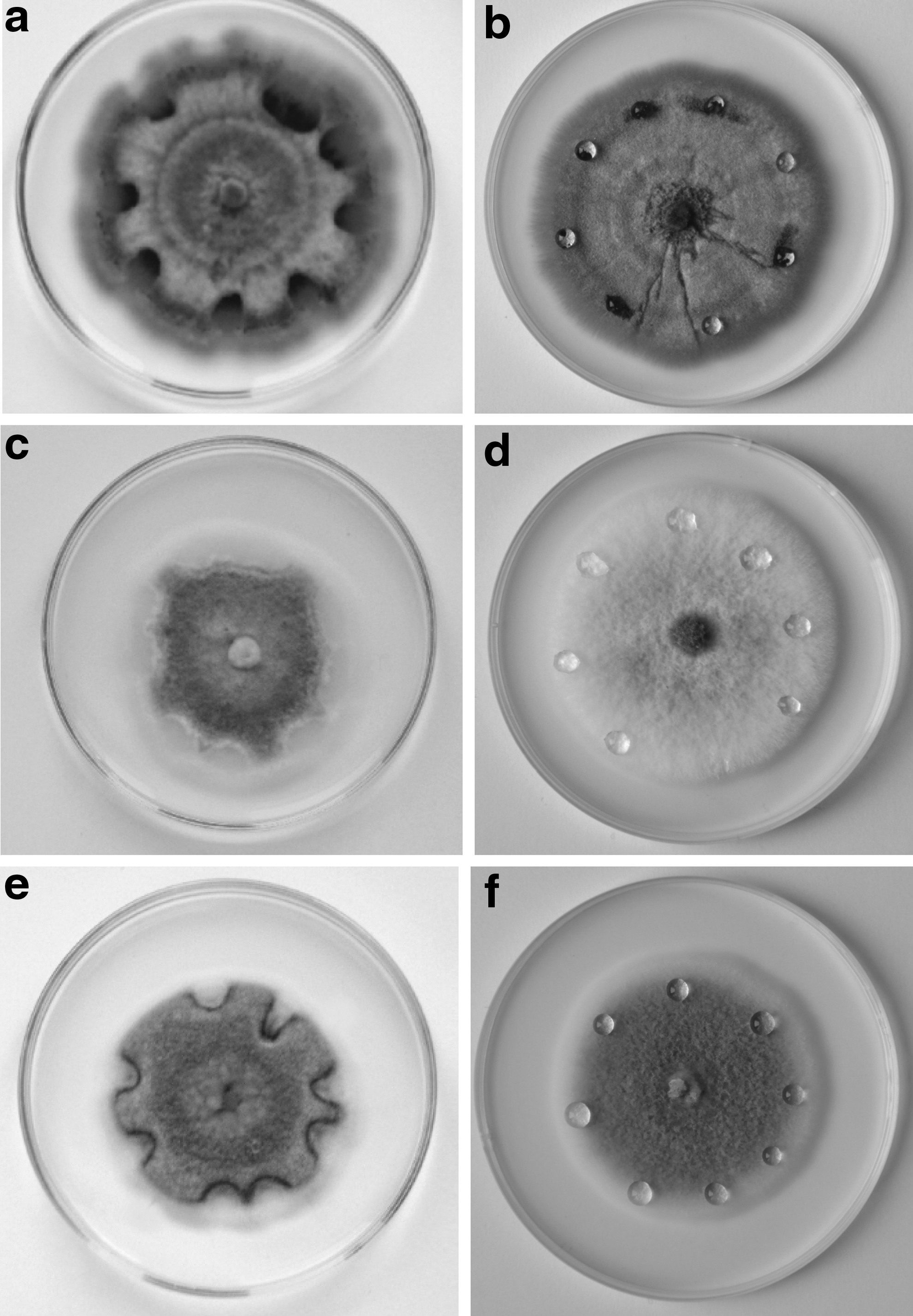
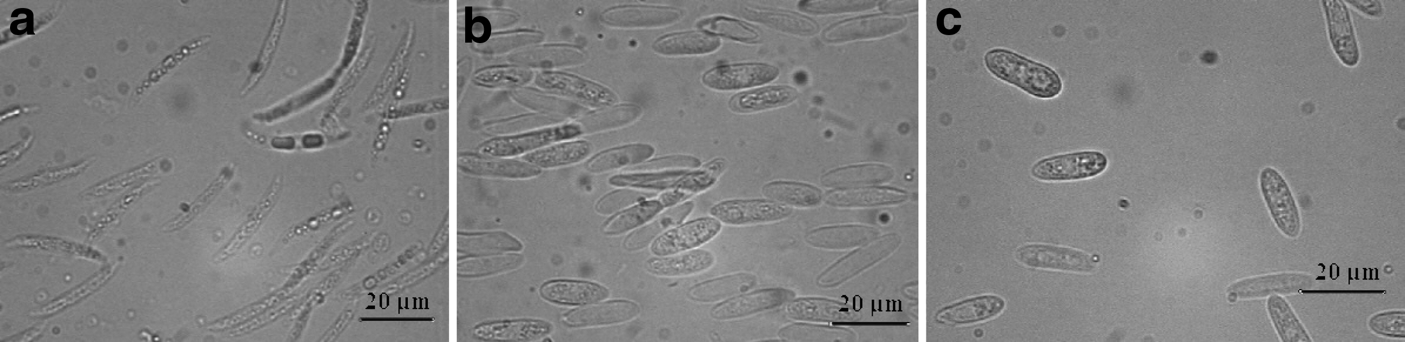
The colonies of C. dematium isolate K426 reached 6.0 cm in diameter after 8 days of growth (Fig. 1a). The mycelium hyphae formed compact, fluffy colonies of gray or dark gray color. The studied isolate intensely stained the medium pink (Fig. 1a). The acervuli of 495×372 μm in diameter formed dark brown setae around the ostiole and on the surface (Fig. 1b). Conidia were released from the acervuli in large numbers in the form of thick, viscous beige drops. The conidia were hyaline, 1-celled, sickle-shaped with sharp ends, with a visible drop in the middle, which refract light. The dimension of conidia on PDA medium ranged from 17.19 to 24.83×3.82–5.72 μm (Fig. 1c). Numerous brownish-black appressoria arranged at the ends or along the hyphae were a characteristic feature of the fungus (Fig. 1d). The studied isolates did not produce sclerotia.
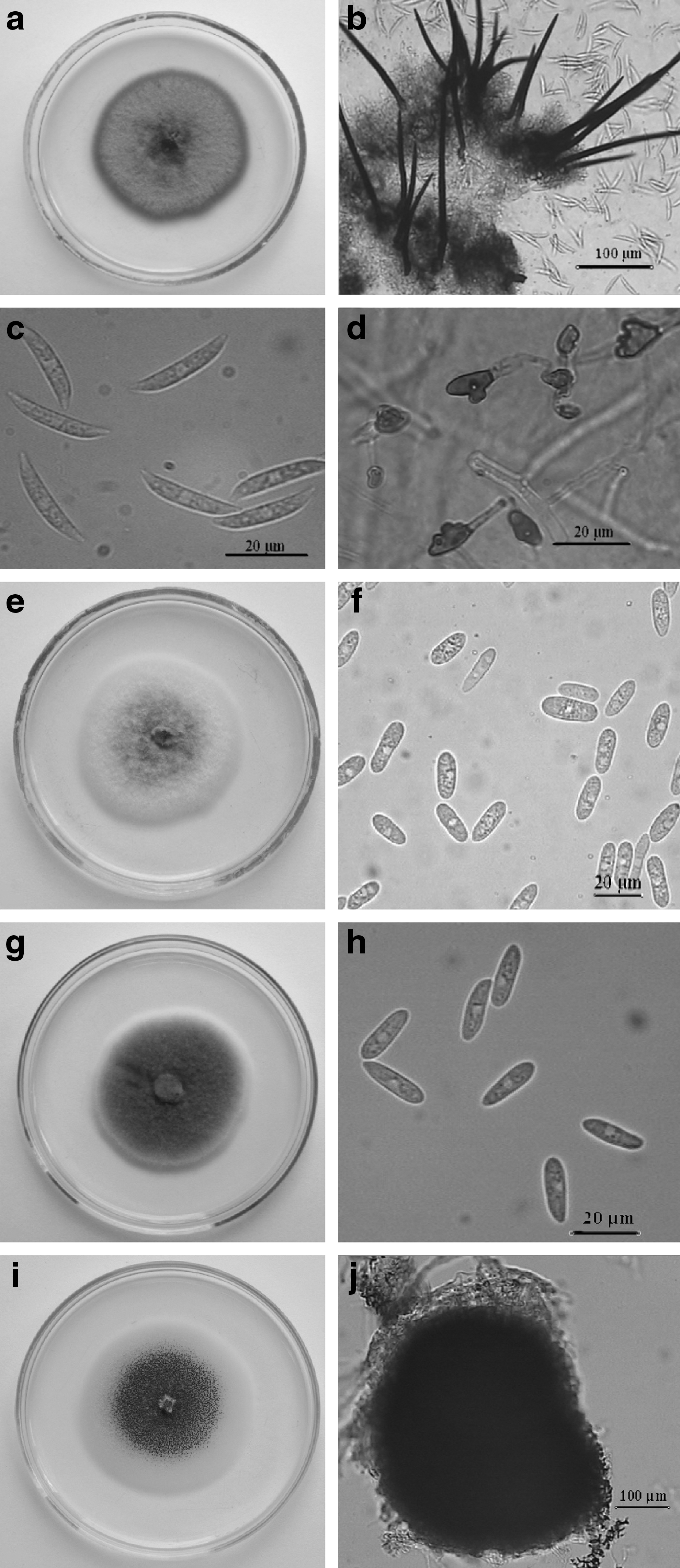
Colonies and morphological structures of Colletotrichum spp.: Colletotrichum dematium K 426,
The colonies of C. gloeosporioides isolate A 271 had 5.5 cm in diameter after 8 days of growth (Fig. 1e). At the beginning colorless, with time the aerial mycelium was getting gray. On their margins, the colonies were flat, but slightly fluffy in the central part (Fig. 1e). Acervuli, with the dimensions from 135 to 378 μm, formed in large numbers, singly or in clusters, over the entire surface of the colony. Thick salmon-colored drops, containing huge amounts of conidia were released in great numbers from the ostiole of the acervuli (Fig. 1f). These conidia were 1-celled, hyaline, elongated with narrow walls, and with one or both ends rounded. The dimensions of conidia on PDA were 11.1–23.35×3.5–6.4 μm (Fig. 1f). The tested isolate did not produce either setae or sclerotia.
The colonies of C. acutatum isolate T5 reached 5.5 cm in diameter after 8 days of growth (Fig. 1g). The appearance of the colonies of the tested isolates was similar to those of C. gloeosporioides. Initially loose and bright, after 3–4 days of growth the aerial mycelium became dark gray, cottony or fluffy, forming colonies up to 3–4 mm in height. Very numerous acervuli were observed especially in the central part of the colony. Conidia were released from the acervuli in the form of mucous orange-colored drops. The conidia were oval or spindle-shaped, straight with the dimensions of 8.05–15.5×2.8–4 μm on PDA medium (Fig. 1h). There was no presence of setae or sclerotia.
The colonies of C. coccodes isolate BPR 316 reached 6.5 mm in diameter after 8 days of growth. The aerial mycelium was very poor, slightly visible, whitish, trailing on the surface of the culture medium. The studied isolate started to form microsclerotia just after 3–4 days of growth. The number of microsclerotia increased steadily until they evenly covered the entire surface of the colony (Fig. 1i). Sclerotia formed individually. They were black, roundish or comma-shaped, macroscopically visible, and reaching a size from 100 to 500 μm (Fig. 1j). The tested isolate did not produce conidia until the 14th day of culture.
Effect of the substance on fungi through culture medium
The growth of colonies of the studied fungal species was inhibited in 100% on the medium containing povidone iodine at the concentration of 1%, 2%, and 5% compared to control (Table 1 and Fig. 2a–c). Complete inhibition of the growth of the fungal colonies occurred both after 4 and 8 days of the contact of mycelium with the medium containing the preparation (Table 1).
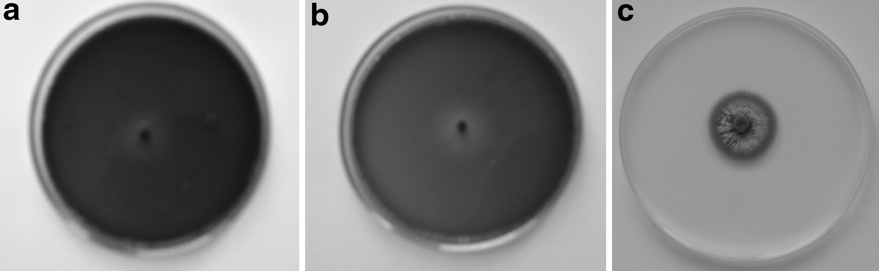
Growth inhibition of C. dematium K 426 on PDA with an addition of povidone iodine 2%
Values marked with the same letter do not differ significantly.
LSD, low significant difference.
The percentage of inhibition of fungal colonies growth ranged from 87.08% to 98.71% on the medium with fluconazole at the concentration of 1%, depending on the fungal species and the duration of the substance action (Table 1). The inhibitory effect of C. dematium, C. gloeosporioides, and C. acutatum on colony growth was significantly higher after 8 days than after 4 days of cultivation (Table 1). The percentage of inhibition of 4- and 8-day-old colonies of C. coccodes by fluconazole at the concentration of 1% was not significantly different from the percentage of inhibition of 8-day-old colonies of the other fungal species. On the other side, it was significantly higher than the inhibition of 4-day-old colonies of C. dematium, C. gloeosporioides, and C. acutatum (Table 1). The percentage of inhibition of 4- and 8-day-old colonies of C. coccodes by fluconazole at the concentration of 1% was not significantly different from the percentage of inhibition of the colonies growth of the same fungus species caused by povidone iodine at the concentration of 1%, 2%, and 5% (Table 1). In the case of the other fungal species, the percentage of inhibition of colony growth caused by fluconazole was significantly lower than that caused by povidone iodine at the concentration of 1%, 2%, and 5% (Table 1).
As regards the substances applied using the method of poisoning culture medium, only povidone iodine was fungicidal to C. dematium, C. gloeosporioides, and C. acutatum at each of the concentration (Table 2). Microscopic examination of the mycelium in contact with medium containing povidone iodine for 4 and 8 days showed strong degenerative changes in mycelium hyphae of the studied isolates. It was the detachment of the cytoplasm from the cell wall, abnormal grains, lysis of hyphae, and the formation of short and thick branches or strands. This deformation resulted in the inability to form acervuli, conidia, and young mycelial hyphae (Fig. 3). The effect of povidone iodine was fungistatic to C. coccodes at the concentration of 1%, 2%, and 5% (Table 2).
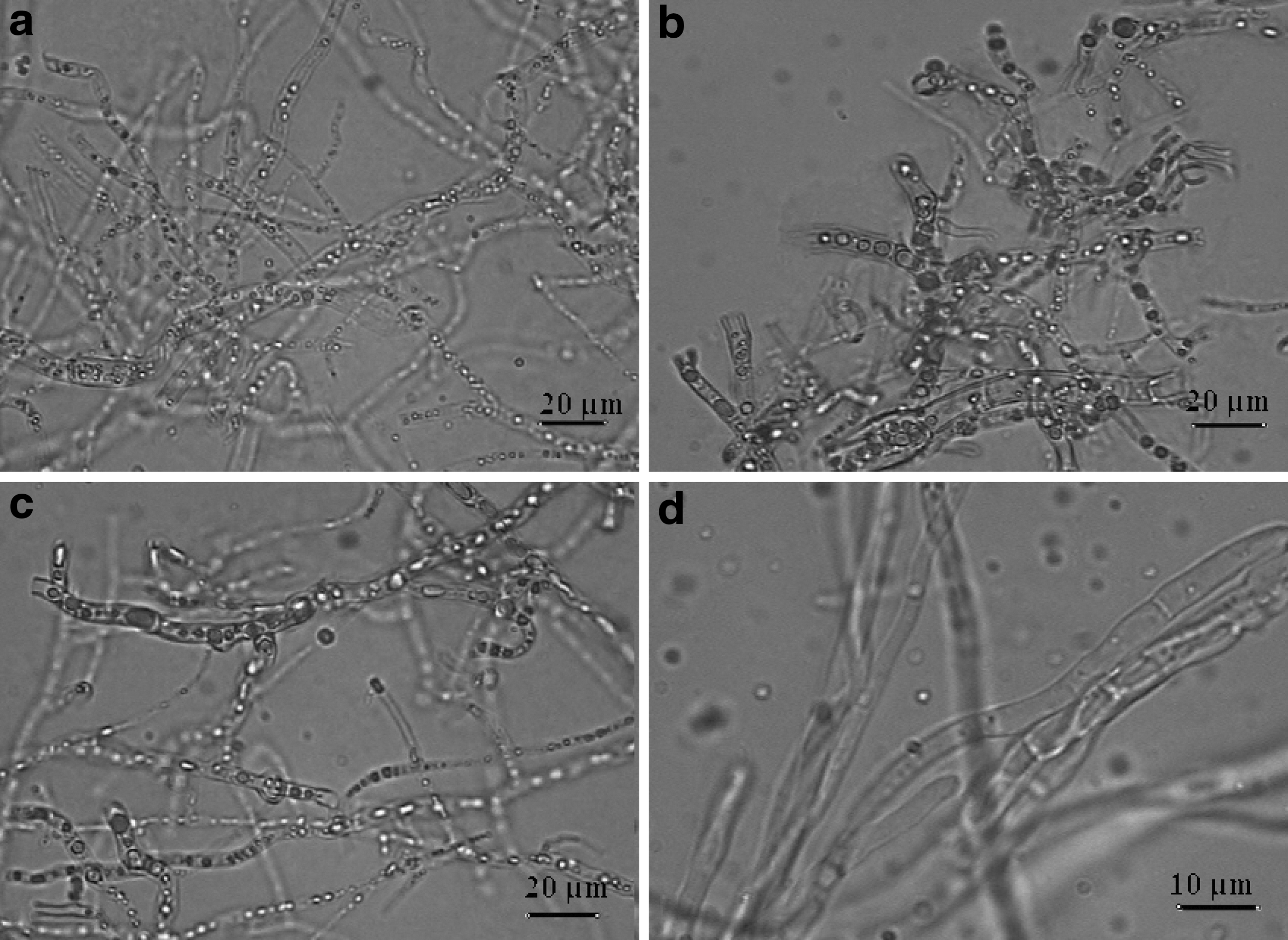
Degeneration of C. dematium K 426 hyphae on PDA after 8 days of effect of povidone iodine 5%
—, Fungicidal activity; +, fungistatic activity.
The effect of 1% fluconazole on Colletotrichum spp., regardless of the fungal species, was fungistatic (Table 2). Hyphae with the signs of degenerations and numerous leaving hyphae uniformly filled with cytoplasm, young acervuli, and germinating conidia were found in the mycelium in contact with fluconazole-containing medium for 4 and 8 days (Fig. 4).
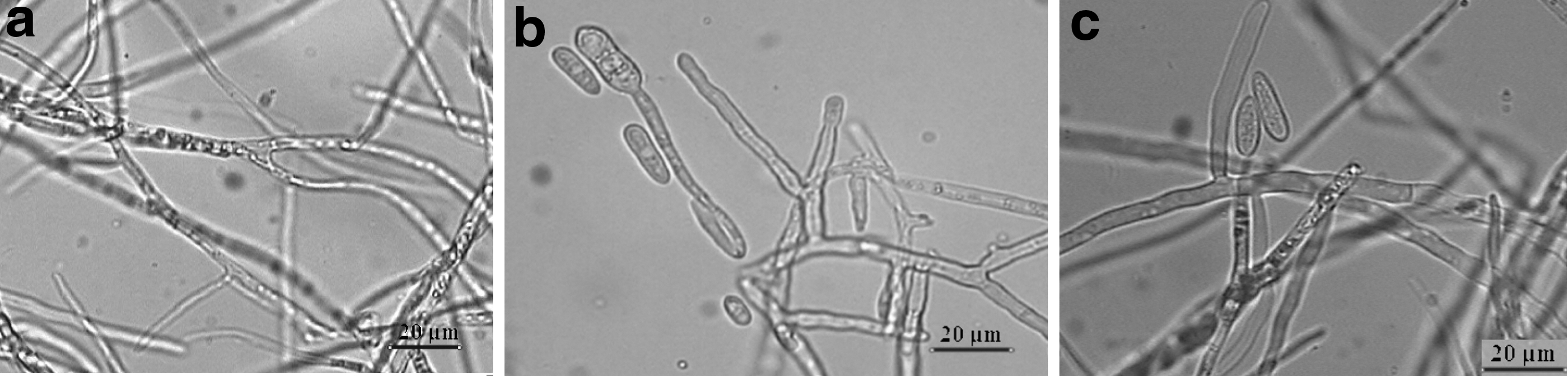
Hyphae
Effect of stippling the substance onto fungal colonies
After 24 h from the application of povidone iodine at the concentration of 1%, 2%, and 5% to the colonies of C. dematium, C. gloeosporioides, and C. acutatum, a local disappearance of aerial mycelium was observed contrary to control colonies, where normal saline was applicated (Fig. 5). The disapperance of mycelium continued as a result of the repeated daily stippling. Only in the case of C. coccodes, the species forming microsclerotia, the colonies were not damaged.
Disappearance of colonies: C. dematium K 426
After 24 h from the application of fluconazole drops at the concentration of 1% on C. dematium, C. gloeosporioides, and C. acutatum colonies, a slight disappearance of aerial mycelium was observed at these points. In spite of stippling fluconazole in the next few days, the inhibitory effect of the preparation did not increase. After the application of 1% fluconazole droplets to the colonies of C. coccodes the inhibitory effect of preparation was not observed.
After 48 h, that is, after 2 applications of povidone iodine at the concentration of 2% and 5% the mycelium hyphae of C. dematium, C. gloeosporioides, and C. acutatum showed signs of very strong degeneration. New acervuli and conidia did not form from such mycelium, in contrast with the control colony. After the application of 1% povidone iodine drops, the fungal hyphae degenerated, but living, growing hyphae were also observed, as in the control colony. Moreover, new forming conidia, fine-grained and evenly filled with cytoplasm were also observed.
After 48 h from the point application of fluconazole at the concentration of 1% to the colonies of the studied isolates, degenerated hyphae were observed apart from normal, healthy hyphae. In the case of C. coccodes, the point application of the preparation, particularly using of povidone iodine, only caused softening and disintegration of microsclerotia without damage to their viability.
After a single application of drops of the substance on sporulating acervuli of Colletotrichum spp., no negative changes in the structure of conidia were observed. However, after the application of the preparation during the 4 following days, 80% degeneration of conidia occurred in C. dematium and about 50% in C. gloeosporioides and C. acutatum. The spores were deformed, had a coarse-grained cytoplasm, and in many cases the loss of cell wall was observed (Fig. 6). However, after stop of stippling for the next 4 days, a quite quick recovery followed and new living conidia, with the ability to germinate started to form again.
Degeneration of conidia C. dematium K 426
Discussion
The selected species of Colletotrichum allowed us to investigate the effect of the studied substances on the morphological structures formed by these fungi. Under these particular conditions, fluconazole and povidone iodine showed a significant inhibitory effect on Colletotrichum spp. The inhibitory effect of the preparations consisted in complete or local degeneration of hyphae, conidia, and overwintering forms, that is, sclerotia, of the studied fungi.
Povidone iodine proved to be more toxic to Colletotrichum spp. than fluconazole. Under the conditions of continuous contact of the substance with the fungi through the culture medium, we observed the fungicidal effect regardless of the tested concentration of the substance. Preparations capable of destroying mycelium are very valuable for practical reasons, because they prevent mycelium regeneration and provide a permanent effect of treatment. It should be stressed that povidone iodine is fungicidal to aerial mycelium and substrate mycelium of C. dematium, C. gloeosporioides, and C. acutatum. In the case of overwintering forms C. coccodes—sclerotia, which can survive up to 7 years in nature, 11 povidone iodine demonstrated only a fungistatic effect. This allows us to presume that this species, having the ability to cause corneal inflammatory reactions without any ocular trauma 28 and exhibiting drug resistance, as shown in our study, is very dangerous for human tissues and exceptionally difficult to eradicate.
Our study has shown the high sensitivity of Colletotrichum genera, especially C. dematium, C. gloeosporioides, and C. acutatum, to povidone iodine at different concentrations. It appears that the 2% concentration of povidone iodine can be recommended for further testing.
Our investigations have also revealed that fluconazole, contrary to povidone iodine, is not fungicidal to Colletotrichum spp. even at 1% concentration. The local application of the pharmacological substances on the fungal colonies for 5 days was not enough to completely destroy the mycelium.
Fernandez et al., 1 analyzed corneal inflammation caused by Colletotrichum spp. and noticed insusceptibility to 5% natamycin in 3 isolates and in 1 isolate complete resistance to fluconazole. Similar resistance to fluconazole and natamycin has been described by Shiraishi et al., 3 in the case of corneal inflammation caused by C. gloeosporioides. This information is in agreement with our observations.
According to Mitani et al., 2 and Yamamoto et al., 5 successful treatment of fungal corneal inflammation was observed after 2-month application of fluconazole in tablets, administrated topically and in subconjunctival injections. Fluconazole is an antimycotic azole that does not affect the human immunological system, but its effect is only fungistatic. 24 Some authors have used a concentration of fluconazole higher than 1%. In another experiment, Yilmaz and Maden, 29 described quite good results in 13 cases of corneal mycosis treated with subconjunctival injections of 2% fluconazole over a period of 5–13 days of treatment.
In corneal mycosis caused by Candida spp., Aspergillus spp., and Penicillium spp. Madhy et al. 30 used 0.2% amphotericine B topically and 2% fluconazole in subconjunctival injections. In 75% of cases, they found corneal ulcers to be completely healed after a period of 4–6 weeks of treatment.
In this study, the stippling of the substances in the form of drops onto the fungal colonies or sporulating acervuli was a good method that allowed us to determine the inhibitory effect of the preparations very quickly, already after 24 h from the application. Such application of the treatment substance caused the degeneration and atrophy of the aerial mycelium of the Colletotrichum colonies, if only the treatment was repeated systematically for several days.
Fluconazole at the concentration 1% tested in the form of drops and even povidone iodine–especially at a 1% concentration, showed a weaker destructive effect on C. coccodes sclerotia compared with their application through culture medium. Similar results were obtained by Guarraro et al., 31 who found significant insusceptibility of C. coccodes to antimycotic drugs, especially azoles such as fluconazole.
The inhibitory effect of pharmacological substances on fungi depends on the type of application of the treatment substance, its concentration, and the nature of the growth and properties of the fungus. It appears to be crucial that the treatment substance should be in continuous contact with the fungal inoculum. It was shown in the cases of povidone iodine applied through culture medium and by regular stippling onto fungi colonies in a repeated way. Among the 2 tested methods, the stippling appears to be very quick and reliable to test many pharmacological substances.
Our observations and results show that the complete elimination of Colletotrichum spp. is a very difficult and long-lasting process that requires the treatment substance to be applied regularly.
The human eye appears to be a very good environment for fungi, because of its high moisture and temperature,7,8 which are very similar to requirements of Colletotrichum spp. (especially Colletotrichum dematium, unpublished data). C. dematium is a very common in nature all over the world and therefore this may suggest that these fungi will play an increasing role in medicine.
Conclusions
Povidone iodine at the concentration of 1%, 2%, and 5% in culture medium is fungicidal to Colletotrichum spp, while 1% fluconazole is fungistatic. Applied locally, 2% and 5% povidone iodine causes strong degeneration and disappearance of aerial mycelium after 24 h. However, 1% povidone iodine and 1% fluconazole are not effective in complete eradication of mycelium of Colletotrichum spp. C. coccodes exhibits the highest degree of resistance to antimycotic treatment. The method of stippling of a preparation onto fungal colonies is a very quick and reliable method to test many pharmacological substances.
Footnotes
Author Disclosure Statement
No competing financial interests exist.