




Abstract
Abstract
Purpose:
To design an MG132-sustained drug delivery capsular ring (SDDCR) and investigate its effect on the inhibition of posterior capsule opacification (PCO) in a rabbit model.
Methods:
The SDDCRs were prepared by forming a slice of film made by the mixture of poly lactic-co-glycolic acid (PLGA) and MG132 on the surface of capsular tension rings (CTRs). The drug-loading capacity, entrapment efficiency, and in vitro release of the drug-containing film were detected. Eighteen New Zealand white rabbits were operated with phacoemulsification and MG132-SDDCRs/PLGA-CTRs/CTRs implantation in the single eye. The images of the anterior segments were acquired at certain days, and the epithelial–mesenchymal transition (EMT) markers were detected by western blot and immunofluorescence.
Results:
The drug-loading capacity and entrapment efficiency of MG132-SDDCRs were 1.15% ± 0.04% and 66.16% ± 0.027%, respectively, and the drug released well within a month. The PCO degree of the MG132-SDDCR group was significantly lower than the other groups. The expression of alpha-smooth muscle actin, fibronectin, vimentin, and collagen-I was lower, and the expression of E-cadherin (E-cad) was higher in the MG132-SDDCR group than the other groups.
Conclusions:
MG132-SDDCRs could be established successfully. The PCO process was prevented, and the expression of EMT markers was inhibited by the implantation of MG132-SDDCRs, indicating that this could be a potential treatment against PCO.
Introduction
P
PCO is to some extent postponed by modifying the IOL surface material, 5 improving the surgical methods,6,7 antimetabolites,8,9 gene therapy,10,11 and the application of lens capsular tension rings (CTRs),12,13 but so far none of them can solve the long-term problem of PCO.1,3,8 The pathological basis of PCO is the proliferation, migration, and epithelial–mesenchymal transition (EMT) of the residual lens epithelial cells (LECs) after surgeries, which cover the posterior capsule together with the fibers.14–16 Transforming growth factor-β (TGF-β) plays a vital part in promoting the expression of EMT markers, such as alpha-smooth muscle actin (α-SMA), fibronectin (FN), and vimentin, and promotes the proliferation of collagen. 14 Compared with some other therapeutic strategies, drugs aiming the corresponding targets of the LECs own the advantages of preventing the proliferation, migration, and EMT of the LECs directly and specifically. However, the drug concentration is difficult to be maintained through traditional methods of administration such as drug-administrated infusion, anterior injection, and subconjunctival injection ascribed to aqueous humor circulation. Therefore, establishing a reliable sustained drug delivery system (SDDS) is of great importance to the prevention of PCO.
Tetz et al. first implanted an IOL-bound SDDS carrying the drug daunorubicin or indomethacin into rabbit eyes and demonstrated that the PCO formation was reduced by ∼50%. 17 Compared with IOL-bound SDDS, sustained drug delivery capsular rings (SDDCRs) might have certain advantages because the CTRs themselves can inhibit the migration of the LECs at the equator area. Moreover, the delivery might be more stable on the CTRs because of the smaller surface area. Pandey et al. reported that an intracapsular ring releasing 5-fluorouracil (5-FU) might prevent central PCO by mechanically blocking the migration of the LECs. 18 Other studies also showed some similar results, indicating that this might be an improvement for drug therapy in the prevention of PCO.19,20
The peptide-aldehyde proteasome inhibitor MG132 (carbobenzoxyl-
Methods
Preparation of SDDCRs
Five milligrams of MG132 and 300 mg of poly lactic-co-glycolic acid (PLGA) (PLA/PGA = 80/20, Mw = 80,000 g/mol) were precisely weighed and dissolved in 2 mL acetone (MG132 con. 5.23 mM, PLGA con. 15%w/v) at room temperature (RT). The CTRs were quickly immersed into the MG132-PLGA acetone solution, and then a slice of film was formed on the surface. The filmed CTRs were dried at RT for 30 min and then put into the vacuum centrifuge for 24 h (Centrifuge: Eppendorf company, Germany. Vacuum pump: Sanli chemical instrument company, Shenzhen, China). After the acetone was volatilized, the SDDCRs were sterilized under ethylene oxide and packaged for use.
Detection of the drug-loading capacity, entrapment efficiency, and in vitro release
Because the actual quality of the drug-loaded film on the CTRs was difficult to be weighed, additional drug-loaded films were prepared for the detection of the drug-loading capacity and entrapment efficiency. First, 50 μL MG132-PLGA acetone solution was pipetted and then dropped onto the surface of distilled water to form the drug-loaded film. Then the film was taken out, washed by distilled water, dried at RT for 30 min, put into vacuum centrifuge for 12 h, and then weighed by a high precision electronic balance (Acculab ALC-110.4; Sartorius Stedim Biotech, Germany). The film was then dissolved completely into 100 μL acetone at RT. After the solution was filtered through 0.22 μm millipore filter, the concentration of MG132 in the filtrate was detected by BCA-100 Protein Assay kit (Bocai Biotech, Shanghai, China) under the manufacture's instruction. Then the drug-loading capacity and entrapment efficiency were calculated according to the following formula:
To evaluate the in vitro release of the drug-loaded film, the CTRs (n = 3) were immersed in 300 μL phosphate-buffered saline (PBS, pH = 7.4) at 37°C. The concentration of MG132 was detected by the BCA-100 kit after 1, 3, and 5 days, and 1, 2, 3, 4, and 5 weeks with the solution centrifuged, the supernate was pipetted out and the fresh PBS was added in.
Establishment of the rabbit PCO model
Eighteen 3–4 months old New Zealand white rabbits (purchased from the Zhongshan Ophthalmic Center, Sun Yat-sen University) were used in this study. All rabbits were treated in accordance with the ARVO and Zhongshan Ophthalmic center statements for the use of animals in ophthalmic and vision research. The rabbits were divided into the experimental group (n = 6), control group (n = 6), and blank control group (n = 6), which were implanted with MG132-SDDCRs, PLGA-CTRs, and CTRs, respectively.
All the rabbits received sham monocular phacoemulsification surgeries performed by one ophthalmologist (Dr. Mingxing Wu). The procedures were as follows: the rabbits first received intramuscular injection with 0.1 mL sumianxin (a compound of xylazole, EDTA, dihydroetorphine hydrochloride, and haloperidol). Then, subsequent 1.2 mL/kg 1% pentobarbital sodium was injected into the marginal vein of the rabbits to induce anesthesia. The eyeball was cleaned by saline and 0.25% povidone-iodine, topically anesthetized with alcaine, and then a 3.2 mm perilimbal incision was made. Viscoelastic agent was injected and continuous curvilinear capsulorhexis with a diameter of 5 mm was completed. The nucleus was extracted by the phaco handpiece (Alcon Infinity System) under continuous irrigation with 0.2% heparin sodium after hydrodissection and hydrodelineation. The remaining cortex was removed by irrigation/aspiration, and then the SDDCR/PLGA-CTR/CTR was implanted into the capsular bag. Postoperative medication included atropine eyedrops, tobramycin and dexamethasone compound eyedrops, and tobramycin and dexamethasone compound ointment for a week. The rabbits were sacrificed by air embolism at a certain stage. After the eyeballs were separated under direct vision, the capsular bags were separated completely under dissecting microscopy for subsequent experiments.
Assessment of PCO degree and toxicity to anterior segment
The anterior segment photography of the rabbits was performed with a slit lamp (TOPCON SL0D8Z) with a digital camera on the 1st, 2nd, 5th, 10th, 13th, 17th, and 22nd day after the surgeries. The PCO degree was quantified by POCOman, a program designed by Lloyd Bender (Thomas' Hospital, London, United Kingdom). 27 Posterior capsule opacification was evaluated by 3 grades: grade 1 (light): small fibers; grade 2 (medium): more fibers with pearl-like structures; and grade 3 (severe): complete opacity. The final PCO value was a weighted average of the 3 grades scored from 0 (complete transparency) to 3 (complete opaque).
The toxicity of the SDDCRs was assessed by corneal edema and inflammation evaluated by anterior segment photography and the IOP measured by a springback tonometry (iCARE company, Finland).
Western blot
Posterior capsular bags from the group MG132-SDDCR (n = 3) and group PLGA-CTR (n = 3) were used for detecting the EMT markers (α-SMA, FN, vimentin, Collagen-I, and E-cad) by western blot. The capsular bags were separated integrally with the surrounding tissues and capsular rings removed clearly under the dissecting microscope. The bags were then washed with PBS and smashed in an ultrasonic cell disruption system together with 100 μL precooled RIPA lysis buffer. The specimens were centrifuged at 4°C at 8,000 rpm for 10 min, and the supernatant was collected. Protein concentrations were detected with a BCA kit. The specimens were mixed with sodium dodecyl sulfate-polyacrylamide gel electrophoresis (SDS-PAGE) loading buffer, boiled for 10 min, separated by SDS-PAGE, and then transferred to a polyvinylidene fluoride membrane. PBS with 0.1% Tween-20 (TBST) containing 5% defatted milk was used to block nonspecific binding sites for 1 h. The membrane was incubated with primary antibodies at 4°C overnight and washed with PBST (0.1% Tween-20 in PBS) 3 times for 5 min. The membrane was then incubated with horseradish peroxidase-conjugated secondary antibody for 1 h at RT and then washed with PBST 3 times for 5 min. The membrane was then observed under a FluorChem FC3 western system (ProteinSimple company).
Immunofluorescence
Another 2 groups of rabbits (n = 3) were used to evaluate the expression of α-SMA, using an immunofluorescence assay. The methods were as follows: the capsular bags were separated, cleaned and dried for 5 min at RT, and then fixed with 100% precooled formaldehyde at 4°C for 10 min. After washed with PBS 3 times for 5 min, the specimens were blocked in 5% bovine serum albumin at RT for 1 h and were then incubated with anti-α-SMA antibodies at 4°C overnight. They were then washed with PBS twice for 5 min. The specimens were incubated with fluorescent secondary antibodies for 1 h at RT and washed with PBS twice for 5 min. The cell nuclei were stained with propidium iodide for 5 min. After washed twice with PBS, the specimens were covered by cover slips and viewed with a laser scanning confocal microscope.
Statistical analysis
Mann–Whitney U test was used to analyze the PCO degree and the IOP values of different groups. P < 0.05 was considered as significantly different.
Results
SDDCRs were fulfilling in drug-loading capacity, entrapment efficiency, and in vitro release
The SDDCRs showed similar shape, tenacity, and plasticity to normal CTRs, which made them easy to implant into the capsular bags (Fig. 1). The drug-loading capacity of the drug-loaded film was 1.15% ± 0.04%, and the entrapment efficiency was 66.16% ± 0.03%. MG132 was detected from the 3rd day to the 35th day with a peak of 3.443 ± 0.56 ng/μL at the 14th day in the in vitro release experiment (Fig. 2).

The SDDCRs showed similar characteristics to normal CTRs. The SDDCRS and the CTRs were photographed by a camera naturally
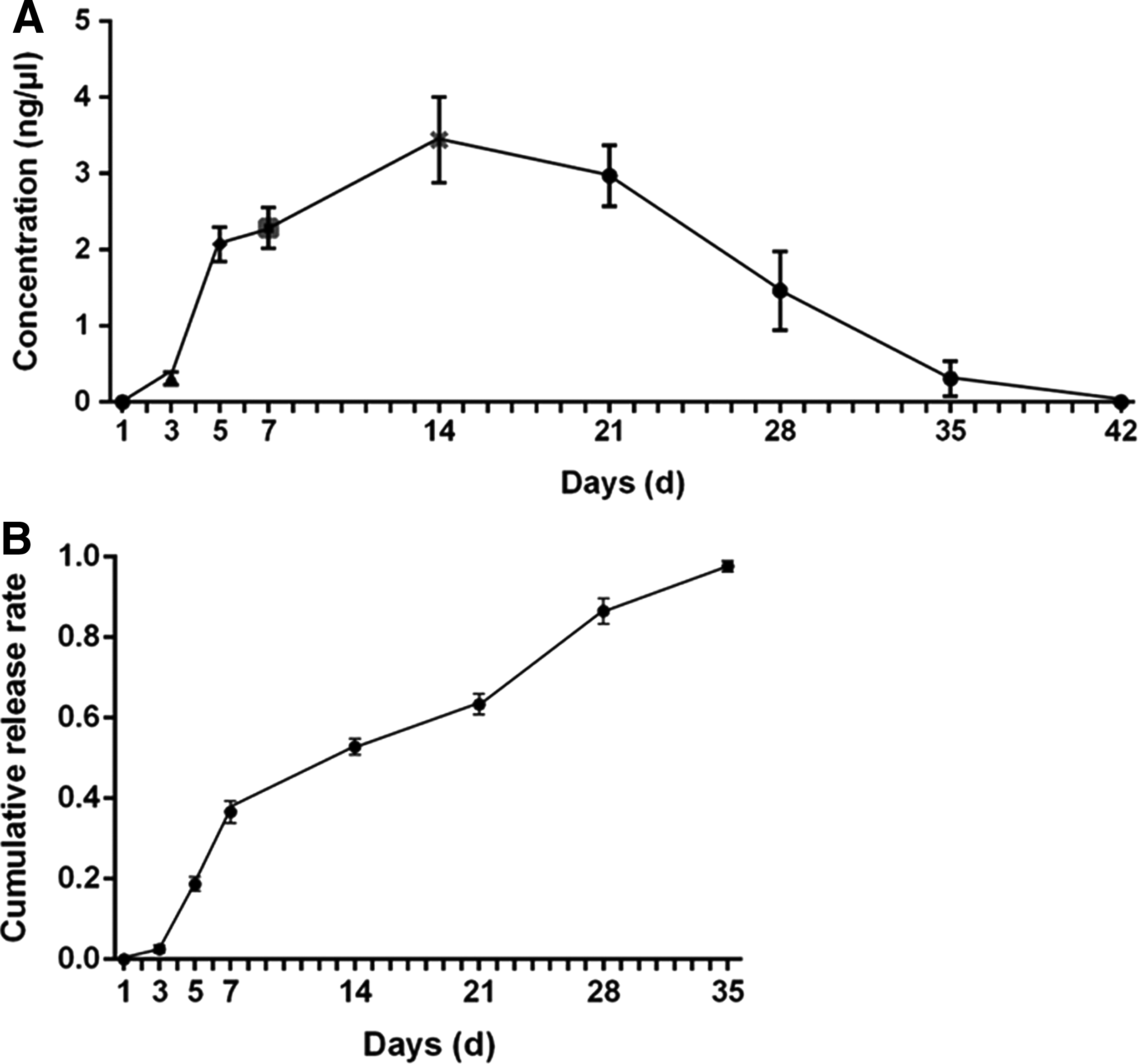
MG132-PLGA drug-loading film showed well in vitro release. The concentration of MG132 per day
The PCO score was lower in the MG132-SDDCR group than control
Neither the MG132-SDDCR group nor the control groups showed PCO till the 5th day after surgeries, and no noticeable proliferation of LECs was found in all groups. Both the PLGA-CTR and the CTR groups showed severe PCO 10 days after surgeries, manifesting as bunchy or crumby cortex regeneration and multiple irregular lens fibers, migrating from the equator area to the posterior central area. Some also exhibited as Elschnig pearl-like corpuscles at the posterior surface. In the MG132-SDDCR group, however, the cortex regeneration was mainly restricted to the equator region, rarely found crossing the capsulorhexis edge (Fig. 3).
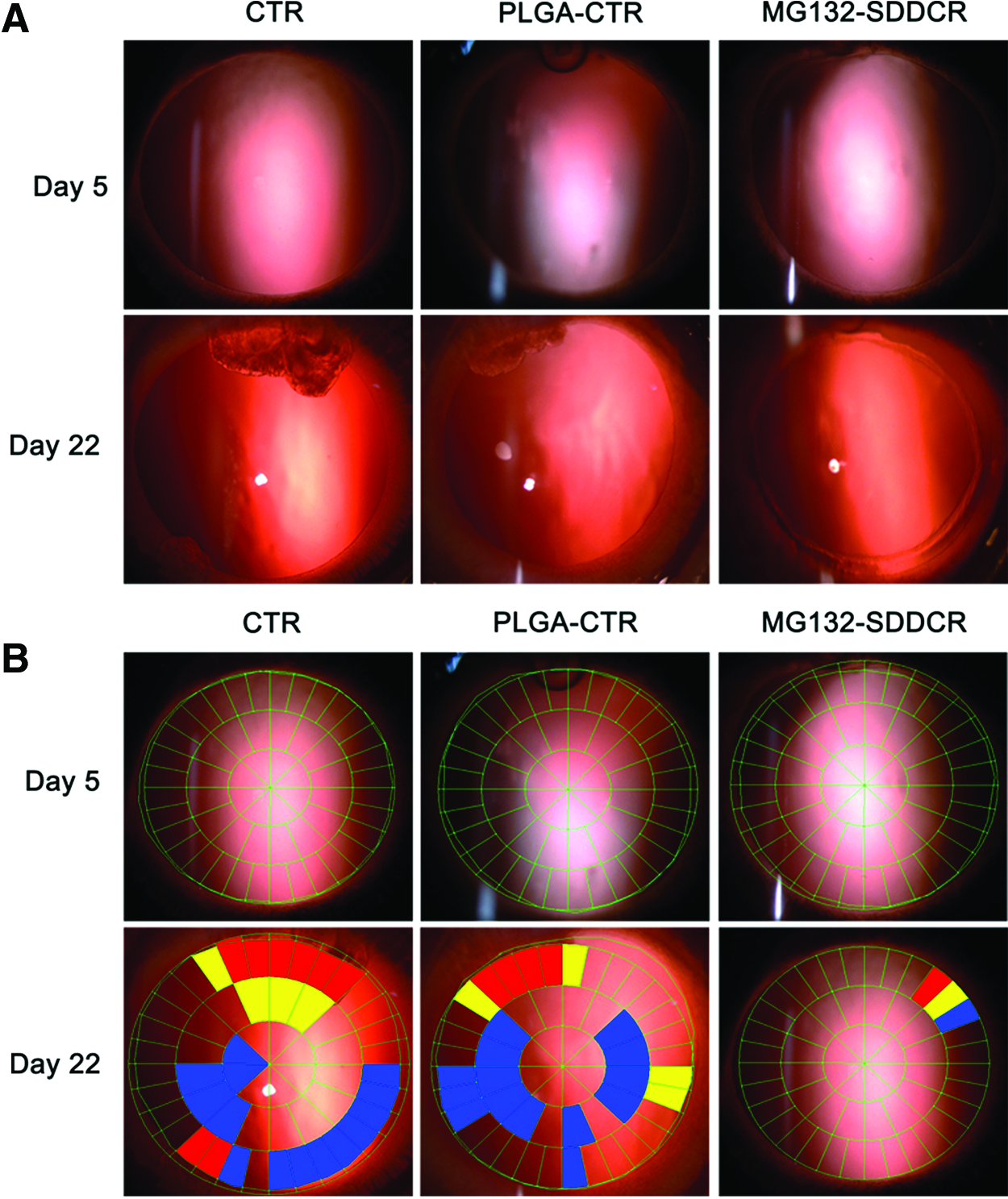
PCO was lighter in group MG132-SDDCR than the controls. Group CTR and group PLGA-CTR showed more severe cortex proliferation than group MG132-SDDCR, especially in the area between the 2 round ends of the CTRs at the 22nd day after the surgeries
POCOman software showed that the PCO score of the MG132-SDDCR group was lower than the control groups at the 10th, 13th, and 22nd day (P < 0.05). Also, there were no differences between the PLGA-CTR group and the CTR group during the observation period (Fig. 4).

PCO score was lower in group MG132-SDDCR. According to POCOman, the PCO degree was significantly lower in the group MG132-SDDCR than the control groups at the 10th, 13th, and 22nd day after the surgeries. *P < 0.05; **P < 0.01.
EMT was inhibited in the MG132-SDDCR group according to western blot and immunofluorescence
The markers of the mesenchymal cell phenotype (α-SMA, FN, vimentin, and collagen-I) showed an obvious lower expression in the MG132-SDDCR group than the PLGA-CTR group, (P < 0.05) and the expression of the epithelial cell marker E-cad was higher in the MG132-SDDCR group (P < 0.05) (Fig. 5). It was showed by immunofluorescence that in the PLGA-CTR group, multilayer and disorderly arrayed LECs were observed at the posterior area, with the nuclei elongated to the fusiform shape. However, monolayer LECs with round and regular nuclei were arraying loosely in the posterior area in the MG132-SDDCR group. The expression of α-SMA could barely be observed in the MG132-SDDCR group (Fig. 6).
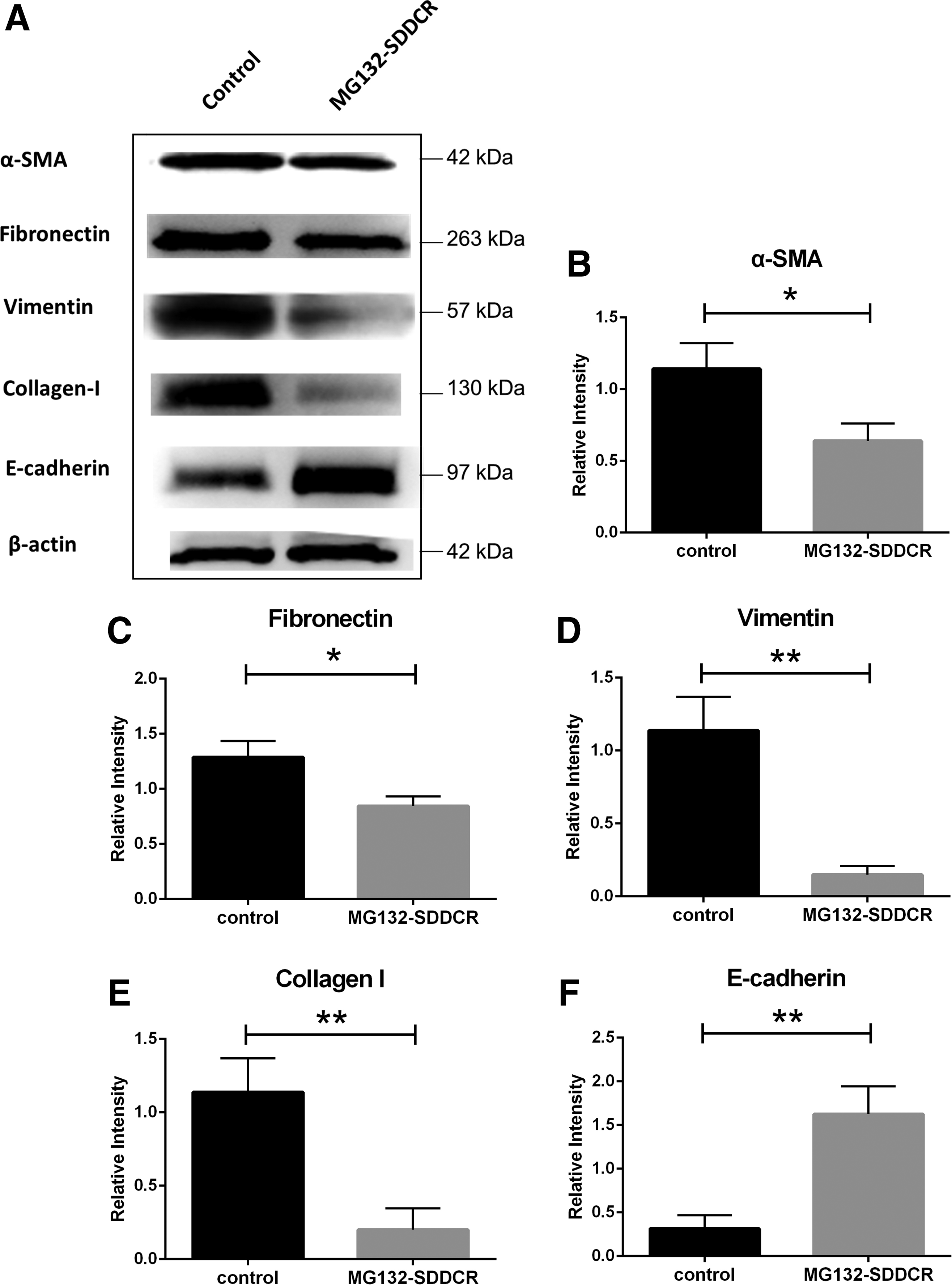
EMT process was inhibited in the group MG132-SDDCR. Western blot showed that the expression of the EMT markers (α-SMA, fibronectin, vimentin, and collagen-I) was significantly decreased in the group MG132-SDDCR than the control groups and the expression of epithelial marker (E-cadherin) was significantly higher. *P < 0.05; **P < 0.01. α-SMA, alpha-smooth muscle actin; EMT, epithelial–mesenchymal transition.
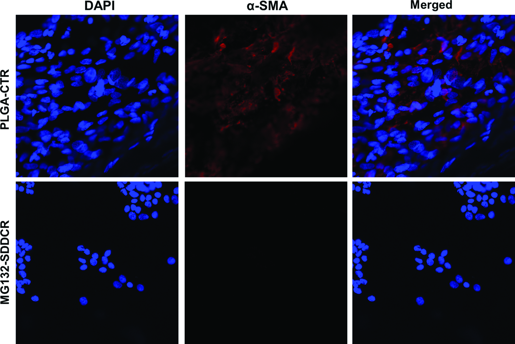
According to the immunofluorescence of the stretched preparation of the posterior capsules, the nuclei of the LECs were denser and arrayed much more disordered in the control group than the MG132-SDDCR group. α-SMA was obviously expressed in the control, but could barely be found in the MG132-SDDCR group. LECs, lens epithelial cells. Color images available online at www.liebertpub.com/jop
The SDDCRs had little toxicity to the anterior segment
Only 2 rabbits from the MG132-SDDCR group were recorded to have a higher IOP at the 1st day after surgeries (P < 0.05) and resumed to normal at the 2nd day. No other high IOP was observed in all groups during the observation period (Fig. 7). There were no differences between the 3 groups except for the first day. No obvious corneal opacity was recorded except for a slight cloudy edema surrounding the incision area in MG132-SDDCR group. Inflammation of the anterior chamber faded away from the 5th day in all groups, and there were no significant differences between the 3 groups considering inflammation.

There were no significant differences in IOP between the 3 groups except for the first day. Color images available online at www.liebertpub.com/jop
Discussion
CTRs are widely applied in modern cataract surgeries mainly to deal with lens subluxation and zonular laxity to maintain the tension and structure of capsular bags. 28 Kugelberg et al. found that CTR with a larger diameter might prevent the migration of LECs from the equator area to the posterior area in a rabbit model, probably due to its mechanical barrier function. 29 Based on that finding, some researchers applied the CTRs to young patients and patients with other ocular diseases only to find that most of them still need Nd:YAG laser posterior capsulotomy.30,31 Therefore, drug-loaded CTRs might be a more scientific solution with regard to the long-term prevention of PCO.
PLGA is a prevalent biocompatible material in tissue engineering such as artificial bone tissue, biovalves, and cytoskeleton material. Besides, PLGA is usually produced into different kinds of macromolecule SDDSs, including drug-loaded nanoparticles and films. 26 PLGA owns the characteristics that the degradation rate and fragility decreases with higher ratio of PLA/PGA, and the viscosity increases with higher molecular weight. Thus, PLGA with a ratio (PLA/PGA) of 80:20 and a molecular weight of 80 kDa was chosen. In this study, CTRs were coated with the film of PLGA encapsulating MG132 to form the MG132-SDDCRs.
In this study, the mass ratio of MG132 and PLGA was 1:60 and the theoretic largest drug-loading capacity was 1.67%, and the actual drug-loading capacity was close to it. The results were fulfilling possibly because MG132 and PLGA dissolved well in the solvent. MG132 strongly promotes cell apoptosis, thus its working concentration should be strictly limited. It was reported that the proliferation of a LEC line was noticeably inhibited by 2 ng/μL MG132. 32 In this study, MG132 was detected ∼1 month with a peak concentration of 3.443 ± 0.56 ng/μL at the 14th day. MG132 was difficult to be detected at the first 3 days probably because the drug was mainly encapsulated inside PLGA and remained little on the surface. With the degradation of PLGA, the drug was slowly released to the medium, thus the initial burst release was avoided. MG132 was released stably in vitro during the observation period, however, for fear of the influence to the intraocular environment, the in vivo release was not performed. The actual drug concentration may be affected by aqueous humor circulation or other intraocular factors.
It was showed by anterior segment photography that in the control groups, the fibers and cortex proliferated apparently from the 10th day after the surgeries, especially between the 2 round ends of the CTRs where the mechanical barrier function was relatively weak. The posterior capsules of the MG132-SDDCR group, on the contrary, remained flat and smooth in about 2 weeks and began to opacify from the 17th to 22nd day. Most of the fibers did not cross the capsulorhexis edge. There was little proliferation between the 2 ends of the CTR probably due to the larger amount of MG132, which induced LECs apoptosis in this area.
POCOman is a semiquantitative software to assess PCO score. According to POCOman, the PCO degree was significantly lower in the MG132-SDDCR group than the control at the 10th, 13th, and 22nd day, indicating that the SDDCRs were able to delay the PCO process during the observation period. There were no significant differences between the PLGA-CTR group and the CTR group for PCO degree, indicating that PLGA could neither promote nor prevent the PCO degree due to its well biocompatibility.
It is essential that an effective anti-PCO pharmacotherapy is able to inhibit the proliferation, migration, and EMT of the LECs under the molecular biological level. Awasthi and Wagner found that MG132 inhibited the proliferation of LECs even in the presence of TGF-β, fibroblast growth factor-2, and hepatocyte growth factor possibly by inhibiting the degradation of p21 and p27, and promoting the cell-cycle exit. 33 In the study, cells in the MG132-SDDCR group showed more regular cell shape and less disordered array, and expressed less mesenchymal cell marker such as α-SMA and FN, indicating that the phenotypic switch of epithelial cells to mesenchymal cells was inhibited by MG132.
To evaluate the safety of MG132-SDDCRs, we assessed the corneal edema level, IOP, and the anterior chamber inflammation. Two rabbits from the MG132-SDDCR group had a higher IOP only at the first day, and it might be caused by excessive injection of balanced salt solution when the anterior chambers were formed. No severe anterior chamber toxicity was observed probably due to the limited drug concentration of MG132 in the anterior chamber, which was bound to be lower than the highest concentration in the in vitro release experiment—3.443 ± 0.56 ng/μL. It is very possible that such MG132-SDDCR is a safe intraocular drug-release system.
This study has its limitation as well. The actual intraocular drug concentration remains unknown, and the observation time was still relatively short, in which PCO is a long-term problem. The production of MG132-SDCCRs could be further improved, which release longer and more stable. Despite these aspects, PCO in the rabbit model was successfully postponed by the implantation of the MG132-SDDCRs, making it a potential therapeutic strategy and a promising research direction in PCO prevention.
Footnotes
Acknowledgment
The work was supported by the Chinese National Natural Science Foundation (No. 81270982).
Author Disclosure Statement
No competing financial interests exist.