
Abstract
Purpose:
The aim of this study was to design naproxen sodium (NS)-containing, biomimetic, porous poly(lactide-co-glycolide) (PLGA) scaffolds for regeneration of damaged corneal epithelium.
Methods:
NS-incorporated PLGA scaffolds were prepared using the emulsion freeze-drying method and then coated with collagen or poly-
Results:
The drug loading efficiency of scaffolds was found to be higher than 84%, and about 90%–98% of NS was released at the end of 7 days with a fast drug release rate at the initial period of time and then in a slow and sustained manner. The corneal epithelial cells were isolated from New Zealand white rabbits. The obtained cells were seeded onto scaffolds and continued to increase during the time period of the study, indicating that the scaffolds might promote corneal epithelial cell proliferation without causing toxic effects for at least 10 days.
Conclusions:
The NS-loaded PLGA scaffolds exhibited a combination of controlled drug release and biomimetic properties that might be attractive for use in treatment of corneal damage both for controlled release and biomedical applications.
Introduction
The cornea is a transparent tissue of the ocular surface and the entirety of this avascular tissue is important to ensure proper visualization. The cornea is surrounded by stratified epithelium that provides smooth ocular surface continuity. 1 Various diseases, traumas, and severe corneal injuries cause the epithelium to lose its transparent and avascular structure. Until now, the only treatment option for irreversible corneal damage has been stated as corneal transplantation, which requires alternative solutions due to donor inadequacy and complications such as corneal decomposition. 1 At present, research on corneal treatments is advancing toward tissue engineering and this presents an important solution to serious corneal injuries, overcoming the disadvantages of corneal transplantation. 2
Traditional tissue engineering methods are generally performed using isolated cell suspensions or biodegradable scaffolds to promote tissue formation.
3
Recently, a number of studies have been conducted to improve tissue regeneration by regulating cell–scaffold interaction and the need to meet cellular metabolic demands. Cell–scaffold interactions can be accomplished by making changes in the surface characteristics of scaffolds such as hydrophobic/hydrophilic properties, charge feature, surface energy, and surface roughness.4,5 Generally, it is stated that the surface, which has moderate hydrophilicity, cationic charge, high energy, and irregular structure, has a positive effect on cell attachment and growth.4,5 In recent years, biocompatible/biodegradable polymers, such as polyglycolide, poly(
The future generation of engineered tissues involves bioactive molecule-incorporated scaffolds to meet cellular metabolic needs or to organize the cellular function (eg, growth or differentiation factors) or to impact the surrounding tissues (eg, anti-inflammatory drugs or antibiotics).
7
Thus, the strategy is to simulate the matrix and provide crucial information or signaling for cell attachment, proliferation, and differentiation and to fulfill the dynamic mutual requirements for tissue engineering. This confirms the significance of drug delivery in tissue engineering applications.
8
Biodegradable scaffolds can be loaded with drugs that provide cell proliferation or differentiation to activate cellular differentiation and tissue remodeling. It was demonstrated that the prolonged release of dexamethasone (a steroidal anti-inflammatory drug) provided by dexamethasone-containing PLGA scaffolds effectively generated differentiation of bone marrow stem cells to osteoblasts or chondrocytes.
9
Furthermore, some bioactive molecules such as chondroitin sulfate,
6
retinoic acid,
10
proangiogenic growth factors (PDGF-BB, FGF2, and VEGF),
11
In severe corneal injury, the response that starts to clear damaged tissues and foreign substances is raised by stimulation of inflammatory cells and the release of chemical mediators such as acidic lipids, for example, prostaglandins, thromboxanes, and leukotrienes; vasoactive amines; and cytokines, which accelerate new vessel formation. 15 Lymphatic vessels in vascularized corneas have been demonstrated and corneal neovascularization has been reported to be associated with an inflammatory reaction. 16 Neovascularization may lead to a decrease in corneal transparency and eventually significant vision loss. In addition, inhibition of corneal neovascularization following administration of nonsteroidal anti-inflammatory drugs (NSAIDs) has been stated. 17 NSAIDs exhibit their anti-inflammatory action by blocking the cyclooxygenase enzymes (COX 1 and COX 2).
In light of the information above, the aim of this study is to develop and characterize biodegradable, functionalized, and biomimetic scaffolds for efficient regeneration in serious corneal injury, providing sustained release of naproxen sodium (NS), an NSAID, and enhancing cell–scaffold interaction, and thus to offer an alternative treatment to corneal transplantation, which is a very challenging treatment method. To achieve this aim, biodegradable/biocompatible polymer PLGA was used to prepare NS-loaded scaffolds and scaffolds modified with collagen or poly-
Methods
Materials
NS was a generous gift from the Abdi Ibrahim Drug Company (Istanbul, Turkey). PLGA (50:50) and PLGA (75:25) were commercially available as Resomer RG 503 and Resomer RG 753 S, respectively, and purchased from Boehringer Ingelheim (Germany). Poly-
Preparation of NS-loaded PLGA scaffolds
The emulsion freeze-drying method was employed to prepare scaffolds. 18 Briefly, PLGA (Resomer RG 503 or Resomer RG 753 S; 10%, w/v) was dissolved in dichloromethane, and NS (10%, w/v) was dissolved in ultrapure water. The NS aqueous solution was emulsified in the PLGA dichloromethane solution by means of sonication for 30 s at 10 W (Bandelin D-12207, Germany). The emulsion was added to a Teflon mold (5 mm diameter), frozen (−80°C), and freeze-dried for 48 h. Then, the samples were stored in a vacuum oven at ambient temperature (24 h) to evaporate any residual solvent. Cylindrical scaffolds were obtained with this technique. Then, these scaffolds were cut using a surgical scalpel into sections with a thickness of 2 mm and a diameter of 5 mm to obtain final formulations (Fig. 1) (before coating). 19
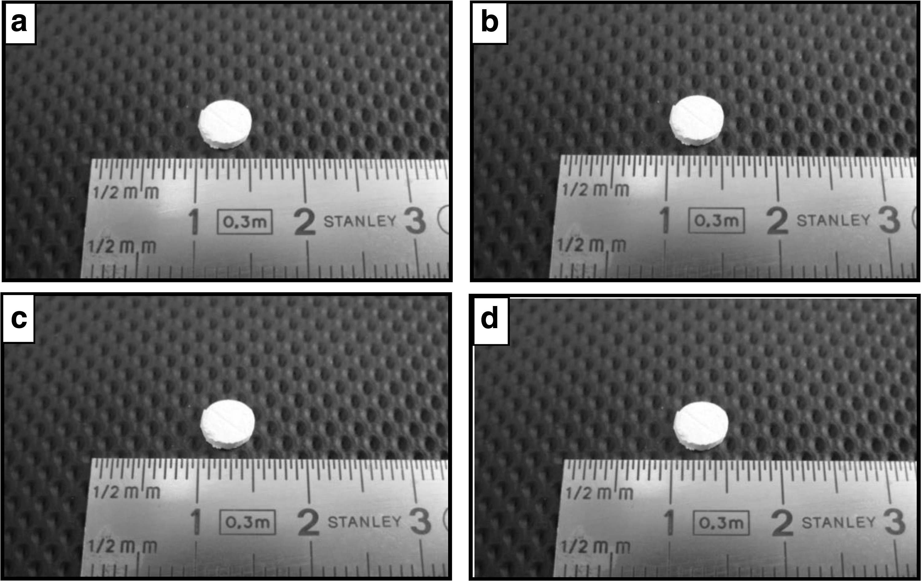
Photograph of blank or NS-loaded PLGA scaffolds;
PLGA/collagen scaffold
To obtain PLGA/collagen hybrid scaffolds, a collagen solution was prepared by dissolving collagen type I in 0.001 N HCl [0.1% (w/v)] at 25°C. Then, PLGA scaffolds were dipped in the collagen solution for 30 min and freeze-dried for 48 h. Following freeze-drying, the collagen layer of the scaffolds was cross-linked by ultraviolet (UV) light irradiation (2 h; 30 min for each side of the scaffold sample). An irradiation time of 30 min was selected on the bases of reports suggesting that after 30 min, the cross-linking density does not increase and that prolonged UV exposure contributes to denaturation of collagen molecules. 20
PLGA/poly-l -lysine scaffold
To attain PLGA/poly-
Scaffold characterization
Morphological analysis
Surface morphologies and cross-sectional structures of blank or NS-loaded PLGA, PLGA/collagen, and PLGA/poly-
Fourier transform infrared analyses
To evaluate the coating efficiency of the blank or NS-loaded hybrid scaffolds coated with collagen or poly-
Pore size and porosity analysis
Porosity measurements of the blank or NS-loaded PLGA, PLGA/collagen, or PLGA/poly-
Water uptake studies
To determine the water uptake and swelling properties of blank or NS-loaded PLGA, PLGA/collagen, or PLGA/poly-

where Wo is the initial weight of the dry scaffold and Wt is the weight of the hydrated scaffold at time point t. 23
In vitro degradation studies
Blank or NS-loaded PLGA, PLGA/collagen, or PLGA/poly-
Drug loading studies
PLGA, PLGA/collagen, or PLGA/poly-
In vitro release studies
NS-loaded PLGA, PLGA/collagen, or PLGA/poly-
Tensile strength
Tensile strengths of PLGA scaffolds were measured using a TA.XT plus Texture Analyser (Stable Micro Systems, UK) at constant temperature and humidity at a rate of 1 mm/s. PLGA scaffolds were gripped in the top and bottom chucks from 2 sides. These 2 chucks connected to the instrument were set at 1 mm. A total of 6 samples were used for each scaffold formulation. The tensile strength analysis was performed with a load cell of 1 kg.
Cell culture studies
Isolation of corneal epithelial cells
The primary culture of corneal epithelial cells was set up from the eyes of New Zealand white rabbits. All protocols were approved by the Hacettepe University Animal Ethics Committee, project number: 2010/1-1. For this purpose, rabbits were sacrificed using sodium pentobarbital solution (100 mg/kg) and rabbits' corneas were excised. 25 The corneal tissues were washed with phosphate buffer containing 1% penicillin/streptomycin and cut into pieces (1 × 1.5 × 2.5 mm). Each tissue piece comprising the epithelium, basement membrane, and stroma was kept in DMEM/Ham's F12 (1:1) supplemented with 1% penicillin/streptomycin containing 1.2 U/mL dispase for 16 h at 4°C, respectively. Small pieces of epithelium were separated from the stroma and located apical side up on a tissue culture plate, including the preculture medium, DMEM/Ham's F-12 nutrient medium supplemented with 1% penicillin/streptomycin. The collected epithelium sheets of epithelium were incubated with 0.25% trypsin/0.01% EDTA at 37°C with gentle agitation for 20 min to generate isolated cells. The cells and medium were centrifuged at 1,200 rpm for 7 min and the preculture medium was removed. 26 The centrifuged cell pellet and cells were redispersed in 30 mL of complete medium (∼250,000 cells/mL) and the complete culture medium contained DMEM/Ham's F-12 supplemented with 20% FBS and 1% penicillin/streptomycin. The cell suspension (0.25 mL) was seeded into each well, followed by addition of 0.5 mL of the complete medium. 27
Immunocytochemistry
Immunocytochemical staining was carried out to assess the expression of different molecular markers required for epithelial cell identification. For cytoplasmic and nuclear protein staining, corneal epithelial cells, which were cultivated on a coverslip with ∼80% confluence, were immobilized with cold methanol for 10 min. Cells were blocked and permeabilized using 3% bovine serum albumin/0.3% Triton X-100/PBS for 30 min. Following primary antibody cytokeratin 14 administration and incubation for 2 h at room temperature, the FITC-conjugated secondary antibody was added and incubated for 30 min. 28 After proper staining with the secondary antibody, the coverslips were reversed (cell side down) and mounted with PBS:glycerol (1:9). The cultivated cells were evaluated under a fluorescence microscope (Leica).
Corneal epithelial cell culture
After isolation of corneal epithelial cells by following procedures previously described, the cells were maintained in a complete cell culture medium containing DMEM/Ham's F-12, supplemented with 20% FBS and 1% penicillin/streptomycin. The media were changed 3 times weekly and cells were cultivated in a humidified condition at 37°C and 5% CO2.
Cell adhesion
NS-loaded PLGA, PLGA/collagen, or PLGA/poly-
Cell morphology
To visualize cells on the scaffolds, the corneal epithelial cells were cultured on blank or NS-loaded PLGA, PLGA/collagen, or PLGA/poly-
Cell viability
In vitro cytotoxic effects of NS-loaded PLGA, PLGA/collagen, or PLGA/poly-
where OD570 (sample) shows the readings from the wells treated with scaffold extracts and the OD570 (control) shows the readings from the wells untreated. 32
Cell proliferation
Cell proliferation was determined quantitatively by MTT assay. NS-loaded PLGA, PLGA/collagen, or PLGA/poly-
Statistical analysis
Statistical analysis of data was performed with a t-test or one-way analysis of variance using Statistica® software and GraphPad Prism 6.
Results
In the emulsion freeze-drying method, after the scaffolds were prepared, they were frozen at −80°C before lyophilization. In preformulation studies, different freezing temperatures (−20°C, −80°C, and liquid nitrogen) were tried and the freezing temperature of −80°C was determined as the most suitable temperature in terms of formability and mechanical strength. A low prefreezing temperature can lead to the accelerated formation of ice crystals and high porosity on lyophilized scaffolds. 33 Accordingly, the macropores will occur in the inside and outside of the scaffolds.
Scaffold characterization
Morphological analysis
Surface morphologies of the scaffolds are demonstrated in Fig. 2. In general, when the scaffolds prepared with the PLGA (50:50) polymer were compared with the PLGA (75:25) scaffolds, it was noted that the porous structure in PLGA (75:25) scaffolds decreased and more dense structures were formed. Despite the reduction of the porous structure, some pores were still observed in the PLGA (75:25) scaffolds.
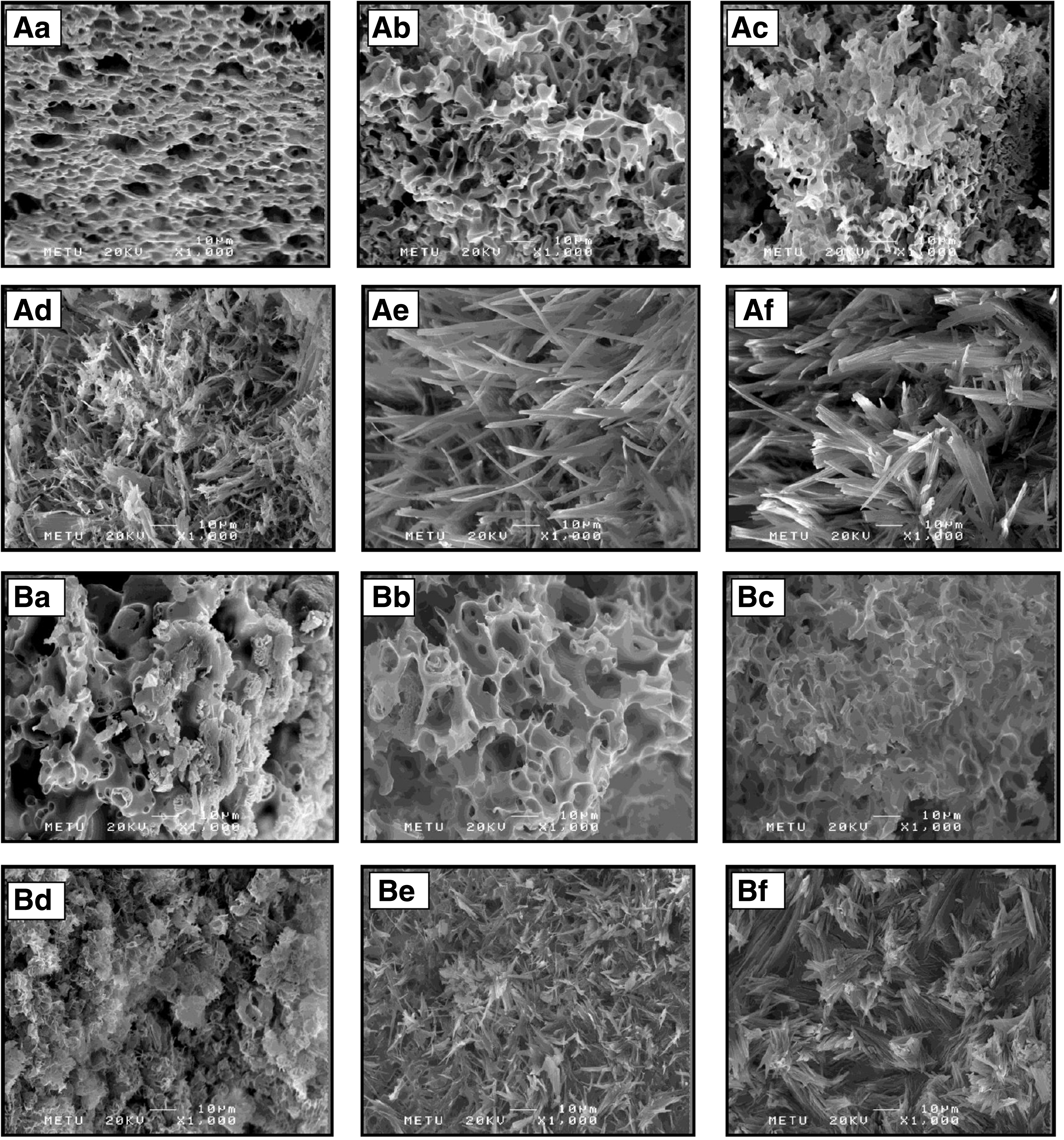
Cross-sectional SEM images of blank or NS-loaded PLGA scaffolds;
As can be seen in Fig. 2Aa–Ac and Ba–Bc, the surface and cross-sectional structures of blank PLGA scaffolds were porous, well interconnected throughout the matrix, and the distribution of pores was almost uniform before the coating procedure. In hybrid blank scaffolds, although the porous architecture of the scaffolds was maintained, pore distribution, pore wall, and pore wall thickness were nonuniform.
As can be seen in Fig. 2Ad–Af and Bd–Bf, the surface and cross-sectional structures of NS-loaded PLGA scaffolds were porous, but the porous structure has decreased slightly compared with the blank formulations and fiber structures that occurred due to addition of NS to formulations before the coating procedure.
FTIR analysis
FTIR spectroscopy was employed to evaluate the coating efficiency of hybrid scaffolds. Figure 3 shows the FTIR absorption spectra for pure collagen, pure poly-
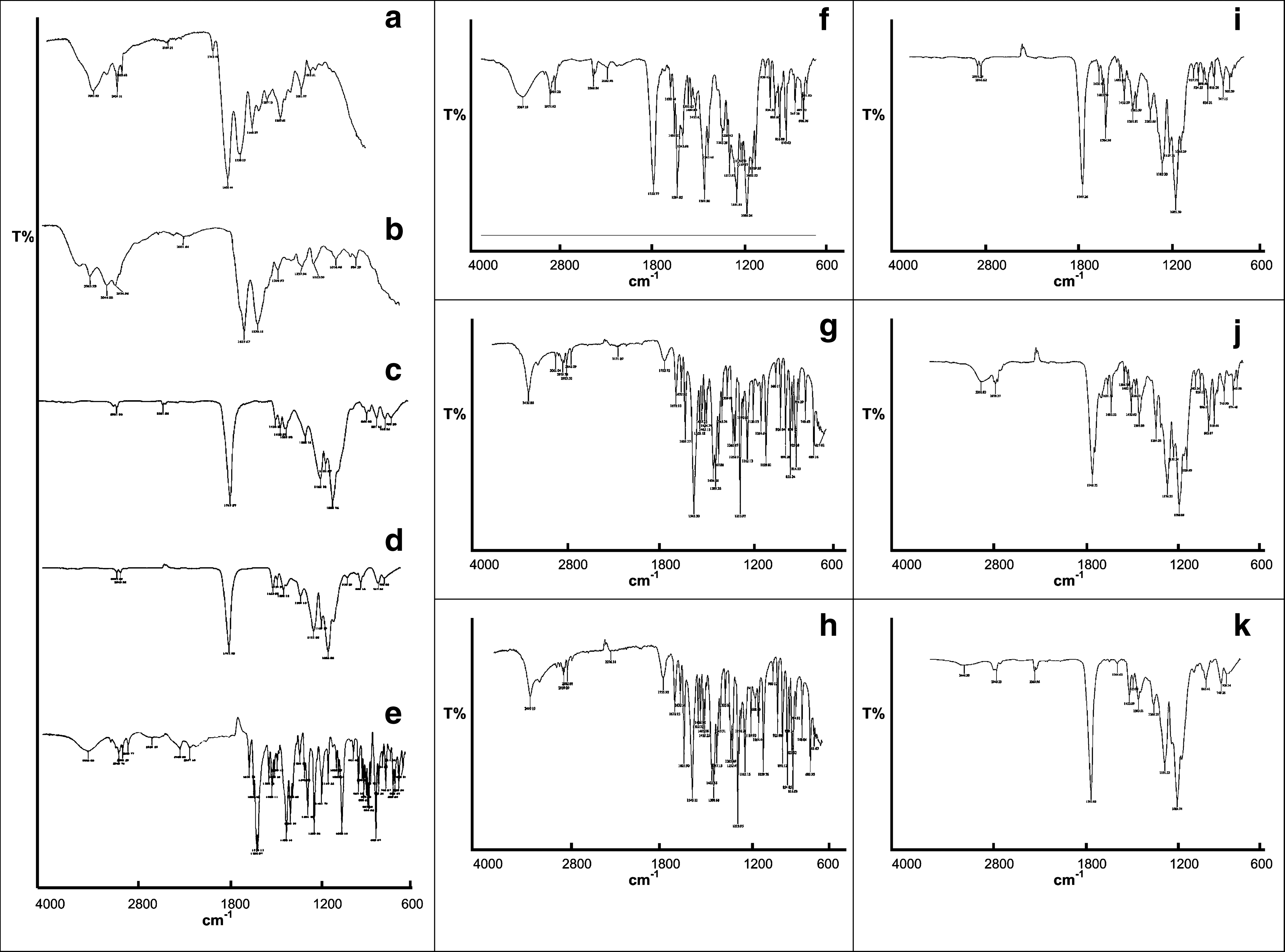
FTIR absorption spectra of
Pore size and porosity analysis
The pores on both the surface and inside of the scaffolds are macroporous and fully connected to each other along the scaffold matrix, with sizes ranging from 150 to 200 μm (Table 1), larger than the size of the cell (about 10 μm). In addition, supporting the morphological structure of the scaffolds determined by SEM (Fig. 2), pore sizes of PLGA (75:25) scaffolds were smaller than PLGA (50:50) scaffolds, although not statistically significant.
Formulation Codes, Pore Diameters, and Tensile Strengths of Poly(Lactide-co-Glycolide) Scaffolds
Mean ± standard deviation (n = 3).
NS, naproxen sodium; PLGA, poly(lactide-co-glycolide).
Water uptake studies
Figure 4a and b presents the percentage of water uptake of the NS-loaded and NS unloaded PLGA scaffolds with different copolymer ratios as a function of time. Statistically significant differences in water uptake ratios were observed at all time points between PLGA (50:50) and PLGA (75:25) scaffolds (P < 0.05). It was clearly observed that scaffolds prepared using PLGA (50:50) had higher water uptake capacity than PLGA (75:25) scaffolds.
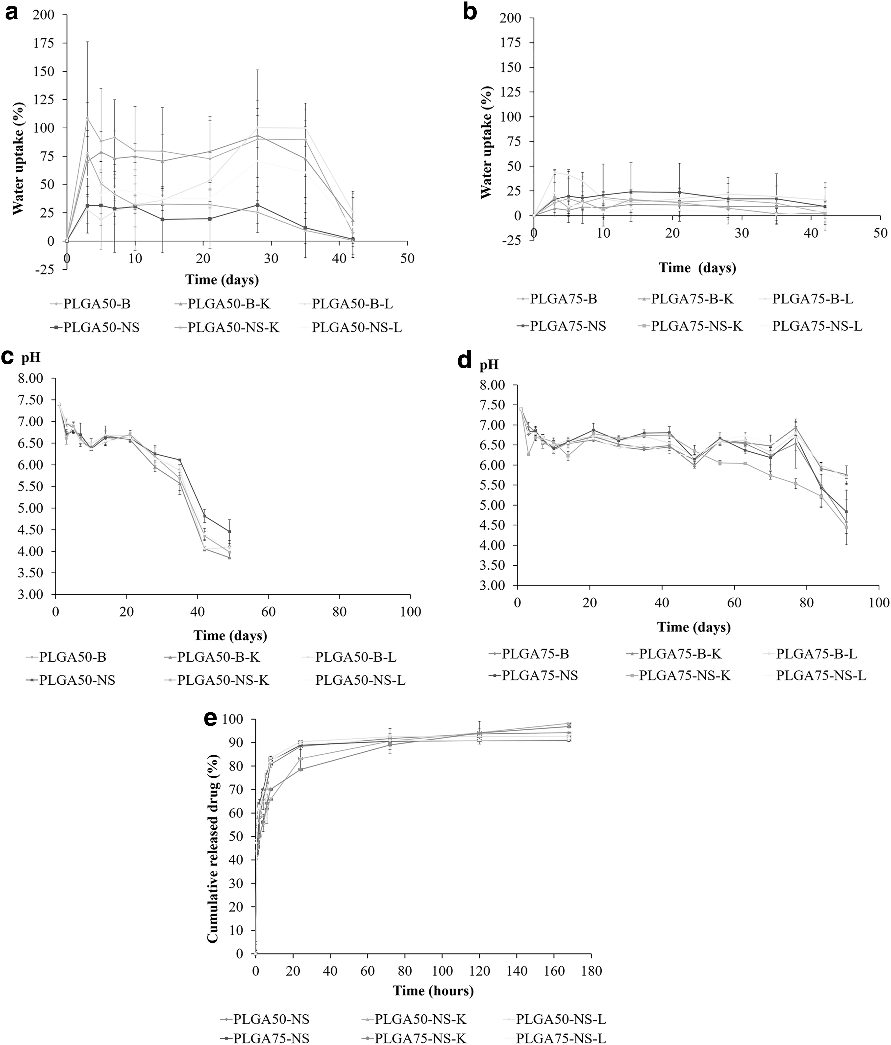
Water uptake
In vitro degradation
Figure 4c and d shows the tendency of the pH of the medium since polymer degradation takes place. The pH of the solution decreased from 7.4 to ∼4.5 and 4.0 with time for PLGA (75:25) scaffolds and PLGA (50:50) scaffolds, respectively. On the other hand, it can be seen from Fig. 4c and d that incorporation of the coating agents (collagen/poly-
Drug loading studies
The drug loading efficiency of the NS-loaded PLGA scaffolds was between 84% and 93% for all formulations. When results were examined, the drug loading efficiency of PLGA/collagen and PLGA/poly-
In vitro release studies
The drug release profiles of NS-loaded PLGA scaffolds are shown in Fig. 4e. About 90%–98% of the drug was released at the end of 7 days for both copolymer ratios and there was no statistically significant difference in the released percentages of NS from PLGA (50:50) and PLGA (75:25) scaffold formulations for all the time points (P > 0.05).
Tensile strength
The tensile strengths of produced scaffolds are demonstrated in Table 1. Although there was a statistically significant difference between tensile strengths of PLGA (50:50) and PLGA (75:25) scaffolds (P < 0.05), no statistically significant difference was observed between tensile strengths of NS-containing PLGA (50:50) and PLGA (75:25) scaffolds (P > 0.05). In addition, the presence of NS in collagen-coated PLGA scaffolds reduced tensile strength significantly (P < 0.05) and coating of NS-containing PLGA (75:25) scaffolds with collagen or poly-
Cell culture studies
Isolation of corneal epithelial cells: immunocytochemistry
As shown in Fig. 5A, the isolated rabbit corneal epithelial cells were stained green after treatment with cytokeratin 14.
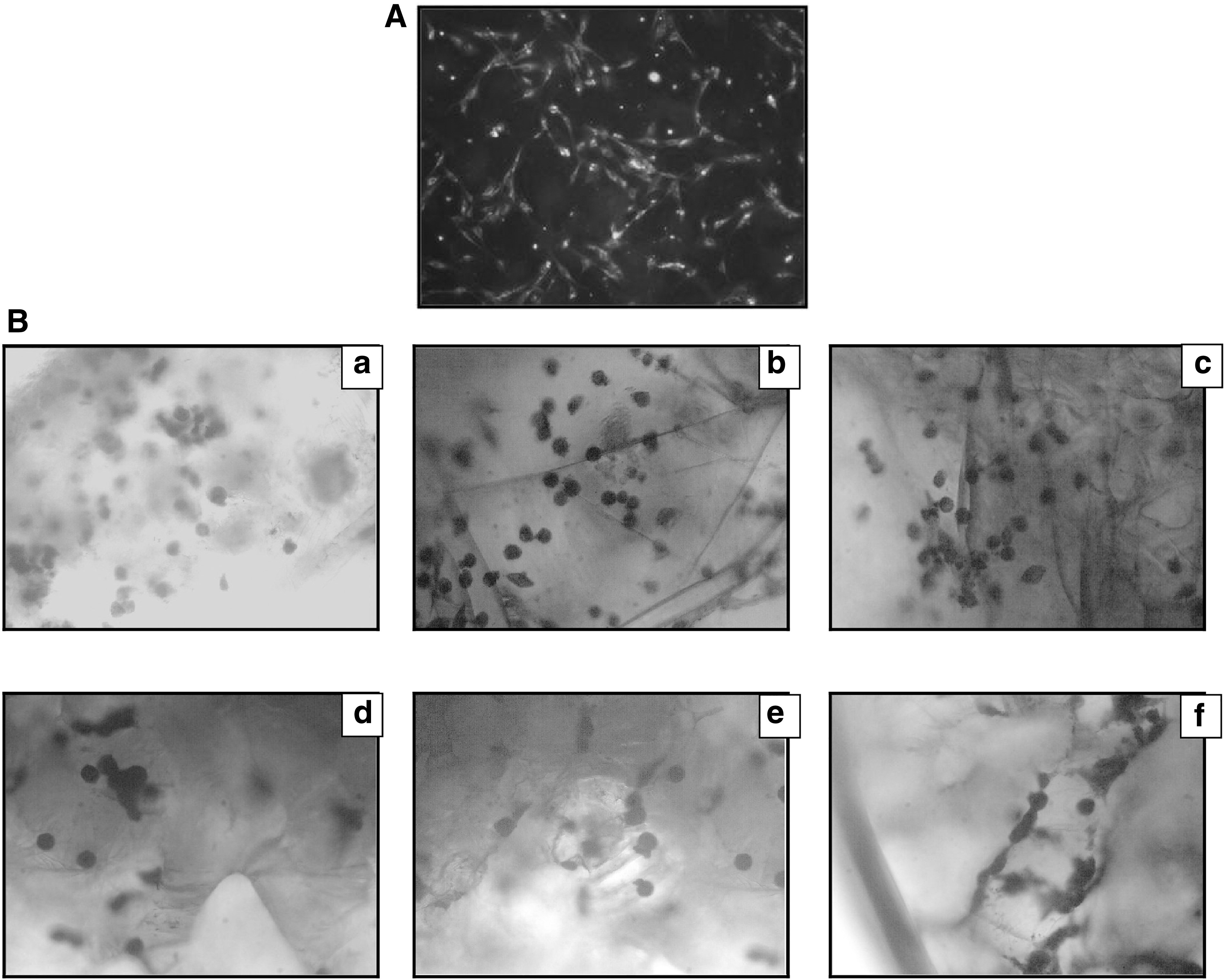
Cell adhesion
For both copolymer ratios, during the 3 days after seeding, cells just attached to the scaffolds and filled the areas of the porous scaffolds and packed together. It has been determined that cells inside and outside of the scaffolds are distributed randomly. As shown in Fig. 5B, the modified PLGA (50:50 and 75:25) hybrid scaffolds enhanced cell adhesion compared with the unmodified one (Fig. 5Ba–f).
Cell morphology
Figure 6 shows the SEM photographs of corneal epithelial cells (arrow displays corneal epithelial cells) growing in the inner side of NS-loaded PLGA scaffolds for both copolymer ratios at 3 days after cell seeding. In the modified scaffolds such as PLGA/collagen and PLGA/poly-
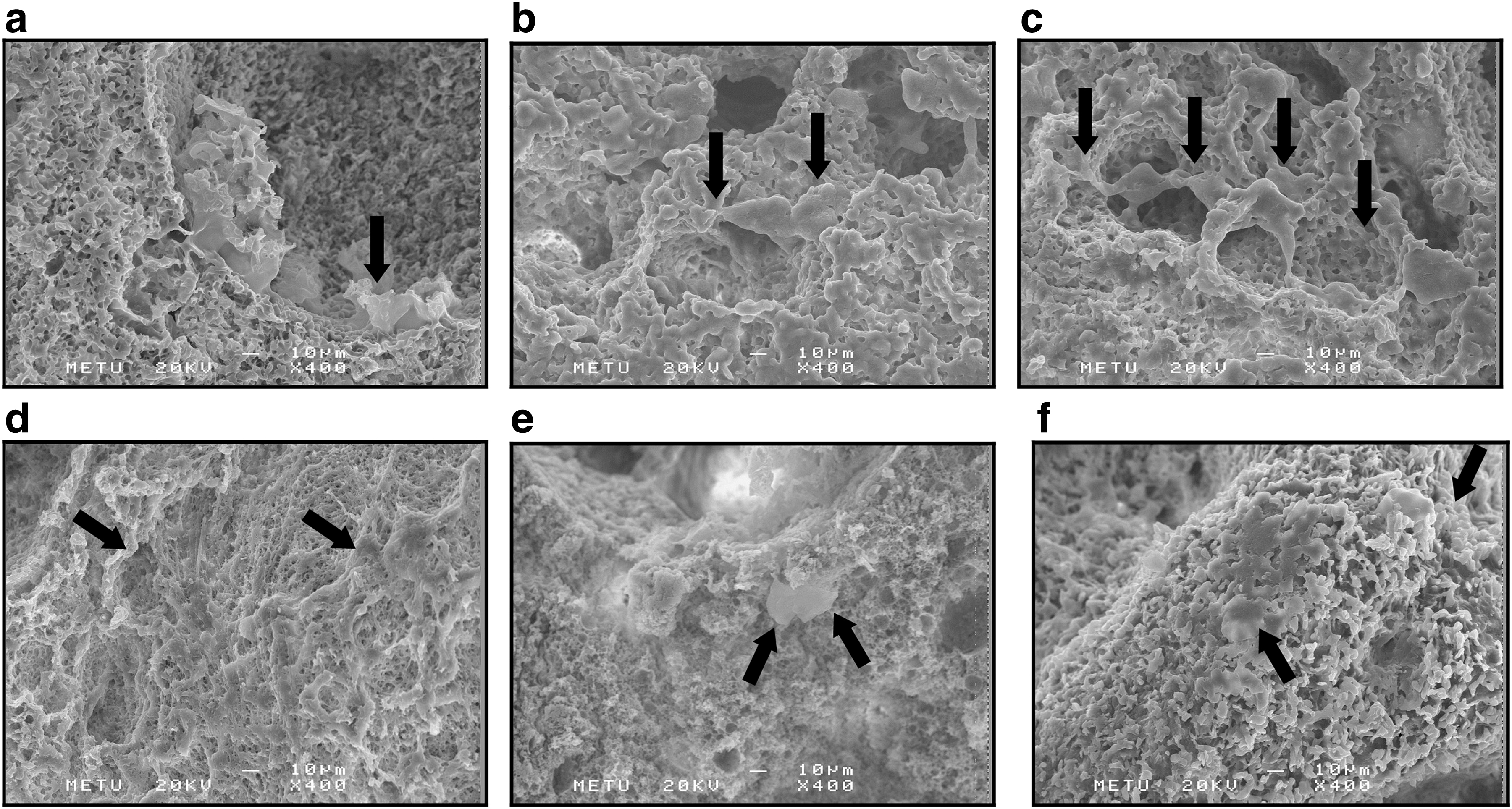
SEM photographs of corneal epithelial cells grown in the interiors of NS-loaded PLGA scaffolds at 3 days after culture (arrow indicates cells attached);
Cell viability
As shown in Fig. 7a and b, in the presence of PLGA (50:50 and 75:25), PLGA/collagen, and PLGA/poly-
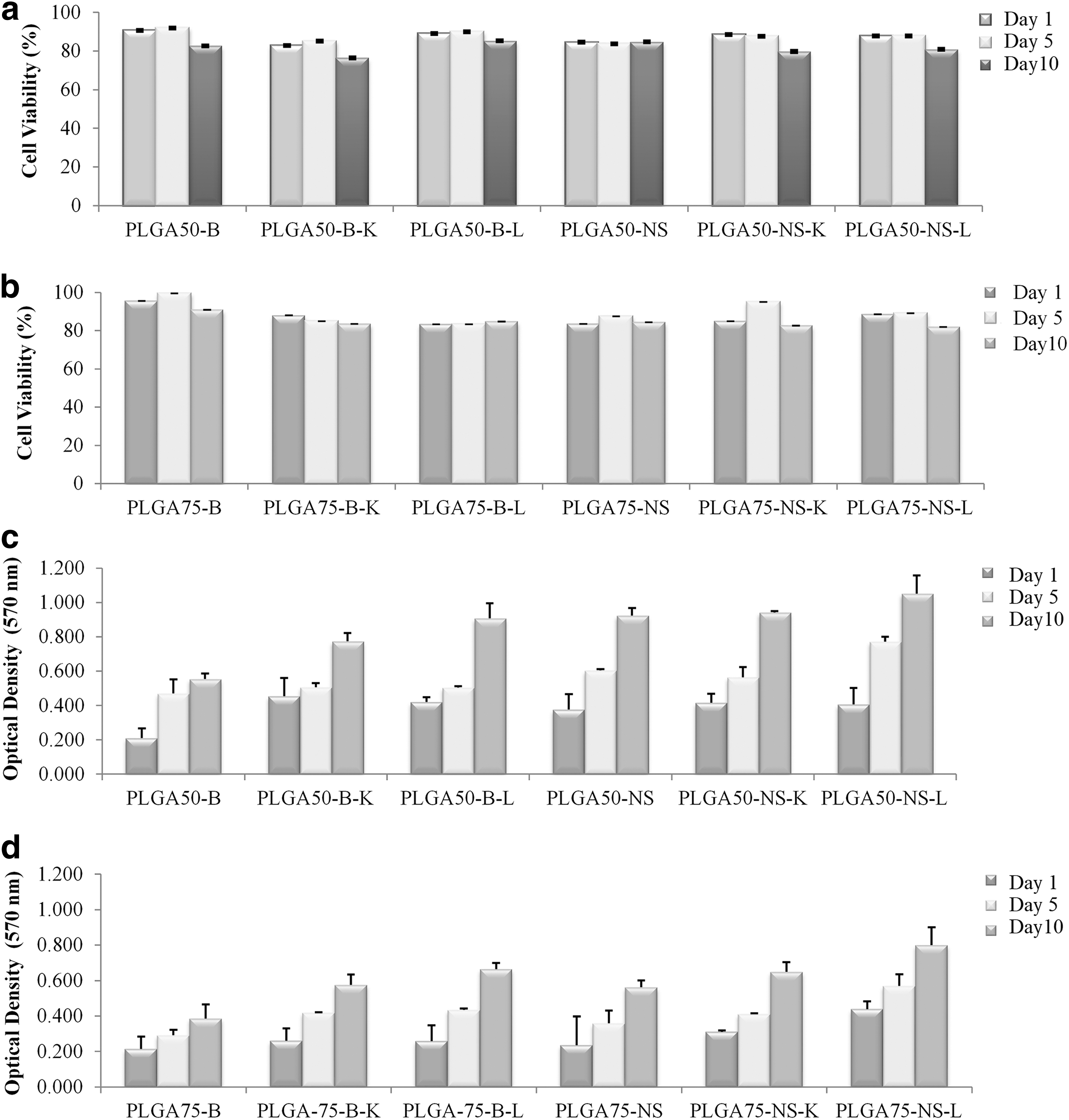
Cytotoxicity of PLGA scaffolds against the corneal epithelial cells (n = 6)
Cell proliferation
The proliferation profile of corneal epithelial cells cultured on PLGA scaffolds was evaluated for up to 10 days. As can be seen in Fig. 7c and d, a number of corneal epithelial cells maintained the increase during the time period studied. Poly-
Discussion
In the present study, blank or NS-incorporated PLGA (with a different copolymer ratio; 50:50 or 75:25) scaffolds were prepared by the emulsion freeze-drying method. This method is widely used for preparation of scaffolds with water-soluble drugs. The main advantages of this technique are that a high temperature and separate leaching step are not needed and the pore size is adjusted with the freezing rate.34,35 Moreover, a water-soluble drug can be easily loaded in the dispersed polar phase. 36
In the morphological analysis, it was observed that scaffolds prepared with the PLGA (75:25) polymer were more dense compared with PLGA (50:50) scaffolds. Similarly, Thomson et al. prepared PLGA scaffolds with different copolymer ratios for regeneration of retinal pigment epithelium and reported that an increase in the PLA ratio in the PLGA polymer caused the decreased porous structure and formation of more dense scaffolds. 37 In hybrid blank scaffolds, the pore distribution, pore wall, and pore wall thickness were not homogeneous; however, the porous architecture of the scaffolds was maintained, which indicates that the hybridization procedure was efficiently applied to the scaffolds. In Fig. 2, it can be seen that porosity decreased by the addition of NS to the formulation. Addition of a hydrophilic active ingredient could fill the macropores of hydrophobic PLGA scaffolds and yield a low porous structure in scaffold formulations. 38 In addition, an interaction between NS and PLGA could cause the formation of fiber architecture. In NS-loaded hybrid scaffolds, although the porous structure decreased (it turned into a fiber structure and more intense architecture has occurred), it was seen that the macropore structure still existed. For this reason, there was still adequate space for nutrients and these areas can be employed to distribute numerous constituents of all types of scaffolds and to meet the needs of the cells.
In the FTIR analysis, for blank PLGA scaffolds (for 2 copolymer ratios), the absorption peak at 2,960–2,850 cm−1 can be assigned to C-H stretching, the peak at 1,760–1,670 cm−1 can be assigned to C = O stretching, and the peak at 1,260–1,000 cm−1 can be assigned to C-O stretching, as is specific for the chemical structure of PLGA. For the blank PLGA/collagen hybrid scaffold, beside the peaks that are specific for the chemical structure of PLGA, the absorption peak at 3,600–3,200 cm−1 can be assigned to O-H stretching and the peak at 1,650–1,580 cm−1 can be assigned to N-H bending, as is specific for the chemical structure of collagen. For the blank PLGA/poly-
For NS-loaded PLGA scaffolds (for 2 copolymer ratios), the absorption peak at 2,960–2,850 cm−1 can be assigned to C-H stretching, the peak at 1,760–1,670 cm−1 can be assigned to C = O stretching, and the peak at 1,260–1,000 cm−1 can be assigned to C-O stretching, as is specific for the chemical structure of PLGA. The absorption peak at 3,600–3,200 cm−1 can be assigned to O-H stretching, the peak at 1,600–1,500 cm−1 can be assigned to C = C stretching, the peak at 995–910 cm−1 can be assigned to CH = CH bending, and the peak at 810–750 cm−1 can be assigned to C-H bending in the aromatic ring, as is specific for the chemical structure of NS. For NS-loaded PLGA/collagen and PLGA/poly-
The NS-loaded PLGA scaffolds produced using the emulsion freeze-drying method revealed very porous and open-cellular pore shapes accompanying a nearly similar surface and inner porosities, and the presence of a coating agent (collagen/poly-
Water uptake capacity of the scaffold is an important aspect affecting the dispersion of the cell suspension all over the scaffold during seeding and the nutrient and oxygen delivery to the cells. Additionally, water uptake of the scaffold mostly affects the drug release rate with diffusion of the drug out of the scaffold. Following implantation of scaffolds, interaction with fluids, such as absorbing water molecules, causes their degradation process, which can induce increased scaffold flexibility and dimensional changes. Moreover, higher water absorption can cause an increase in the hydrolysis rate of the polymer.
42
In our study, PLGA (50:50) scaffolds had higher water uptake ratios, which could be attributed to the hydrophilic nature of PLGA (50:50). The results were in accordance with the literature.
43
Furthermore, the employing of coating agents (collagen/poly-
For the porous polymeric scaffolds, control of the degradation rate is one of the crucial parameters for successful tissue/organ regeneration. The degradation profile of a scaffold has effects on some processes such as cell growth, tissue regeneration, and sustenance of differentiation. 46 For tissue engineering scaffolds, it is important to provide sufficient cell numbers and cell–cell interactions after the implantation into the damaged area. 47 In this case, it is desired that the degradation rate of the scaffolds must be proportional to the tissue regeneration period. On the other hand, extremely high degradation rates may induce chronic inflammatory reactions, so the scaffolds must degrade within a specific time after the tissue regeneration is complete. 48 Differentiation of the acidity of the medium is one of the consequences of polymer hydrolysis. In our study, as a result of polymer degradation, pH decreased from 7.4 to 4.45–3.85 for PLGA (50:50) scaffolds and 7.4 to 5.71–4.45 for PLGA (75:25) scaffolds (Fig. 4). The decrease in the pH value of the solution showed that the NS-loaded PLGA (50:50) scaffolds started to degrade in 49 days, while this period was 84 days for PLGA (75:25) scaffolds due to their hydrophobic nature. In a study conducted by Chew et al., the degradation properties of PLGA (50:50) and PLGA (75:25) scaffolds were examined and, similar to our results, it was shown that the PLGA (75:25) scaffold had slower degradation, which was associated with having more lactic acid than PLGA (50:50) and therefore more difficult degradation of ester linkages. 49
When the drug-loading capacity was examined, no significant difference between PLGA (50:50) and PLGA (75:25) scaffolds was obtained (P > 0.05). In PLGA (50:50) scaffolds, the drug-loading capacity was significantly reduced by coating with collagen or poly-
The drug release rate from PLGA scaffolds was very rapid at the beginning and following this initial burst, the drug was released continuously, and the rate reduced for all PLGA scaffolds with time (Fig. 4e). In addition, it was observed that PLGA/collagen scaffolds released NS at a slower rate at initial time points than the other scaffolds, but at the end of 168 h, the cumulative released NS (%) of PLGA/collagen scaffolds was higher than the others. These release profiles can be explained by the porous structure that occurs at the end of the freezing and freeze-drying procedure during the fabrication process. 50 During the lyophilization process, the porous structure created by ice crystals caused the PLGA scaffolds' rapid rehydration in the release medium and so the release medium quickly penetrates into the scaffolds and leads to an increase in the amount of released NS. In addition, the initially observed burst effect can be explained with the NS hydrophilicity. It is thought that the loading of NS to the formulations increased water penetration into the scaffolds, which led to an increase in the rate of drug release. 51 Furthermore, although the physical interaction between hydrophilic NS and hydrophobic PLGA is limited, the major amount of drug has been localized to the surface of the scaffold and this causes the initial burst effect. 52 As summarized above, the release profiles of PLGA scaffolds may allow controlled delivery of the drug to provide an anti-inflammatory effect at the damaged tissue and to aid in long-term healing. 53 In clinics, the inflammation occurring as a result of corneal damage is requested to be treated in a continuous and controlled manner so as to be successful in the treatment of inflammation.
The tensile strength studies showed that PLGA (75:25) scaffolds had higher tensile strength than PLGA (50:50) scaffolds due to their thicker structure. 54 However, addition of NS to the formulations caused this difference to disappear, indicating that NS masked the different mechanical properties of PLGA (50:50) and PLGA (75:25). The tensile strengths of PLGA (50:50) scaffolds were between 0.156 × 10⁶ and 0.343 × 10⁶ N/m2 and PLGA (75:25) scaffolds were between 0.134 × 10⁶ and 0.553 × 10⁶ N/m2, which are compatible with literature. 55 One of the tools for examining the various molecular markers suggested for recognition of epithelial cells is immunocytochemical staining. In our study, the isolated rabbit corneal epithelial cells were stained green after treatment with cytokeratin 14 (Fig. 5A), which implies that the cells obtained during the isolation process were corneal epithelial cells.56,57
Corneal epithelial cells were seeded on the NS-loaded PLGA scaffolds and their adhesion to the PLGA scaffolds has been evaluated. Even on washing with PBS, a high percentage of the attached cells were obtained through the scaffolds and the cells adhere well to the scaffolds. High cell adhesion through the modified PLGA scaffolds can be explained by the increased wettability and cell adhesion feature of the added collagen and poly-
For accomplishing drug delivery and tissue engineering applications, it is desirable that PLGA scaffolds have minimum toxicity. To examine the possible cytotoxic effects of PLGA scaffolds, the viability of corneal epithelial cells was evaluated in the presence of different types of PLGA scaffolds using the MTT assay. The cell viability results demonstrated that PLGA, PLGA/collagen, and PLGA/poly-
The MTT assay was performed to examine the capability of corneal epithelial cells to grow on PLGA scaffolds. MTT analysis results on days 1, 5, and 10 showed that corneal epithelial cells increased with time, indicating that the NS-loaded PLGA (50:50 and 75:25) scaffolds are able to support corneal epithelial cell proliferation without causing toxic effects for at least 10 days. Poly-
Conclusions
Corneal blindness due to serious corneal damage is one of the serious health problems. Tissue engineering approaches attract much attention among researchers for treatment of serious corneal damage. PLGA scaffolds might have the potential to deliver corneal epithelial cells to the damaged area and ensure organization and maintenance of differentiation. Furthermore, in designing PLGA scaffolds for corneal epithelial cells, incorporation and release of an anti-inflammatory drug (NS) could be beneficial while treating the inflammation occurring in the damaged area for the success of the tissue-engineered scaffold. As a result, these developed biodegradable, functionalized, and biomimetic, NS-loaded PLGA scaffolds had potential for use in the treatment of corneal injuries as an alternative to corneal transplantation, showing both controlled release and biomimetic properties.
Footnotes
Author Disclosure Statement
The authors met the ICMJE criteria and received neither honoraria nor payments for authorship. No competing financial interests exist.
Funding Information
This study was supported by Hacettepe University, Scientific Research Projects Coordination Unit (0801301004 and 09D06301002).