




Abstract
Purpose:
Safety and toxicity evaluation of a novel, liposome-encapsulated rapamycin formulation, intended for autoimmune ocular disorders.
Methods:
The formulation was assessed by micronucleus polychromatic erythrocyte production, irritability by Hen's Egg Test-Chorioallantoic Membrane (HET CAM), sterility, and pyrogenicity testing. Subconjunctival (SCJ) and intravitreal (IVT) administration of the formulation were performed to evaluate subacute and acute toxicity, respectively. Differences between groups in biochemical and hematological parameters were evaluated by analysis of variance and t-tests. Numeric score was assigned to histopathological classification. Electroretinography (ERG) testing was also performed. Data were analyzed by a 1 way no parametric Kruskal–Wallis and the Mann–Whitney tests. Significance was considered when P < 0.05.
Results:
No significant toxicity directly related to the preparation was detected. Micronucleus count, mucous irritation score, and pyrogenicity results were negative. Pathology demonstrated no damage related to the formulation after SCJ injection. After IVT injection, only lens injury associated with technique was observed. Retinal function was also conserved in ERG.
Conclusions:
The preparation evaluated offers a good toxicity and safety profile when injected in a SCJ or IVT manner in an animal model. A clinical trial conducted in humans is highly warranted, as it could reveal an alternative immunosuppressive treatment for ophthalmological immune-mediated pathologies.
Introduction
Rapamycin, also called sirolimus, is a macrolide with antifungal, antineoplastic, and immunosuppressive properties discovered in 1975. 1 It was approved by the U.S. Food and Drug Administration (FDA) in 1999 for the prophylaxis of organ rejection in human patients older than 13 years at a dose of 2 mg/day.2,3 It is practically insoluble in water (2.6 μg/mL), yields high liposolubility (log PO/W 5.77), very unstable in ionic medium, and has demonstrated a high rate of degradation under ultraviolet (UV) light.4–6 Despite its liposolubility, the drug has low corneal penetration and has therefore not been able to show a high effectivity rate for the treatment of autoimmune ocular surface disorders. 7
Some of the advantages of rapamycin as an immunosuppressant derive from its unique mechanism of action, its improved side effect profile, and its ability to synergize with other immunosuppressive agents (calcineurin inhibitors) due to the shared metabolism by CYP 3A. 8 Because of this, its potential clinical uses range from organ transplantation to ocular disorders, including noninfectious uveitis, diabetic macular edema, autoimmune non-necrotizing anterior sclerosis, and Sjögren syndrome.9–12
Rapamycin pharmacokinetics display large inter- and intrapatient variability, due to different disease stages and use of concurrent immunosuppressants or other interacting drugs, as well as polymorphisms in the genetic configuration of CYP 3A. Due to the long half-life of rapamycin, dose adjustments should ideally be based on levels obtained 5–7 days after initiation of therapy or dosage change. 13 Unfortunately, although rapamycin-induced toxicity has been reported extensively in patients with organ rejection, there are few reports concerning specific ocular toxicity. Adult horse and rabbit models with different ophthalmologic disorders have been used for this purpose. Administration through intravitreal (IVT) and subconjunctival (SCJ) injection at doses ranging from 20 μg to 10 mg has reported no evidence of tissue damage or toxicity.14,15 The safety profile of rapamycin in adult human patients has also been evaluated; no toxicity was reported at doses ranging from 44 to 1,760 μg.10,11
Carriers can be used to enhance the intraocular concentrations of drugs, as well as minimize the dose, and therefore side effects of medications. One type of such carriers is liposomes. Liposomes are microscopic vesicles composed of lipid bilayers surrounding an aqueous core.16,17 They can improve the solubilization and absorption of a lipophilic drug encapsulated inside the vesicle, as well as the bioavailability in the ocular environment while protecting it from degradation.18–20 Since liposomes have a similar composition as cell membranes, they are expected to be biocompatible and biodegradable. 21
Furthermore, liposomes tend to accumulate in inflamed targets, especially those including cholesterol, promoting high local drug concentrations with the administration of a smaller dose; once there they can act as a depot releasing constantly the drug so they are expected to be able to reduce nonspecific side effects and toxicity of drugs.22,23
Recently, other authors pointed out the importance of toxicity studies regarding a sirolimus liposomal formulation; this preparation demonstrated not to be toxic by MTT assay, TUNEL assay, and ocular histopathology. 24 However, the toxicity of different components in the formulation and possible byproducts or traces related to each manufacture technique make important the evaluation of each formulation. In addition, metabolic effects may pass undetected by in vitro assays that also do not yield information about functional preservation of the tissue.
Hence, the complete study of the toxicologic profile of new pharmaceutical formulations is always important in the drug development process. Taking into consideration the scarcity of in vivo studies demonstrating the assumed nontoxicity of liposomes prepared through nonconventional solvent free methodologies, only tested in vitro in nonocular cell lines by other authors, 25 the aim of this study is to describe the results of an in vitro and in vivo toxicity evaluation of liposomes loaded with rapamycin using a proprietary methodology, administered through SCJ and IVT injections to support their potential as a treatment of ocular inflammatory autoimmune disorders.
Methods
Materials
Rapamycin loaded liposomes (RL) and placebo liposomes (L) were provided by Laboratorio Santgar (Mexico City, MX) and produced using a proprietary methodology.26,27 Test formulations were kept under refrigeration (2°C–8°C) at the experimental facility until use.
For the micronucleus test, male Hsd:ICR Specific Pathogen Free (SPF) certified mice were purchased from UNAM-ENVIGO Center of Laboratory Animal Production (Coyoacan, MX). Light microscopy DM2500 (Leica Biosystems, Nussloch, DE) was used for micronucleus determinations, as well as slide observation, in the subacute toxicity study (STS).
For the Hen's Egg Test-Chorioallantoic Membrane (HET CAM) assay, 9-day-old SPF hen's embryos were purchased from ALPES, S.A. de C.V. (Puebla, MX). Sodium dodecyl sulfate (SDS) from Sigma-Aldrich (Saint Louis, MO) was used as HET CAM positive control. Physiologic saline solution (PhS) purchased from PISA, S.A. de C.V. (Guadalajara, MX) was used as a negative control in this study, as well as in the Intravitreal acute retinal toxicity study (IARTS) and the STS.
For pyrogenicity assay and STS, healthy certified male New Zealand rabbits were purchased from Science Animals (Mexico City, MX). Independently, 3 animals were purchased for pyrogenicity assay and 12 animals for STS. Rectal temperature measurements were done using a Sejoy MT 401 (Hangzhou Sejoy, CN) calibrated rectal thermometer for pyrogenicity assessment.
SCJ injections in STS, as well as IVT injections in IARTS, were performed using new 30G needles, attached to 1 mL sterile disposable syringes (Becton Dickinson and Co., Franklin Lakes, NJ). Proparacaine HCl 0.5% veterinary ophthalmic solution (Paracaina®) provided by Laboratorio Santgar was used as local anesthetic in STS and IARTS. For STS, blood samples were collected using both empty Microtainer® and Microtainer containing EDTA K2. (Becton Dickinson and Co.). Hematologic samples were processed in a BCVet analyzer (KONTROLAB International Corp., Rome, IT). Hematological slides were stained using a semiautomatic system Hematek® (Siemens Healthcare GmbH, Munich, DE). Biochemical analysis was performed by the automated chemistry analyzer CST-240 (DIRUI Industrial Co., Ltd., Changchun, CN).
For IARTS, 15 healthy certified New Zealand rabbits were purchased from Soluciones MG (Mexico City, MX). Fundus photographs were recorded using a Kowa Genesis-D Handheld Retinal Camera (Kowa American Corporation, Torrance, CA), Cyclopentolate 1%, Phenylephrine HCl 2.5%, and Tropicamide 1% veterinary ophthalmic solution (Midriavet®) provided by Laboratorio Santgar was used as local mydriatic. Electroretinograms were recorded with a BMP 200 Retinographics electrodiagnostic system (Dioptrix, Toulouse, FR). For IVT injections, animals were anesthetized with isoflurane Sofloran Vet® (PiSA Agropecuaria, S.A. de C.V. Tula, MX) and sodium pentobarbital Sedalpharma® (Pet's Pharma, Nezahualcoyotl, MX). In all cases, animal euthanasia was induced with an overdose of sodium pentobarbital Pisabental® (PiSA Agropecuaria, S.A. de C.V. Tula, MX) according to local normativity.
Rapamycin loaded liposome preparation
The preparation of the proprietary liposomes has been previously described.26,27 The liposomes were composed of phosphatidylcholine and cholesterol; the lipid dispersions were hydrated with phosphate buffered saline and heated to 70°C while stirring at 750 rpm in a N2 atmosphere for 30 min. The mixture was then left at room temperature for 30 min. The dispersion was filtered through a 0.45 μm membrane and collected in amber vials. The vials were freeze dried, and the final lyophilizate was sterilized by gamma irradiation at a 10 kGy dose. Vials were kept under refrigeration until use. Before administration, the lyophilizate was reconstituted with sufficient water for injection in aseptic conditions.
This method of rapamycin loaded liposome preparation is proprietary to the team and has an FDA-issued patent. It is of critical importance to highlight the differences between this formulation and the one used by other investigators. 24 In this work, the formulation was not prepared using thin lipid film hydration method, does not use chloroform or ethanol in the mixture, nor needs to be placed under vacuum for solvent elimination. Furthermore, the aqueous phase does not contain TES buffer or 6% trehalose, but it does contain cholesterol and an antimicrobial preservative. Rapamycin concentration and drug-phospholipid ratio is also a distinctive. To our knowledge, there are no toxicity studies regarding Rapamycin liposomes exposed to temperatures up to 70°C, including a preservative in the formulation.
Methods
All the following tests were conducted under a Quality Management System that assures the accomplishment of Good Laboratory Practices (GLPs). The experimental facility, Preclinical Research Center of National Autonomous University of Mexico (UNIPREC-UNAM) had registration at the EMA/OCDE (BPL-002/15) accreditation unit, for GLPs endorsement. In addition, the center was approved by local Mexican authority (SENASICA AUT-B-B-0919-056) for animal experimentation.
Experimental protocols were approved by the Institutional Committee for the Care and Use of Laboratory Animals (CICUAL) of the UNAM Faculty of Chemistry. All animals were treated in accordance with local and international guidelines followed by CICUAL for the ethical use of laboratory animals in research.
Care for animals used in this research is in concordance with the Association for Research in Vision and Ophthalmology (ARVO) statement for the Use of Animals in Ophthalmic and Vision Research.
Evaluation of genotoxicity by in vivo micronucleus test
The animal model and sample size were selected according to OECD guidelines for Mammalian Erythrocyte Micronucleus Test. 28 Fifteen male mice Hsd:ICR SPF, weighing between 25 and 30 g, were randomized into 3 groups: (1) positive control using cyclophosphamide, (2) RL, and (3) L. Doses measuring 40 mL/kg were administered through intraperitoneal injection. Posterior to this, 3 blood samples were taken from caudal vein as follows: before the administration and 36 and 72 h after administration. Samples were fixed with ethanol at 70% for 10 min, stained with Giemsa 10% solution, and observed with light microscopy by a trained specialist. Quantification of polychromatic erythrocytes for each thousand erythrocytes and micronucleated polychromatic erythrocytes (MPE) for each 2,000 polychromatic erythrocytes were determined for each sample time.
Results were analyzed by 2-way analysis of variance (ANOVA) with a post hoc Tukey test. A P value of <0.05 was considered significant using SigmaPlot software version 13.
HET CAM mucous irritation potential analysis
Mucous irritation potential of a drug can be evaluated by observing its effects on the chorioallantoic membrane of a fertilized, incubated hen's egg. Ten day old SPF chicken embryos were used. On the 10th day, eggshells were carefully removed, and the exposed membranes were inoculated in triplicate with 300 μL of SDS 1% for positive control, Phs 0.9% for negative control, and RL & L for test product. Afterward, for the following 5 min after inoculation, membranes were monitored for hemorrhage, vascular lysis, and coagulation reaction. Time to each reaction was recorded in seconds. The Irritation Score was calculated according to DB-ALM:INVITTOX protocol No. 96, as follows:

Where H: hemorrhage; L: vascular lysis; C: coagulation; sec: initial time in seconds. Assignation to severe grades on each reaction was: 0 = no reaction, 1 = low, 2 = moderate, and 3 = severe reaction.
Determination of pyrogenicity
The increase in temperature due to pyrogens was analyzed in vivo. The test was performed according to compendial methodology described in the Pharmacopoeia of the United Mexican States (FEUM). 30 Test dilutions (1:10) were prepared by diluting test formulations with Phs 0.9% in aseptic conditions. A measure of 1.5 mL was administrated intravenously in the marginal ear vein of 3 healthy male New Zealand rabbits weighting 1.5 to 3.0 kg. Rectal temperature was measured using a digital thermometer. Basal temperature was recorded in the normal range (38°C–39.8°C) before administration. Postadministration measurements were recorded each 30 min for 3 h.
Evaluation of subacute toxicity in vivo in male New Zealand rabbits after SCJ injection
We performed a set of in vitro toxicity tests that will be later discussed, to warrant in vivo experimentation. A STS was performed accordingly with U.S. FDA Center for Veterinary Medicine Guidance for Industry #185 (CVM GFI #185) 31 with some modifications in which scientific rationale will be later discussed.
Sample size was calculated using G-power software 3.1 version (Heinrich-Heine-Universität Düsseldorf, DE) with a power of 80%. Twelve young male adult New Zealand rabbits weighting 2.0 kg (±0.5 kg) received SCJ rapamycin doses, empty liposomes, and injectable water as control in groups of 3 rabbits each (Table 1). Doses were administrated once weekly for 3 weeks (0, 1, 7, and 14 days). First, anesthesia was performed by instilling 2 drops of proparacaine 0.5% in the ocular surface of the rabbits. One minute after instillation, bulbar dorsal conjunctiva was slightly raised, and the corresponding preparation was slowly injected at the superior fornix until formation of a ballooning effect was observed in the inner conjunctiva.
Subacute Toxicity Study Experimental Design
Rapamycin liposomes group;
Placebo liposomes group;
Control group.
Ophthalmologic examination was performed after each injection by trained veterinarians using slit lamp and fundoscopy. Changes in gait, posture, and behavior were also monitored daily at the same hour of the day by trained personnel at the bioterium looking for tonic or clonic involuntary movements, signs of aggressivity, and presence of stereotypes such as excessive grooming or circling. The trained veterinarians also looked for changes in skin, fur, eyes, and mucous membranes of the individuals.
Assessment of metabolic changes by weight monitoring
Body weight of each subject was recorded at 1, 7, 14, and 21 days. Interaction between days and groups, comparisons between groups, and initial and final weight were analyzed with the ANOVA test (P < 0.05).
Biochemical assay
Blood samples were collected from marginal vein before treatment and on the 10th and 22nd days of follow-up. Previously, subjects were fasted for 4–6 h to reduce postprandial biochemical changes and stress. For hematologic tests, 400 μL were collected in Microtainers® containing EDTA as anticoagulant, and for biochemical assays, 700 μL were collected in empty Microtainers. Nonanticoagulated blood samples were centrifuged after blood clot was formed to obtain blood serum. Samples were analyzed using an automated chemistry analyzer to observe any renal or hepatic alterations caused by formulation treatment. Metabolic panel was measured.
Hematology tests
Complete blood count was performed through an automated hematological analyzer. Subsequently, blood smears were performed and examined through the microscope for white blood cell differential, platelet estimate, and erythrocyte morphologic examination.
Necropsy
On days 21 and 22, animals were euthanized through intravenous anesthetic overdose. All rabbits were submitted to a complete necropsy.
Samples were placed in 10% buffered formalin, dehydrated, and embedded in paraffin. Afterward, 3-μm thick slices were obtained. Slices were stained with hematoxylin and eosin (H&E) and Gram stain for further microscopic examination by a veterinary pathologist.
Evaluation of acute retinal toxicity in vivo in New Zealand rabbits after IVT injection
To assess the potential retinal toxicity of rapamycin liposomes, an evaluation of retinal function and histology was performed after administration of 2 doses (40 and 440 μg) of liposomal rapamycin applied through IVT injection.
Sample size was calculated using the same parameters as the SCJ injection group. As a result, 15 young adult New Zealand rabbits weighting 2.0 kg (±0.5 kg) were selected to receive IVT injections of liposomes loaded with rapamycin, empty liposomes, and Phs as negative control (Table 2).
Acute Retinal Toxicity Study Experimental Design
The injection procedure was conducted as follows: Anesthesia was induced by inhalation of isoflurane according to standard protocol of UNIPREC-UNAM; also a drop of topical anesthetic was applied. After 1 minute, 0.1 mL of the test product was injected through a 30G needle 2 mm posterior to the limbus directed toward the center of the vitreous cavity. Povidone-iodine solution was instilled to prevent microbial infection. Finally, an ophthalmologic evaluation was performed to detect immediate adverse reactions. An ophthalmologic evaluation was conducted every 24 h for 14 days by trained veterinarians using slit lamp and fundoscopy. Gait and behavioral changes were monitored as described above.
Electroretinography and fundus pictures
Basal electroretinographic measurements and fundus pictures were registered before injection procedures. First, mydriasis was induced with topical tropicamide. After a dark adaptation period of 1 h and topical anesthesia, an electroretinography (ERG) of each eye was recorded. The active electrode with golden ring was placed on the cornea, the reference electrode was introduced subcutaneously near the lateral canthus, and the ground electrode subcutaneously in the back of the neck; scotopic flash electroretinogram was recorded. Seven days after the experiment, a second ERG was recorded, and new fundus pictures were taken.
Histopathology
On day 14, animals were euthanized by intravenous anesthetic overdose. In the postmortem study, a macroscopic evaluation of the ocular structures was executed, and those with evidence of pathology were considered for microscopical examination. Ocular globes were collected for histopathology, including optic nerve and corneas. Samples were fixed and prepared in the same manner as the other experiment.
Sterility test
Sterility of the test product was assessed according to compendial methodology described in the Pharmacopoeia of the United Mexican States (FEUM). 30 Fluid thioglycollate medium (MFT) and Trypticase soy casein digest medium (CST) were prepared under aseptic conditions and sterilized in autoclave for 15 min at 118°C–121°C. The mediums were aliquoted in test tubes for inoculation. Both mediums were inoculated with 1 mL of sample test; a negative control was also incubated in parallel with a positive control. For the positive control samples, an MFT portion was inoculated with ∼100 CFU of Clostridium sporogenes and Staphylococcus aureus and CST with the same amount of Candida albicans and Bacillus subtilis.
MFT tubes were incubated at 30°C–35°C and CST tubes at 20°C–25°C for 15 days. The media tubes were examined for macroscopic evidence of microbial growth at intervals and at the end of the 15 days.
Additional statistical analysis
Shapiro–Wilk and Kolmogorov–Smirnov test were applied to all results to verify for normality. Pearson or Spearman Correlation test was carried out to determine if variables or individuals were similar. The rest of testing is described in the respective sections. All results were evaluated at P < 0.05 using SigmaStat® version 13.0 software. Plots were edited by OriginLab 2016® or Microsoft Excel 365®.
Results
Evaluation of genotoxicity by in vivo micronucleus test (MPE)
According to Fig. 1, there was no increase of MPE in groups treated with rapamycin liposomal formulation or empty liposomes compared with positive control, where there was an evident increase of MPE at 36 h.
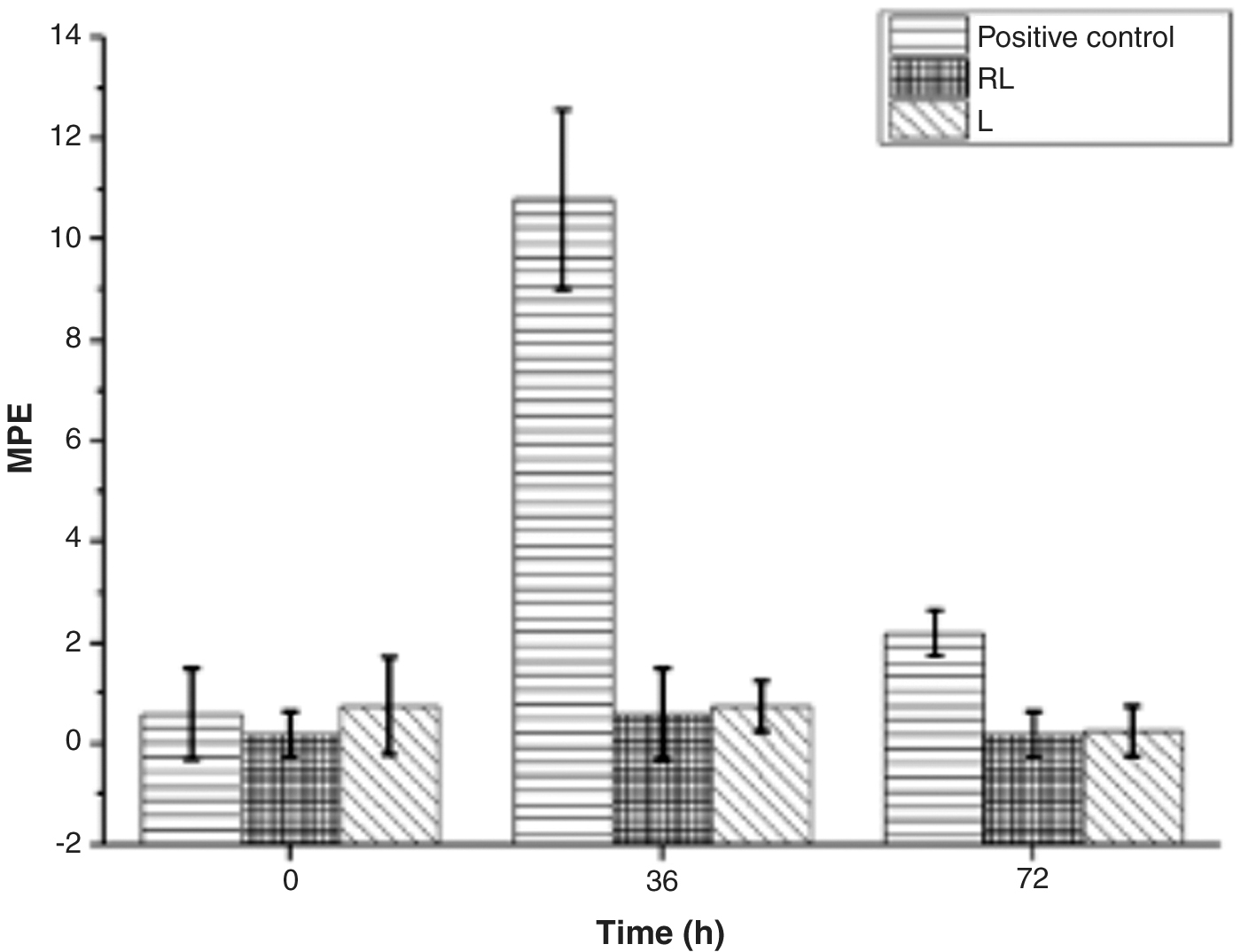
MPE produced, in peripheral blood of mice administrated with RL (rapamycin loaded liposomes) and L (empty liposomes). MPE, micronucleated polychromatic erythrocytes.
HET CAM mucous irritation potential analysis
Irritation score of the formulations and control was <0.9, Table 3, and thus corresponds to no reaction according to standardized guidelines, 29 while positive control showed a moderate reaction. These results were observed in egg-chorioallantoic membranes, Fig. 2, which showed increased membrane vascular lysis, hemorrhage, and coagulation when SDS was placed in contact, in contrast with RL and L formulations that had no effect.

Representative Injuries of different tissues.
Irritation Score of Formulations and Controls in HET CAM Test
SDS 1%: 1% solution of sodium dodecyl sulfate
L, placebo liposomes; HEM CAM, Hen's Egg Test Chorioallantoic Membrane; RL, rapamycin loaded liposomes; SDS, sodium dodecyl sulfate.
Determination of pyrogenicity
According to the following exclusion criteria, established in the Mexican Pharmacopeia FEUM, 30 pyrogen absence is warranted if rabbit temperatures differ more than 2°C between 2 temperatures consecutively measured at the acclimatization period, no rabbit temperature is higher than 39.8°C or lower than 38.0°C, no rabbit temperature increases individually above 0.5°C compared to basal temperature during the test, no rabbit shows an increase in temperature above 0.6°C compared to its individual basal temperature, and the sum of the maximum increment observed in the 3 rabbits does not exceed 1.4°C.
Rapamycin loaded liposomal formulations met the mentioned requirements and can therefore be considered pyrogen free (Table 4).
Temperature (°C) Results of Pyrogenicity Evaluation of Rapamycin Loaded Liposomes
Subacute toxicity in vivo in male New Zealand rabbits after SCJ injection
Metabolic changes
No deaths or apparent adverse clinical signs were found in any group throughout the study period. There was no statistical difference in mean body weights in the comparisons made between groups. As it is shown in the results presented in Table 5, there was no discrepancy.
Analysis of Variance of Comparisons Between Groups, Final and Initial Body Weight and Interaction Day Group
Clinical evaluation and histopathologic analysis
All the specimens were observed macroscopically at least twice a day through the entire duration of the study. Upon examination, they presented adequate general body condition. However, in all groups lacrimal glands reached 2 times their normal size. This effect can probably be attributed to a transitory parasympathetic stimulation effect caused by the injection on the lacrimal gland processed by afferent fibers in the conjunctiva and sclera of the eye. This is supported by the fact that the effect was seen in both treatment and control groups and by that after the second SCJ injection, rabbits #1, 2, and 11 presented tearing that resolved within 2 days.
In addition, the regulation of the lacrimal gland is associated with the levels of androgens, so the increased size of the tear glands can be also influenced by the amount of circulating androgens in the stage of sexual maturation since we worked with young adult rabbits. Rabbit #1 also presented swollen eyelids and pain, which also resolved within 2 days postadministration. After euthanasia of the specimens, a complete necropsy was performed. The internal general inspection detected mild-to-moderate pulmonary, hepatic, and renal congestion in all groups. Figure 2 presents some of the observed alterations after histopathological analysis of the samples. Histopathological observations are summarized in Fig. 3; P values of group comparison for each lesion demonstrate that no statistically significant differences between groups, including controls, were found.
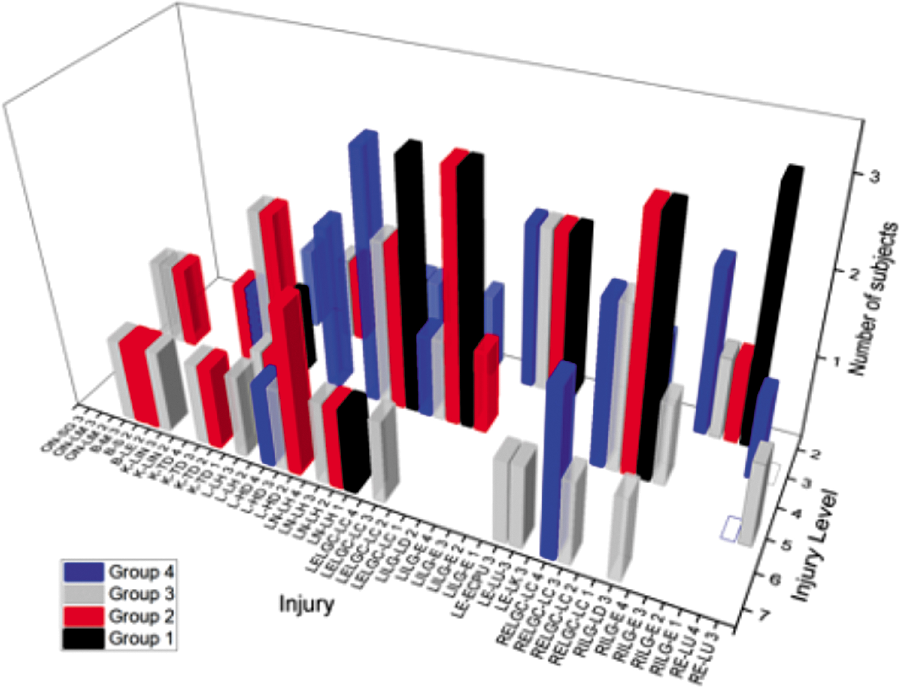
Histopathological observations at necropsy of rabbits after subacute toxicity exposure.
Biochemical assay
Results are summarized in Table 6. No toxicity-indicative differences were found between groups, sampling time or interaction between group and sampling time in all experiments.
Analysis Of Variance Analysis of Serum Biochemical Parameters
Glucose (GL), creatinine (CR), urea, alanine amino transferase (ALAT), alanine aspartate aminotransferase (ASAT), alkaline phosphatase (AP), phosphorus (Ph), total bilirubin (TB).
In bold characters are the values of p for each of the ERG measurements.
P ≤ 0.05.
Hematologic testing
Results are summarized in Table 7. No relevant differences between groups were noted in sampling time or interaction group-sampling time.
Analysis of Variance Analysis of Hematology Parameters
Hematocrit (Hat), hemoglobin (Ham), erythrocytes (Ert), platelets (Pla), leukocytes (Leu), neutrophils (Neu), lymphocytes (Lym), monocytes (Mon), eosinophils (Eos), and basophils (Bas).
In bold characters are the values of p for each of the ERG measurements.
P ≤ 0.05.
Evaluation of acute retinal toxicity in vivo in New Zealand rabbits after IVT injection
All the specimens were observed macroscopically at least once a day through the entire duration of the study by trained veterinary ophthalmologists. Upon examination, they presented adequate general condition. Fundus pictures showed no evident macroscopical alterations in retinal structure; Fig. 4 presents representative pictures of basal appearance versus postinjection aspect of retinas.
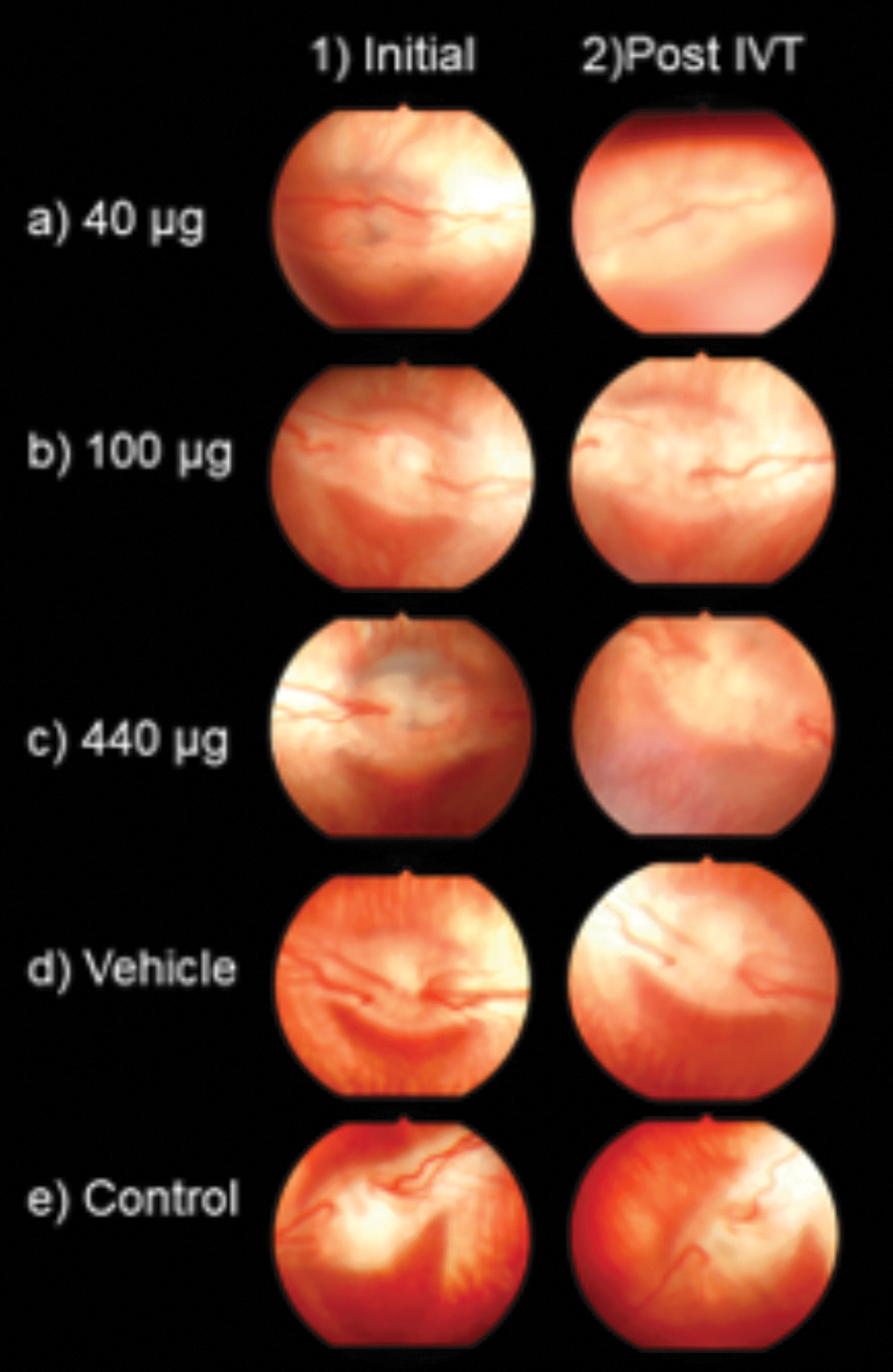
Representative fundus photographs. (a1) OD Rabbit #1 basal, (a2) OD Rabbit #1 7 days after IVT injection of 40 μg dose, (b1) OS Rabbit #4 basal, (b2) OS Rabbit #4 7 days after IVT injection of 100 μg dose, (c1) OS Rabbit #5 basal, (c2) OS Rabbit #5 7 days after IVT injection of 440 μg dose, (d1) OS Rabbit #8 basal, (d2) OS Rabbit #8 7 days after IVT injection of vehicle, (e1) OD Rabbit #9 basal, (e2) OD Rabbit #9 7 days after IVT injection of physiologic saline. IVT, intravitreal.
Electroretinography
No reduction in amplitude, increased implicit time, or alteration in waveform was observed between the basal measurements and postinjection response or in test groups versus control group. Mann–Whitney U test (P < 0.05) was performed to determine significance. Implicit times of b-wave measured in test groups compared against control group response 7 days after treatment are presented in Fig. 5; statistical analysis revealed no changes associated to test products. Representative ERG waveforms of each group 7 days after IVT injection are presented in Fig. 6.
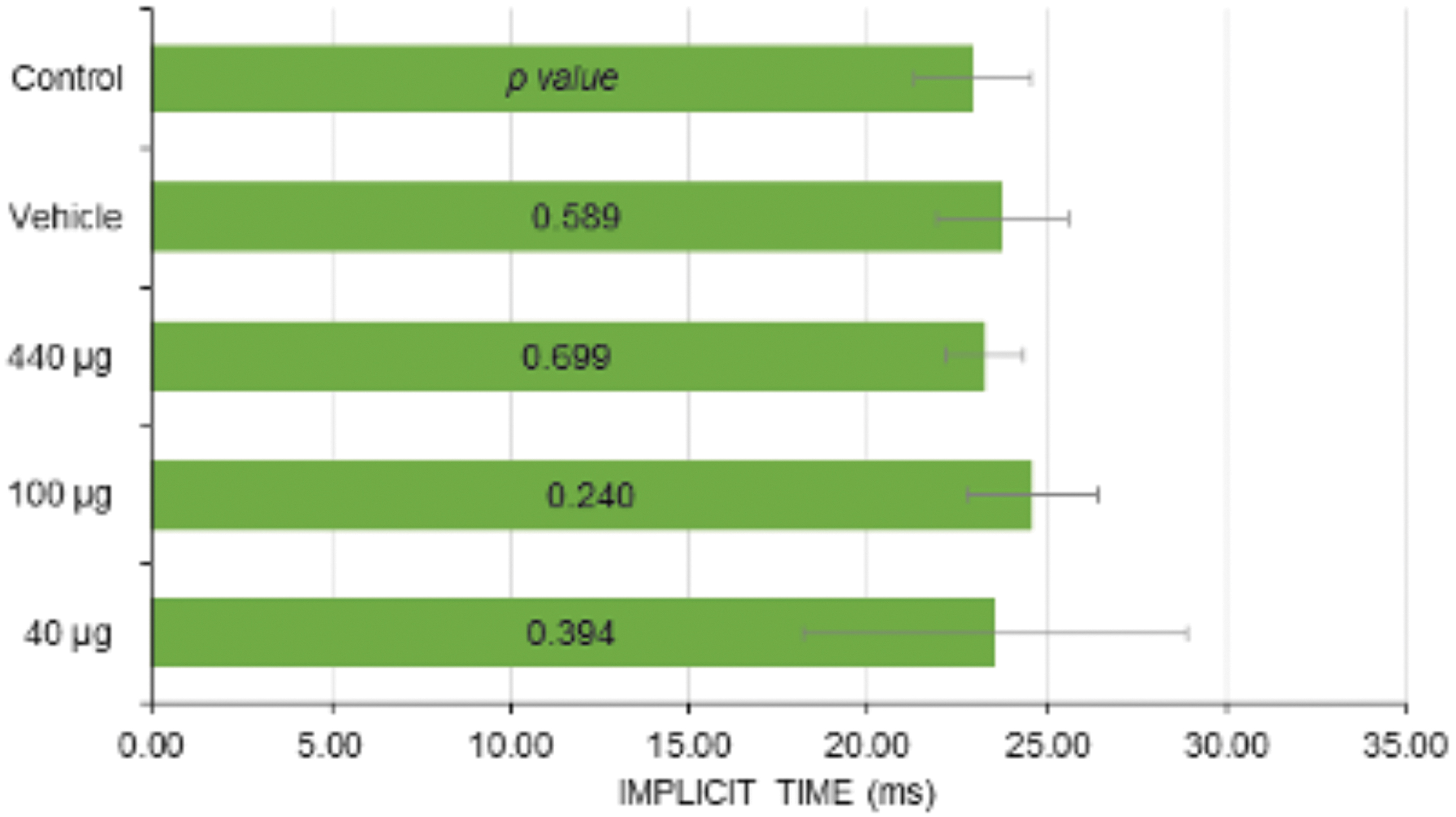
Mean implicit times of b-wave after IVT injection in comparison with control group. Mann–Whitney test analysis result with significance P < 0.05 is presented (Data are mean with standard deviation marked in error bars. n = 6 eyes per group).

Representative scotopic ERG waveforms 7 days after a single 0.1 mL IVT injection in comparison with basal waveform before treatment. Mann–Whitney analysis result with significance *P < 0.05 is presented (n = 6 eyes per group). ERG, electroretinography.
Statistical difference was tested between b-wave amplitude 7 days after treatment compared with the basal measurement of each group (Fig. 7). No significant detriment in amplitude was observed in any group; the group administered with 40 μg of rapamycin resulted in statistically different amplitude due to an increase in amplitude after IVT injection. Amplitude detected 7 days postinjection of test groups was also compared with that of the control group (Fig. 7). As well as comparative results versus basal measurements, only the group administered with 40 μg of rapamycin was statistically different from control group due to increased amplitude observed in this group after treatment.
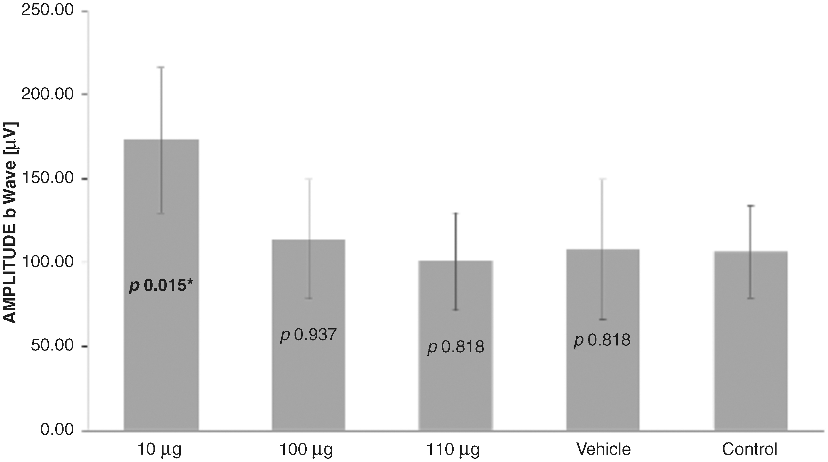
Mean scotopic b-wave amplitudes after IVT injection in comparison with control group. Mann–Whitney test analysis result with significance *P < 0.05 is presented (Data are mean with standard deviation marked in error bars, n = 6 eyes per group).
Histopathology
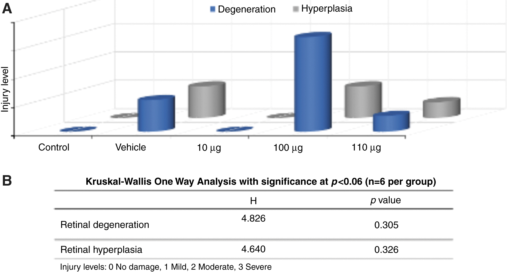
No evident pathological alterations were found in the macroscopic observation of retinas. Nevertheless, some specimens showed signs of traumatic lens fragmentation. Figure 2 shows microscopic findings. No histopathological injury was found in the control group or the group treated with 40 μg of rapamycin. In the rest of the groups, retinal degeneration and hyperplasia were observed at least in 1 sample. To determine the significance of these findings, a 1 way nonparametric Kruskal–Wallis ANOVA was performed with significance at P < 0.05, Fig. 8, Table 8. However, no significant differences were found between groups. Representative histologic micrographs of microscopic observations are shown in Fig. 9.
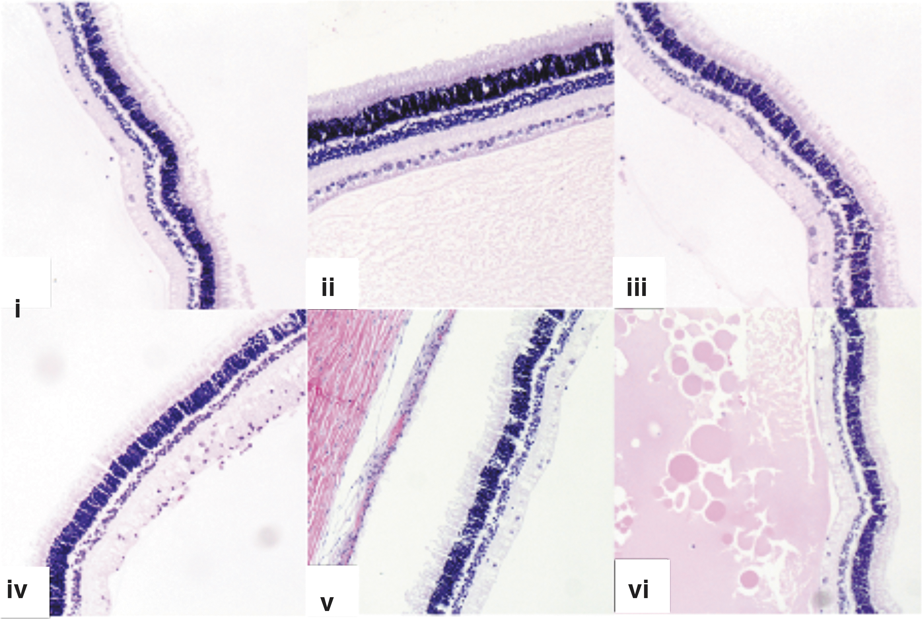
Selection of histopathologic micrographs (10 × ) of retina slices stained with H&E 14 days after IVT injection. (i) OD Rabbit #15, physiologic saline; (ii) OD Rabbit #7, vehicle; (iii) OS Rabbit #2, 40 μg; (iv) OD Rabbit #11, 100 μg; (v) OD Rabbit #12, 440 μg; (vi) OS Rabbit #3, 100 μg.
One Way Nonparametric Kruskal–Wallis Analysis of Variance Analysis
Injury levels: 2 = Mild diffuse, 3 = Mild focal, 4 = Mild Multifocal, 5 = Moderate diffuse, 6 = Moderate focal, 7 = Moderate multifocal, 8 = Severe diffuse, 9 = Severe focal, 10 = Severe multifocal.
P < 0.05.
Sterility
No evidence of microbial growth was found in the test sample tubes or negative controls. Validity of the test was assured by the performance of negative and positive controls. Growth promotion in the media used was verified by the development of visible growth in positive controls. No turbidity or creamy consistency residue was developed in media inoculated with rapamycin loaded liposomes or empty liposomes; this was determined by visual comparison against negative and positive controls incubated in parallel with test samples.
Discussion
Evaluation of genotoxicity by in vivo micronucleus test
Increased cancer risk is a serious adverse effect among patients undergoing immunosuppressive therapy. Micronucleus frequency has been reported to be significantly higher in pediatric patients with immunosuppressive therapy after kidney transplant. 32 Although the dose challenged in this article is local and much lower, this novel RL formulation is being proposed for a chronic disease that may imply long-term therapy, which is why assessment of genetic damage is mandatory.
The results presented in Fig. 1 indicate a lack of genotoxicity effect, which is consistent with studies reporting that unlike other immunosuppressants, when used specifically for ocular inflammation, rapamycin has been reported to inhibit immunosuppression-induced neoplasia. 33 This could be explained by the different mechanism of action of rapamycin versus other immunosuppressants. Oncogenic transformation, one of the most worrying consequences of DNA damage, is favored by the loss of cell cycle control and the activation of growth promoting pathways, such as the signaling path involving the mammalian Target of Rapamycin (mTOR).
Rapamycin and other inhibitors of mTOR decelerate cell proliferation and contribute to avoid oncogenic transformation by suppressing the signals required for cell cycle progression, cell growth, and proliferation. 34 Thus, RL, as well as its liposomal vehicle, are not expected to produce genetic damage when administered in mammals.
Evaluation of mucous irritability potential by HET CAM analysis
The occurrence of vascular injury or coagulation in egg-chorioallantoic membranes in response to a compound has a good correlation with the Draize rabbit test. 35 This test provides information about immediate effects after administration of a product, mainly by vascular alterations, but also by protein interactions making it an adequate prescreen method of eye injury hazard potential. The results indicate that the product is not expected to produce irritation effects on ocular membrane when tested in vivo. Testing of the vehicle irritation potential and the combination with rapamycin are yet to be proved. These results justify the innocuity of the product to be tested in live animals, as well as confirm biocompatibility of the selected nanocarrier.
Determination of pyrogenicity
Pyrogens are substances that can produce fever when present as contaminants in a drug. Most pyrogens are biological substances derived from microorganisms that trigger an immune response; they can be life threatening to patients because the produced systemic reaction can go from fever to neurologic effects, shock, and death. These results are of great importance since some of the most recognized drawbacks for the application of liposomal technology in medicine are presence of organic solvent residues, difficult pyrogen control and sterility assurance, poor stability, and adequate size distribution.
It is worth mentioning that the formulations were also tested for sterility with good compliance with FEUM standards (data not published), along with absence of pyrogenic reaction. This means that proprietary methodology used for the preparation of the samples tested in this work is suitable for commercial production.
Aside of ruling out endotoxin contamination, absence of pyrogenicity gives indirect information on acceptable size distribution of RL, since it has been reported that liposomes larger than 200 nm tend to cause nonendotoxin-dependent rise in temperature. 36 Lack of pyrogenic reaction suggests a suitable size distribution of the RL liposomes for intraocular injection.
Subacute toxicity in vivo in male New Zealand rabbits after SCJ injection
Subacute systemic toxicity is defined as the adverse effects occurring after multiple or continuous exposure between 24 h and 28 days. For this kind of studies, the FDA recommends testing the maximum dose intended for therapeutic use, and 3 and 5 times that dose to identify effects associated with overdosing and increased duration of administration. 31 In addition, in this early stage of our development process it will help to establish a safe dose range for future optimal dose exploration.
In this study the dose range tested was limited to only 3 times the dose intended for therapeutic use, and the administration frequency was doubled. Rationale for these modifications relies on the limitations of the route of administration. The maximum volume generally recognized as safe for SCJ injection is 0.5 mL; for this study the highest dose was rounded to 0.45 mL that represents 3 times the therapeutic dose. Regarding administration frequency, instead of testing a higher dose, weekly administration was established in the protocol that frequency is twice as frequent as the dose regimen considered for therapeutic use.
Metabolic changes
Although comparisons between final and initial weight show that a slight alteration was observed, it is attributable to the innate growth of the rabbits. Hence, no significant metabolic changes were noted.
Clinical evaluation and histopathologic analysis
Macroscopic evaluation only revealed mild alterations in 3 subjects after the second injection. Necropsy did not present relevant disturbances. The microscopic examination of histologic slices after necropsy revealed odd pathology results for each tissue, (Fig. 2). For both right and left eyes, lymphoplasmacytic uveitis was detected mainly in control subjects. Due to these alterations, especially in rabbit #1, all the slices were stained with Gram. One subject (rabbit 1, group 3) was detected with presence of Encephalitozoon cuniculi spores. This subject exhibited multifocal lymphoplasmacytic infiltrates in stroma and ciliary body of the right eye; this finding was also detected in right lacrimal internal gland, as well as mild multifocal necrosis.
The left eye of this specimen showed similar injury, but the infiltration covered also the iris structure. In the left lens slice multiple spores of Encephalitozoon cuniculi were found, as well as in liver and kidney samples. Optical nerve samples of this subject presented mild multifocal lymphoplasmacytic meningitis.
Based on macroscopic and microscopic findings, it was determined that 6 subjects (2 rabbits from group 2, 3 rabbits from group 3, and 1 rabbit from group 4) presented injuries compatible with encephalitozoonosis but histopathological confirmation of spores was only possible in rabbit #1 from group 3. Most of the injuries observed were similar between groups; this statement was confirmed by statistical analysis that displayed no significant differences among treatment groups so no relationship between treatment and organic damage could be elucidated. Figure 2 summarizes the injuries observed in the microscopic examination of the subjects.
It can be inferred that the highest injury levels were presented in group 3 treated with liposomal vehicle. The decreased quantity and level of injuries detected in animals administered with rapamycin containing formulations compared with empty liposomes may be related to the local immunosuppressant and anti-inflammatory activity of rapamycin. 37 Lymph nodes in all groups exhibited lymphoid hyperplasia at mild multifocal level; these can be appreciated in Fig. 2D where germinal centers of the lymph node present discrete hyperplasia. In addition, as mentioned, all the rabbits showed lymphoplasmacytic dacryoadenitis. These findings are consistent with the presence of Encephalitozoon infection. 38
Histologic examination of the liver detected hepatocellular degeneration in all groups. Group 1 showed only mild hepatocellular degeneration and congestion. Furthermore, subjects of groups 2, 3, and 4 presented moderate periportal lymphoplasmacytic hepatitis, Fig. 2F. Possibly, hepatitis could be associated with lymphoid activity and hepatocellular degeneration increase, and necrosis could be related to Encephalitozoon cuniculi infection, since these lesions were found near the parasite spores in rabbit #1. 39 It is difficult to attribute these injuries to the administration of the test product since the most affected rabbits were in the control and vehicle groups; also different kind of efforts was taken to ensure the sterility of the product as discussed below in section 4.6.
In addition, it was not possible to determine a statistically significant difference between treatment groups. Mild-to-moderate multifocal tubular degeneration was observed in the kidneys of all groups. In addition, lymphocytic interstitial nephritis in groups 2 and 3 was presented with 1 subject at level 4 and other at level 7, which is represented in Fig. 2G. Both degeneration and nephritis could be a consequence of the parasitic infection. No trend was observed in kidney injuries between groups due to rapamycin administration. 39
Lymphocytic encephalitis and satellitosis were exhibited in the brain sample of 1 subject of group 2, and granulomatous meningoencephalitis was displayed in 1 subject of group 3. All cases were at moderate multifocal injury level. Figure 2H displays the encephalic section of rabbit #1 confirmed with E. cuniculi infestation, where an inflammatory infiltrate, as well as plasmatic cells surrounding blood vessels, can be observed. The optical nerve exhibited lymphoplasmacytic-meningitis in 1 rabbit of groups 2 and 3; also satellitosis and gliosis were observed in a rabbit from group 3, Fig. 2I. Those pathologies could be linked again to parasitic infection. No damage was observed in groups 1 and 4 either in brain or optical nerve.40–42
Our animals were certified as healthy by the provider at the beginning of the study; however we detected infestation in our test population. Encephalitozoon cuniculi is a common opportunistic protozoan in laboratory animals, which is very hard to identify until symptoms appear. 43 It is an obligate intracellular microsporidian parasite. Immunocompetent animals usually are subclinical carriers, but immunocompromised hosts often present chronic granulomatous inflammation. 44 Regarding this, we could hypothesize that the presence of this infection could have been associated and promoted by immunosuppressive activity of rapamycin; however, no microorganisms could be detected in the subjects from groups 1 and 2, administered with rapamycin loaded formulations.
As discussed above, parasitic infestation was confirmed only for rabbit #1, administered with 450 μL of liposomal vehicle. In addition, all kinds of lesions encountered were statistically similar in all the treatment groups. It has been reported that cerebral lesions can only be observed about 8 weeks after initiation of antibody response to the infestation, which takes place within 3 weeks postinfection. 45 The duration of our experiment was of 3 weeks, with 10 additional days of quarantine where no obvious clinical signs of disease were observed.
Since the age of the animals was of 10 weeks at the beginning of the quarantine period, it is probable that these subjects were subclinical carriers that represented an infection focus of dissemination to the rest of the animals from urine excretion of spores that starts 3 to 5 weeks after antibody response. 45 Thus, the rest of the animals were probably in a primary stage of the infestation at the moment of the necropsy due to urinary horizontal dissemination at quarantine period. This is also consistent with the presence of kidney injury in 8 of 12 subjects, considering that kidney damage is part of the primary stage of the disease. 46
Despite the infestation found in 1 subject, there was no relationship between injuries and treatment according to ANOVA, which means that no effect can be attributable to rapamycin formulations. All the lesions observed were attributable to encephalitozoonosis, and no other significant lesions were assessed in the high or low dose test groups. The fact that the confirmed subject was administered with liposomal vehicle without rapamycin suggests that other variable may have participated in the immunosuppression of the animals. In this manner, it has been studied that in assays with live laboratory animals, alterations due to stress often occur during toxicity studies and may interfere with the interpretation of the results.
As discussed before, some of the evaluated parameters suggested that the animals were affected by stress during the study, which may explain the development of the opportunistic infection. 47 Finally, it is worth mentioning that the etiopathological origin of Encephalitozoon Cuniculi in rabbit lens has been reported in literature, 48 being ingestion of contaminated food or water the most likely origin. Transplacental and respiratory routes are also possible and common in the rabbit-breeding sector.
Biochemical assay
Samples were analyzed to observe any renal or hepatic alteration caused by administration of liposomal formulations with respect to water for injection as a control. Differences in the aforementioned parameters were analyzed at 0, 10, and 22 days after SCJ injection.
Biochemical measured values were between reference levels; therefore, no influence from product administration can be assumed. Differences observed in urea and creatinine levels showing an upward trend in time can be considered normal due to increase in body weight of the rabbits due to normal growth. Cholesterol levels were significantly higher for group 3, treated with placebo liposomes. This difference was found because basal measurements in this group presented the highest values for this analyte; therefore, differences cannot be associated with product administration.
According to literature, administration of rapamycin has been associated with cholesterol serum elevation at rapamycin doses from 1 to 7 mg/day 49 ; however, there was no increase observed in cholesterol levels in rapamycin treated groups. This could be explained by the very low dose that was locally administered, in conjunction with the liposomal carrier that encapsulates the drug and may inhibit systemic effects. 50
Concerning liver function tests, ALAT results showed difference between groups 1 and 2 for all sampling times because group 1 presented higher values for this parameter since basal measurements. Moreover, since the difference was observed throughout the whole study, it is probably not related to hepatic injury. This is also supported by a lack of difference between treatments for ASAT and AP results, which reaffirms that there is no evidence of damage on hepatocellular integrity.
None of the serum biochemical parameters related to liver and kidney function showed results indicative of toxicity due to product administration in low or high dose, as well as the liposomal carrier.
Hematologic parameters showed significant differences in hematocrit and erythrocyte count between groups. This difference was due to higher values of group 4, presented in basal measurements. Bearing in mind that this difference was found in basal measurements from the control group, it has no physiological relevance. This may be explained as an effect secondary to a hemoconcentration state of the animals at the beginning of the study.
Significant differences in leukocyte count were also observed. Specifically, there was an increase in neutrophils in groups 1, 2, and 3. In all groups, an increase in monocytes and a decrease in lymphocytes at the first sampling time were observed. Even though statistically significant differences were observed, measurements were still in normal range. The injection process could have been more stressful for the animals than expected, summed with the expected stress generated by the sampling processes for biochemical and hematological parameters. This is consistent with the fact that lymphocytes and monocyte count alterations were also observed in the control group that was handled in the same manner, Table 7.
Evaluation of acute retinal toxicity in vivo in New Zealand rabbits by IVT injection
IVT injection of rapamycin has shown good tolerability in some animal models so far.9,10,14,15 However, the toxicity evaluation of every new formulation is mandatory being that the excipients of the formulation are not always compatible with the intraocular route. 51 In this study, no statistically significant retinal acute toxicity was observed in electrical function evaluation or histological tissue examination. Fundus pictures showed no evident macroscopic alteration in retinal structure; cataract formation was attributed to accidental lens traumatism.
Electroretinography
Retinal damage can lead to vision loss due to lack of transmission of visual signal and has been in the top 5 most important causes of new drug candidate dismissal during drug development process. 52 Dark-adapted ERG was used as a noninvasive in vivo evaluation of the electrical response of retinal cells. In our study no statistically significant (P < 0.05) reduction in amplitude, increased implicit time, or alteration in waveform was observed between the basal measurements and postinjection response.
These results are of great importance because we are challenging a new formulation of a drug that has been previously proved as safe for IVT injection by many other independent research groups, but also, this same drug has also been found to be toxic to the ocular structures when formulated in certain excipients with specific deterioration of retinal function evidenced by unfavorable ERG results. 53 Interestingly, the b-wave response in the ERG of the group administered with 40 μg of rapamycin was statistically different from control group and of its original basal measurements due to increased amplitude observed in this group after treatment. These may suggest a potential mechanism to improve impaired visual signaling in the retina.
This amplitude augmenting effect has been previously reported at least once to our knowledge by Brandao-De Paiva et al., with the use of sustained-release rapamycin systems and was considered transient and clinically irrelevant. 54 In addition, a protective effect of rapamycin on visual impairment during inflammation has been reported as the attenuation in a- and b-wave reduction due to induced inflammation. 55 Further studies to explore its effects on human retinal signal transmission would be interesting.
Clinical evaluation and histopathologic analysis
Light microscopy showed normal tissue organization and cellularity of retinas in most subjects. Although nonstatistically significant alterations were observed, some subjects showed mild-to-severe injuries, specifically tissue degeneration and hyperplasia. In addition, as can be appreciated in Fig. 2, lens fragmentation and presence of Morgagnian globules were due to vacuole formation. This was consistent with slit-lamp examination and is probably related to histologic alterations observed since those subjects with cataract formation presented higher levels of histologic injury.
Limitations of this study include the small number of eyes included in each experiment and unexpected development of a subclinical disease common in laboratory animals. Further research to elucidate intraocular pharmacokinetics of the formulation and ocular biodistribution of the drug will be determining to achieve aim of clinical testing for the treatment of eye immune mediated diseases. A 40 μg liposomal rapamycin dose appears to have the best toxicity profile to be used by intraocular route; further research of its clinical effectivity is warranted.
Sterility test
Sterility assessment of the formulation was of critical importance due to a subclinical infection observed in STS experiment. Microbiological cultures yielded no growth of microorganisms in the compendial sterility test that we performed. It is worth mentioning that the etiopathological origin of Encephalitozoon Cuniculi in rabbit lens has been reported in literature, 48 being ingestion of contaminated food or water the most likely origin. These variables were controlled inside the bioterium through a strict quality system of the facility regarding quality control of animal food.
Transplacental and respiratory routes are also possible, and it is a possibility that an individual was an asymptomatic carrier that was not detected by the provider at the moment of clinical examination. This is also consistent with the type and chronology of injuries observed and the time progression of infection reported in the literature. 43 We consider unlikely that our formulation and controls were the source of this infection since the preparation of the liposomal samples implied a filtration step through a 0.45 μm membrane and the size of E. cuniculi spores is 3 times bigger (1.5–3.0 μm) and the actual parasite up to 60–120 μm. 56 In addition, the formulations were sterilized by gamma radiation at a dose of 10 kGy; E. cuniculi spores are reported to be inactivated at a 3 kGy dose. 57
Regardless, we performed cultivation of the formulation to assess sterility, yielding negative results to microorganisms. However, control group administered with water for injection (certified by commercial provider as sterile) also developed lesions. All the above, in conjunction with the facts that no signs or injuries related to parasite infection, were observed in the animals of our pyrogenicity test (purchased with same provider 3 months before), or the animals of the IVT administration experiment (intentionally bought with a different provider) led us to think that the source of the infection was at the provider facility and affected only the colony of animals used for STS experiment.
In this context, we found that this subclinical disease has been previously recognized as a common interference in laboratory research in Latin America, with an asymptomatic subclinical presentation58–61 ; this unfortunate and unexpected drawback makes us aware of the necessity of establishing a more strict control of laboratory animals in Latino America, as serological or PCR screening for this pathogen before experimentation; some authors recommend the use of pellet medicated with fenbendazole as a preventive diet at the beginning of the study. 62
Conclusions
Upon detailed examination of the formulation, no toxic effect was found in the different trials. Counting of micronucleus was normal, and the overall irritation score showed no significant differences compared with the negative control. No presence of rapamycin in peripheral blood was found, and no genotoxic damage was noted. Furthermore, the different histological, metabolic, biochemical, and hematological evaluations demonstrated no rapamycin-related harmful effects after SCJ injection. In addition, conservation of retina function after IVT injection was demonstrated. The liposome-encapsulated rapamycin formulation challenged in this article is safe to use in a dose range from 40 to 440 μg/eye in the different animal models experimented by SCJ or IVT injection. Furthermore, it shows great potential to be proven in a human clinical trial.
Footnotes
Author Disclosure Statement
M.J.B.-B. and PhD G.A.G.-S. own the patent used in this work. The rest of the authors declare no commercial relationships.
Funding Information
Part of this work was supported by CONACyT's Innovation Stimulus Program under grant 231008.