




Abstract
Purpose:
This study aims to evaluate the effect of citicoline administration in suppressing retinal damage due to methanol intoxication. This study hypothesizes that citicoline will minimize the loss of retinal ganglion cells (RGCs), minimize disruption of photoreceptors, suppress ganglion layer edema, increase expression of bcl-2 as the antiapoptotic protein, and decrease expression of caspase-3 as the proapoptotic protein.
Methods:
Fifteen Sprague-Dawley rats were divided into 5 groups, including the control group (A); methanol groups, observed on day 3 (B1) and day 7 (B2); and methanol+citicoline groups, observed on day 3 (C1) and day 7 (C2). Rats in groups B and C were placed in an inhalation chamber filled with N2O:O2 during the experiment, then methanol was administered orally. Citicoline, 1 g/kg every 24 h, was orally administered for group C. Enucleation was performed and retinas of rats were prepared for histology and immunohistochemistry examination to evaluate photoreceptor morphology and RGC density, as well as bcl-2 and caspase-3 expression.
Results:
RGC density of citicoline-treated intoxicated rats was higher than no-citicoline methanol-intoxicated rats on both day 3 (P < 0.001) and day 7 (P < 0.001). The ganglion layer thickness of citicoline-treated intoxicated rats was thinner than no-citicoline intoxicated rats, which means citicoline-treated rats had milder ganglion layer edema. Citicoline-treated rats showed higher bcl-2 and lower caspase-3 expression than no-citicoline rats. No differences were found in photoreceptor findings among groups.
Conclusions:
This study demonstrated citicoline's potential benefits for management of ocular methanol intoxication. However, more preclinical and clinical trials are needed to obtain a preferred dosage and timing of citicoline administration.
Introduction
Methanol (CH3
After ingestion, about 90% of methanol undergoes oxidation to formaldehyde, which then rapidly degrades to formic acid, a toxic metabolite.1,2 Some tissue are specifically vulnerable to formic acid toxicity, including the retina, optic nerve, and basal ganglia. 2 The causative factors of this vulnerability include high metabolic activity and oxygen demand in these cells and also a low oxidation rate of formic acid in these cells, which facilitates accumulation of toxins and high uptake of formic acid into the mitochondria.2,4–6
Histopathologically, Seme et al. 7 and Eells et al. 8 showed the most evident retinal abnormalities due to methanol in the outer retina, including photoreceptor outer segment swelling, disruption of mitochondrial photoreceptors, and photoreceptor nucleus fragmentation. Nanditya et al. 5 reported a decreased retinal ganglion cell (RGC) density in methanol-intoxicated rats. Wang et al. 9 showed that formic acid activates c-Jun N-terminal kinase (JNK), one of the important apoptotic signals. Related to the mitochondrial-dependent apoptotic pathway, bcl-2 (an antiapoptotic protein) expression was suppressed and caspase-3 (a proapoptotic protein) expression was increased by formic acid. 9
The definitive treatment for methanol-intoxicated optic nerve and retina has not been established. However, the use of neuroprotectors is thought to have potential. 10 Citicoline (cytidine-5′-diphosphocholine) transforms into cytidine monophosphate and phosphocholine with the help of the enzyme, phosphodiesterase, on the cell wall and enters neuronal cells. 11 Because of its effect in increasing membrane phospholipid biosynthesis and maintaining intracellular stability, citicoline is considered to have a protective effect on neuronal cells. 11 Furthermore, citicoline also inhibits enzymatic hydrolysis of cardiolipin by phospholipase A2, thus causing an increase in the cardiolipin level. Cardiolipin is a major component of the mitochondrial inner membrane and essential for electron transport. 12 Schuettauf et al. 13 reported the citicoline effect of suppressing RGC death due to bcl-2 upregulation.
The Sprague-Dawley strain rats are one of the most widely used experimental animals, including in neurobiological and pathological research, because of their calmness and ease of maintenance. 14 Several studies of retinal toxicity due to methanol also used Sprague-Dawley rats as animal models.8,13,15,16
This study aims to evaluate the effect of citicoline administration in suppressing retinal damage due to methanol intoxication in rats. The hypothesis is that citicoline is effective in preventing photoreceptor and RGC death, suppressing retinal edema, increasing bcl-2 expression, and decreasing caspase-3 expression on methanol-intoxicated rat retina.
Methods
This research was conducted after approval from the ethics committee of Faculty of Medicine, Universitas Indonesia, with protocol number 18-07-0747.
Animals
Male Sprague-Dawley rats, ranging in weight from 200 to 350 g, were used in the study. Fifteen Sprague-Dawley rats were divided into 5 groups, including 3 rats in the control group (A); 6 rats in methanol-only groups, divided into group B1, which was observed for 3 days, and group B2, which was observed for 7 days; and 6 rats in methanol+citicoline groups, divided into group C1 and group C2, which were observed for 3 and 7 days, respectively. The control group (A) includes rats that were unexposed to nitrous oxide (N2O), methanol, or citicoline administration.
All animals were supplied with food and water ad libitum and maintained on a 12-h light–12-h dark schedule. Rats were placed in a plexiglass chamber with dimensions of 35 × 55 × 24 cm, with 3–6 rats placed in each chamber. All animal experiments were performed in accordance with the resolution for the Use of Animals in Ophthalmic and Vision Research issued by The Association for Research in Vision and Ophthalmology.
Methanol intoxication protocol
Rats in groups B and C were placed in a plexiglass chamber exposed to N2O:O2 with a flow rate of 2 L/min started 4 h before methanol administration until the experiment is completed (Fig. 1). Nitrous oxide (N2O) exposure is needed in this animal experiment to cause a decrease in tetrahydrofolate levels of rats so that accumulation of formic acid can occur and to facilitate intoxication that is similar to methanol intoxication in humans.15,17 Methanol was administered through oral gavage with an initial dose of 3.2 g/kg and 1 supplemental dose of 1.6 g/kg on the second day.
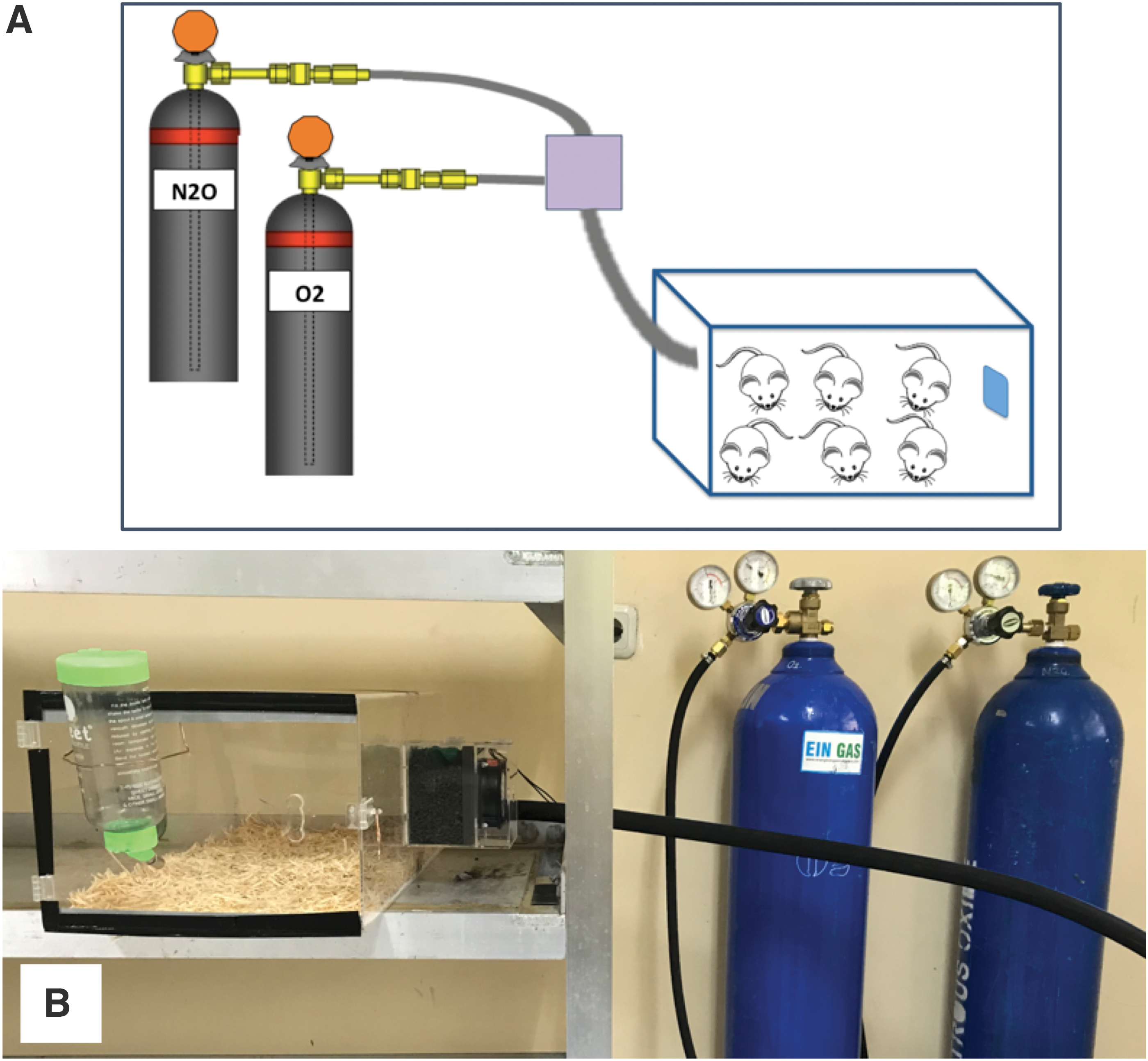
Schematic picture
Citicoline and placebo administration protocol
Citicoline (RG-Choline®) 1 g/kg (in a 5-mL solution) every 24 h was administered through oral gavage for rats in group C. Citicoline administration was started 4 h after methanol administration. However, rats in group B received 5 mL of normal saline as a placebo through oral gavage every 24 h, also started 4 h after methanol administration.
Retinal tissue preparation
Euthanasia was performed by intrahepatic ketamine and xylazine injection. Afterward, retinas from the right eyes of rats were prepared for histopathology and immunohistochemistry examination. Retinal tissue preparation was done by a histology technician in a masked manner.
The sample preparation process includes the following:
A fixation step to maintain the structure of the cellular network. This stage is needed to harden the tissue so that its structure does not change due to further manipulation. This stage is also necessary to withstand the process of tissue autolysis. The substance used for the fixation process in this study was 10% buffered formalin. Immersion in the fixative fluid lasts for 1 night (4–24 h). Dehydration, which is the process of removing water from pieces of tissue, is carried out slowly using serial alcohol. Infiltration and embedding are processes of inserting a substance (paraffin) that acts as support into every gap in the tissue, allowing the tissue to harden in the mold, and then implanting the tissue into paraffin block media. Sectioning includes paraffin block cutting with a microtome to produce a series of thin sections with a certain thickness. Sections were made with a thickness of 4 μm in the area of the posterior pole of the retina, involving the optic nerve and extending along the vertical meridian. Mounting is the process of permanently gluing the tissue on the microscope object glass so that unstained preparations are available. The label is written on the slide.
Histopathology protocols
The staining process was carried out with toluidine blue. Histopathology procedures were performed by a histology technician in a masked manner. The stages of staining included the following:
Preparation of 0.1% toluidine blue staining solution by mixing 0.1 g of toluidine blue (Toluidine blue; Sigma-Aldrich, MO) with 100 mL of distilled water, which was then filtered.
The tissue made into paraffin blocks was cut using a microtome with a thickness of 4 μm, dried at 37°C, and then the sample number was written on each slide.
The preparation was heated on a slide warmer for 60 min at a temperature of 58°C.
Deparaffinization of paraffin sections was performed by soaking 3 times in xylol, each for 5 min.
Alcohol rehydration was performed by using ethanol, 96% alcohol, and 70% alcohol, each for 5 min.
The slide was washed with running water for 5 min.
The preparation was stained with 0.1% toluidine blue dye for 1–2 min.
The preparation was dipped in distilled water for 1–5 min.
Alcohol dehydration was performed by using 70% alcohol, 96% alcohol, and ethanol, each for 5 min.
Clearing was done with xylol I, II, and III, each for 5 min.
The preparation was mounted with resin (Entellan®), then attached to a cover glass.
Immunohistochemistry protocols
From each paraffin block preparation, 2 immunohistochemical preparations were made for bcl-2 antibody and caspase-3 antibody staining. Furthermore, the preparation was heated on a slide warmer for 60 min at a temperature of 60°C. Deparaffinization was performed by soaking 3 times in xylol, each for 5 min, followed by decreased alcohol rehydration (absolute alcohol and 96% and 70% alcohol) for 4 min each, and then washing with running water for 3 min.
Each preparation was blocked with 3% H2O2 in methanol for 30 min. The process of antigen retrieval was performed with Tris-EDTA, pH 9.0, in a decloaking chamber at 96°C for 10 min and then the preparation was cooled at room temperature for 25 min. After that it was washed with PBS, pH 7.4. Blocking was done with Background Sniper for 20 min.
Then, the primary antibody was incubated with bcl-2 at a 1:200 dilution on one preparation and with caspase-3 at a 1:800 dilution on the other preparation for 60 min. The preparation was washed with PBS, pH 7.4, for 2 min and incubated with Trekkie Universal Link secondary antibody for 20 min and then the preparation was washed with PBS, pH 7.4, for 2 min. This was followed by incubation with TrekAvidin-HRP Label for 15 min and washing with PBS, pH 7.4, for 2 min. Incubation was then carried out with a mixture of 1 mL of Betazoid DAB substrate buffer for 5 min with 1 drop of Betazoid DAB chromogen for 2 min, then the preparation was washed with running water and counterstained with hematoxylin for 1 min. The tissue was stained with saturated lithium carbonate (5% in Aquabidest) for 5 s, washed under running water for 2 min, and dehydrated with increasing alcohol for 5 min each. Clearing was done with xylol I, II, and III, each for 5 min. The preparation was mounted with resin (Entellan) and attached on the cover glass. This histopathology procedure was performed by a histology technician in a masked manner.
Histopathology and immunohistochemistry analysis
Toluidine blue stain is used for the histopathological evaluation of photoreceptors and RGCs. Histopathological parameters that were taken into account included the thickness of the photoreceptor inner segment, thickness of the outer retinal layer (including the photoreceptor outer segment layer, inner segment layer, outer nucleus layer, and outer plexiform layer), ganglion layer thickness, total retinal thickness, number of fragmented photoreceptors, and ganglion cell density. Microscopic examination was performed with 400 × lens magnification. Measurement of retinal layer thickness was performed using ImageJ software.
An immunohistochemistry examination was performed to evaluate the expression of bcl-2 and caspase-3 in ganglion cells. Positive expression of bcl-2 or caspase-3 in ganglion cells is shown as brown-colored cytoplasmic cells. The microscopic evaluation was performed with 400 × lens magnification. Histology and immunohistochemistry interpretation and image analysis were performed by an anatomic pathologist in a masked manner.
Statistical analysis
Data analysis was performed using the Statistical Package for the Social Sciences (SPSS)™ 20.0 for Mac. Normality tests were conducted using the Shapiro–Wilk test. Comparison analysis of retinal layer thickness, number of fragmented photoreceptor nuclei, ganglion cell density, and expression levels of bcl-2 and caspase-3 between 2 groups (for example, group A vs. B1, A vs. B2, A vs. C1, and B1 vs. C1) was performed using the independent t-test due to the normal distribution of data, and no sample was paired between groups. In all cases, the minimum level of significance was taken as P < 0.05.
Results
The effects of methanol intoxication and administration of citicoline on retinal histology were assessed by light microscopy after toluidine blue staining. Figure 2 illustrates the retinal morphology in the representative control (A), methanol-only group observed on day 3 (B1), methanol-only group observed on day 7 (B2), methanol+citicoline group observed on day 3 (C1), and methanol+citicoline group observed on day 7 (C2).
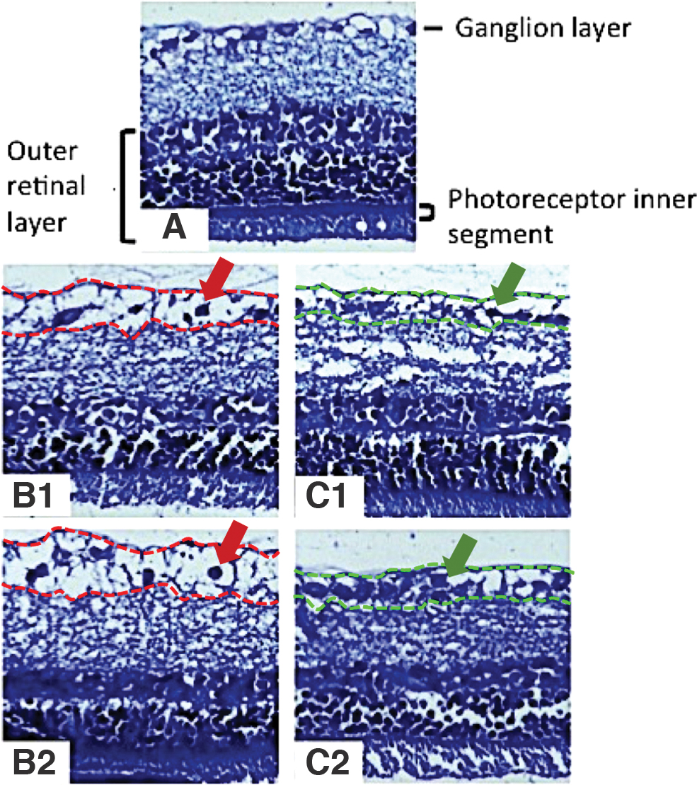
Retinal histopathology with toluidine blue staining in the representative untreated control
Thickened retina represents a retinal edema condition, which is prominently seen in rats from B1 and B2 groups. Only the ganglion layer was thickened in intervention groups (B and C), compared with the control group (Table 1), while other layers did not show any significant differences between intervention and control groups.
Thickness of the Photoreceptor Inner Segment Layer, Outer Retinal Layer, Ganglion Layer, and Total Retinal Layer in All Groups (N = 15 Rats)
t-Test result when compared with group A (two-group comparison); minimum level of significance was taken as P < 0.05.
A, control; B1, methanol-only, observed on day 3; B2, methanol-only, observed on day 7; C1, methanol+citicoline, observed on day 3; C2, methanol+citicoline, observed on day 7.
Ganglion layer thickness differs significantly between methanol-only groups and methanol+citicoline groups (B1 vs. C1 and B2 vs. C2), as shown in Fig. 3.
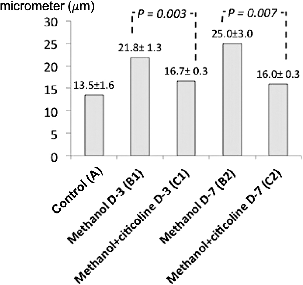
Ganglion layer thickness comparison (N = 15 rats). The ganglion layer thickness differs significantly between methanol groups and methanol–citicoline groups (B1 vs. C1 and B2 vs. C2). A, control; B1, methanol-only group observed on day 3; B2, methanol-only group observed on day 7; C1, methanol+citicoline group observed on day 3; C2, methanol+citicoline group observed on day 7.
Histopathological staining of rat retina showed no fragmentation of photoreceptor nuclei in all groups. However, the RGC density count showed differences between methanol groups and the control group (A vs. B1 and A vs. B2). The mean RGC density in methanol-only groups (B1 and B2) is significantly lower than the control group (A), with a P value of 0.009 and 0.002, respectively. Moreover, the RGC density in methanol+citicoline groups was significantly higher compared with the methanol-only groups on both day 3 and day 7, as shown in Fig. 4.
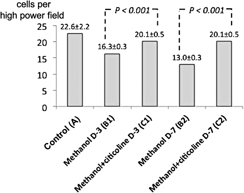
Retinal ganglion cell density (N = 15 rats). RGC density differs significantly between methanol groups and methanol–citicoline groups (B1 vs. C1 and B2 vs. C2). A, control; B1, methanol-only group observed on day 3; B2, methanol-only group observed on day 7; C1, methanol+citicoline group observed on day 3; C2, methanol+citicoline group observed on day 7.
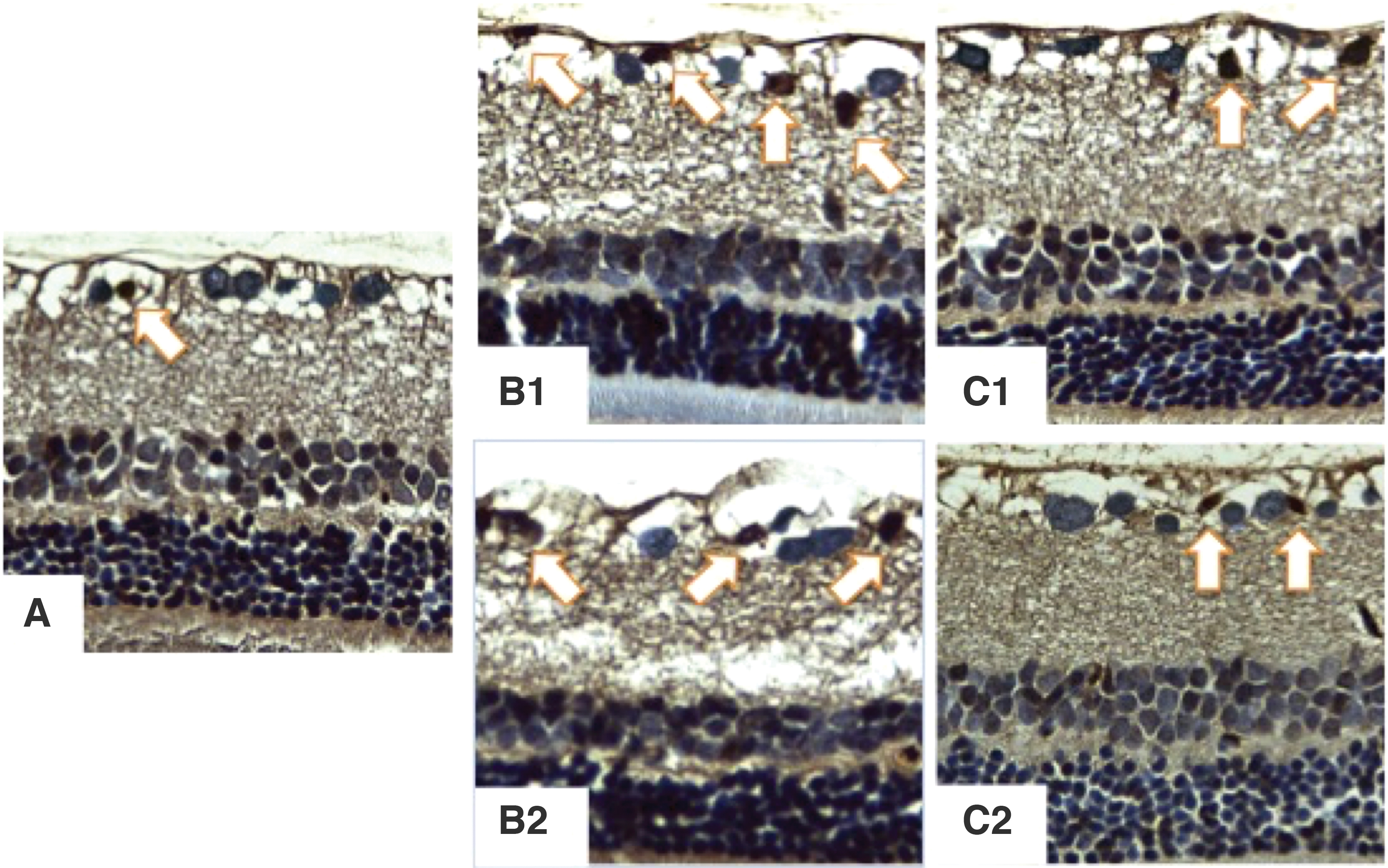
bcl-2 is an antiapoptotic protein that plays a role in the apoptotic mechanism of neuronal cells, including retinal neuronal cells. The bcl-2 expression in this study was assessed for 2 layers in the retina, which are the outer nuclear layer where the photoreceptor nuclei are located and the ganglion layer. Expression of bcl-2 in the ganglion layer was shown as brown cells. Based on the immunohistochemistry examination, expression of bcl-2 was found in the ganglion layer (Fig. 5) and not in the outer nuclear layer.

Bcl-2 expression in all groups. Positive expression shown by white arrows.
The number of cells expressing bcl-2 in the retinal ganglion layer was counted in 3 fields of view (400 × magnification) and results are shown in Table 2. Expression of bcl-2 was found to be higher in the methanol+citicoline group (group C) compared with the methanol-only group (group B) on both day 3 and day 7 (Fig. 6).
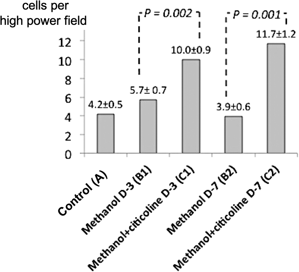
Level of bcl-2 expression (N = 15 rats). The number of cells expressing bcl-2 differs significantly between methanol groups and methanol–citicoline groups (B1 vs. C1 and B2 vs. C2). A, control; B1, methanol-only group observed on day 3; B2, methanol-only group observed on day 7; C1, methanol+citicoline group observed on day 3; C2, methanol+citicoline group observed on day 7.
Comparison of bcl-2 Expression Levels in the Ganglion Layer Between the Treated and Untreated Groups (N = 15 Rats)
t-Test result when compared with group A (two-group comparison); minimum level of significance was taken as P < 0.05.
A, control; B1, methanol-only, observed on day 3; B2, methanol-only, observed on day 7; C1, methanol+citicoline, observed on day 3; C2, methanol+citicoline, observed on day 7.
Caspase-3 is the proapoptotic executor protein in the neuronal cell apoptosis mechanism. The expression of caspase-3 in this study was assessed for 2 layers in the retina, including the outer nuclear layer and the ganglion layer. The expression of caspase-3 in the ganglion layer was shown as brown cells. Based on the immunohistochemical examination, the expression of caspase-3 was found in the ganglion layer (Fig. 7) and not in the outer nuclear layer. The number of cells expressing caspase-3 in the retinal ganglion layer is shown in Table 3.
Caspase-3 expression in all groups. Positive expression shown by white arrows.
Comparison of Caspase-3 Expression Levels in the Ganglion Layer Between the Treated and Untreated Groups (N = 15 Rats)
t-Test result when compared with group A (two-group comparison); minimum level of significance was taken as P < 0.05.
A, control; B1, methanol-only, observed on day 3; B2, methanol-only, observed on day 7; C1, methanol+citicoline, observed on day 3; C2, methanol+citicoline, observed on day 7.
Caspase-3 expression was found to be significantly lower in the methanol+citicoline group (group C) compared with the methanol-only group (group B) on day 3 and day 7, as shown in Fig. 8.
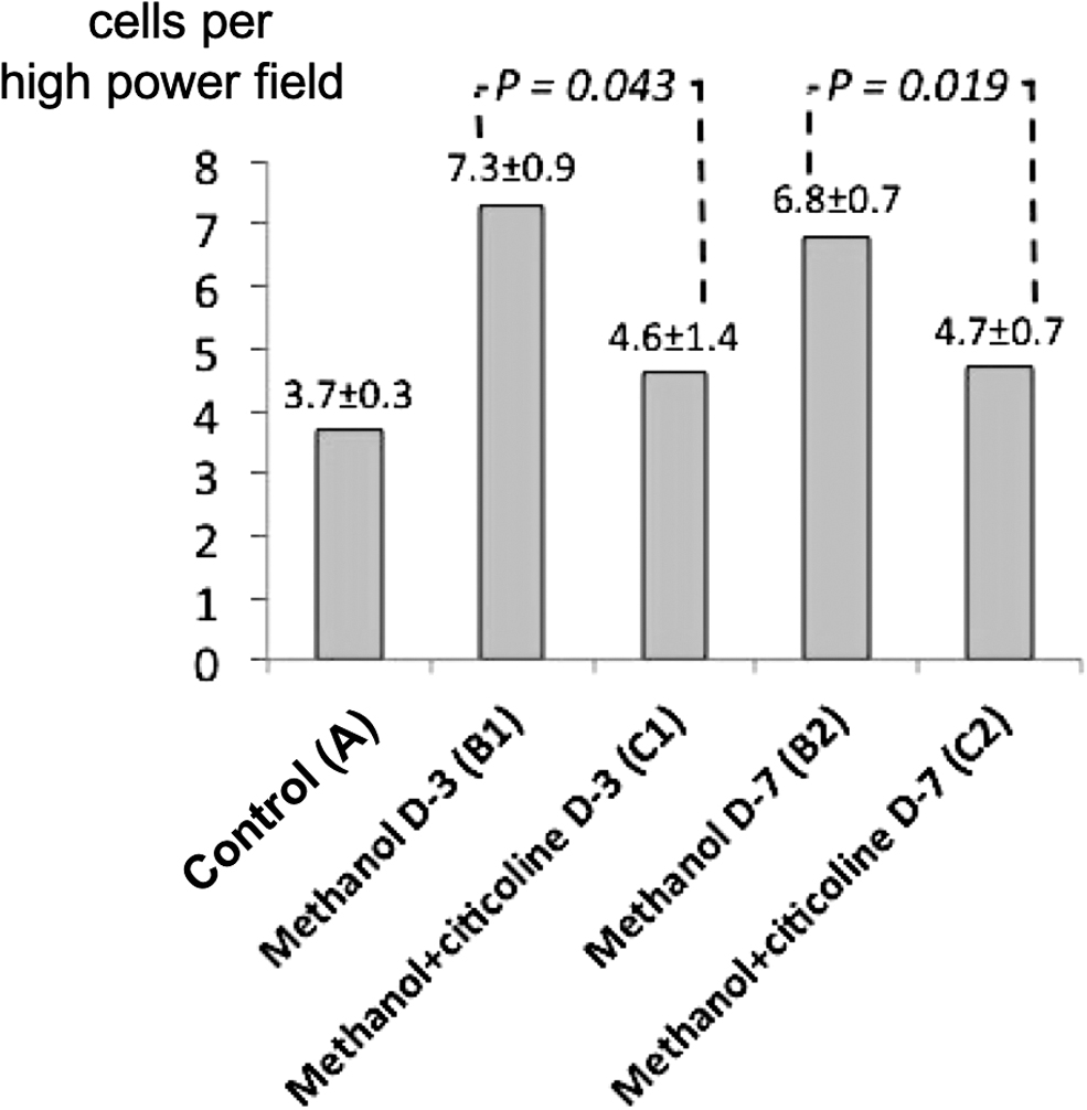
Level of caspase-3 expression (N = 15 rats). The number of cells expressing caspase-3 differs significantly between methanol groups and methanol–citicoline groups (B1 vs. C1 and B2 vs. C2). A, control; B1, methanol-only group observed on day 3; B2, methanol-only group observed on day 7; C1, methanol+citicoline group observed on day 3; C2, methanol+citicoline group observed on day 7.
Discussion
There are several essential findings of this study. First, this study proved the loss of RGCs as the main structural damage that occurs due to methanol intoxication. This loss of RGCs is generally accompanied by axonal damage and both are the major causes of methanol-induced optic neuropathy. 18
Second, edema occurs specifically in the ganglion layer only. A similar finding, ganglion layer edema, was also shown by Nanditya et al. 5 12 h after oral methanol administration. The main pathogenesis of ganglion layer edema in methanol intoxication is related to mitochondrial failure in producing adenosine triphosphate (ATP). The lack of ATP results in infusion of Ca2+ through voltage-dependent Ca2+ channels in ganglion cell membranes. As a result, intracellular glutamate is released, which then accumulates extracellularly. This condition causes activation of ionotropic glutamate receptors, which results in Na+ and Cl- ion influx and induces osmotic cell swelling. 19 The ganglion layer, a layer comprising RGCs, is one of the structures most susceptible to ATP deprivation.
The present study also proved that the apoptotic pathway plays an important role in optic nerve damage due to methanol intoxication; it is shown in this study with the significant increase in caspase-3, an executioner caspase of the apoptotic pathway, after methanol administration.
Previous studies have reported retinal abnormalities after methanol administration in an animal model.5,7,8 However, until now, no study had proven the benefits of citicoline as a neuroprotector in a methanol-intoxicated retina. The most important finding in this study is that it provides proof of concept that citicoline has potential for management of ocular methanol intoxication. Theoretically, citicoline suppresses retinal damage in methanol intoxication through several mechanisms. First, citicoline increases the synthesis of phosphatidylcholine, the main component of cell membranes and organelles, thus it maintains the structural integrity of cell membranes. 19 Second, citicoline supports the process of mitochondrial oxidative phosphorylation to produce ATP. With adequate ATP availability, the influx of Ca2+, Na+, and Cl− into ganglion cells, which results in cell swelling through ion influx, can be prevented. 19 Another important mechanism is through inhibition of phospholipase A2 (PLA2), which increases cardiolipin levels. Cardiolipin plays a crucial role in the oxidative mechanisms of mitochondria.20,21
Another important point is the successful creation of a methanol-intoxicated rat model through an uncomplicated procedure, and retinal intoxication was proven through a relatively simple assessment, which is histological examination. Not only were structural abnormalities found but also the role of the apoptotic process could be demonstrated by immunohistochemical examination. This method of rat model creation and outcome assessment could be replicated in other research in laboratories with limited facilities.
There were some limitations in this study. Biochemical examination confirming blood-circulating formic acid levels could not be performed in this study. Another limitation was related to the thickness of retinal tissue preparation for the microscopic assessment, which is 3–4 μm, thicker than other studies that used 1-μm-thick tissue sections for light microscopy and an even thinner cut for electron microscopic examination. The use of electron microscopic examination may help in finding “unseeable” photoreceptor abnormalities. This study also did not assess the functional status through electroretinography. The absence of morphological abnormalities in photoreceptors due to methanol intoxication in this study did not rule out the possibility of functional abnormalities in photoreceptors.
Although this in vivo study cannot represent the preferred timing of citicoline administration and the minimal effective dose for clinical purposes, the present study can be an initial trigger for further exploration of the clinical use of citicoline in ocular methanol intoxication.
In conclusion, citicoline has potential benefits for management of ocular methanol intoxication, through attenuation of the apoptosis pathway and preservation of RGCs. In the future, more preclinical and clinical trials are needed to obtain the best dosing regimen and preferred timing of citicoline administration in methanol intoxication.
Footnotes
Acknowledgments
The authors thank Rani Indira Sari, MD, and Joshua P.F. Lumbantobing, MD for useful discussion and support. The authors also thank Anggi Arris Faisal, Deny Suprihati S.Si., Candra Kusuma  AK., and Slamet Hartono for technical help in animal handling and sample preparation.
Author Disclosure Statement
No competing financial interests exist.
Funding Information
No funding was received for this article.