




Abstract
Purpose:
We tested the role of microRNA-328 in dry eye disease (DED). Benzalkonium chloride (BAC) has been used to induce DED in animal models. We first demonstrated that both BAC and hyperosmotic stress induced overexpression of miR-328 in corneal cells and then tested whether anti-miR-328 could be a new therapy.
Methods:
BAC was instilled to both eyes of 41 rabbits and 19 mice from day 0 to 21 to induce DED. Animals of each species were divided to receive topical instillation of saline or anti-miR-328 eye drops between day 8 and 21. The DED signs were assessed by corneal fluorescein staining, histological examination, apoptosis of corneal cells, and inflammatory cytokines in rabbit eyes. For mice, only corneal fluorescein staining was assessed for the therapeutic effects. The corneal fluorescein staining scores ranged from 0 of no staining to 4 of coalescent.
Results:
For the rabbits, the staining score was significantly reduced (P = 0.038) after the 14-day anti-miR-328 treatment (n = 42 eyes), but the score was not improved by saline treatment (n = 40 eyes). Furthermore, rabbit eyes treated with anti-miR-328 had thicker corneal epithelium (P = 9.4 × 10⁻⁵), fewer apoptotic cells in corneal epithelium (P = 0.002), and stroma (P = 0.029) compared with the saline-treated eyes. Anti-miR-328 was more effective than saline to reduce the block of orifices of Meibomian glands, although such an effect was only marginally significant (P = 0.059). Similarly, anti-miR-328 was more effective than saline in reducing corneal staining in mouse eyes (P = 0.005).
Conclusion:
Overexpression of miR-328 may contribute to DED. Anti-miR-328 protects corneal cells and promotes re-epithelialization for DED treatment.
Introduction
Dry eye disease (DED) is due to a lack of sufficient lubrication to the eye's aqueous tear film layer, leading to the ocular surface's disruption, eye discomfort, and even visual disturbance. 1 DED is a multifactorial disorder with multiple risk factors, among which Meibomian gland dysfunction (MGD) is an important risk factor.2–4 MGD causes a disruption in the tear film lipid layer that affects the rate of tear evaporation. The pathogenesis of both MGD and DED interact, resulting in a double vicious circle. 5
The prevalence of DED varies among populations and sex. The prevalence of DED in Asians is ∼30%, which is higher than 12% in Caucasians. 6 Women have a higher risk than men. 7 The first-line treatment is frequent administration of nonprescription artificial tear substitutes, but the effect is only temporary and minimal. For more severe cases, prescribed drugs may be needed to alleviate the discomfort.
Benzalkonium chloride (BAC) is a commonly used preservative in the ophthalmic solution. However, it was then reported to increase risk of DED due to its toxicity on the ocular surface, the conjunctiva, the cornea and even deeper ocular structures. 8 In recent years, BAC has emerged as an agent for investigating the pathogenesis of DED in animals. In animal models, topically applied 0.1%–0.2% BAC twice or triple daily over 4–14 days triggers clinical signs of DED, including increased fluorescein staining and corneal irregularity.9,10
MicroRNAs (miRNAs) are noncoding single-stranded RNA molecules of about 21–23 nucleotides in length. A miRNA is first transcribed as pri-miRNA with a cap and poly-A tail and then processed to a short 70-nucleotide stem-loop structure known as pre-miRNA in the cell nucleus. 11 The pre-miRNA is then processed to mature miRNAs in the cytoplasm. A mature miRNA is complementary to a part of 1 or more messenger RNAs (mRNAs). Animal miRNAs are usually complementary to a site in the 3′ untranslated region of mRNA. The annealing of the miRNA to the mRNA causes an inhibition of protein translation, and/or cleavage of the mRNA. MiRNAs can regulate cell growth, differentiation, and apoptosis; therefore, its dysregulation may lead to human diseases.12,13 miRNAs have been reported to be involved in DED as well as inflammatory diseases.14–17
We previously reported that overexpression of miR-328 in the eye is a risk of myopia. 18 Recent studies also showed that overexpressed miR-328 induces cell apoptosis, and reduces cell migration and proliferation.19–22 Since DED is also characterized by loss of corneal epithelium and cell apoptosis, it is of interest to investigate whether overexpression of miR-328 also contributes to DED. 23 In this study, we first demonstrated that overexpressed miR-328 is a risk factor for DED. We then used anti-miR-328 oligonucleotide as a therapeutic agent to treat DED in both mice and rabbits. Our data indicate that suppression of miR-328 may be a novel approach to alleviate DED.
Methods
In vitro studies
The Statens Seruminstitut Rabbit Cornea (SIRC) cell line was purchased from the Bioresource Collection and Research Center (BCRC). The cells were cultured in Dulbecco's modified Eagle medium (DMEM) (5 mM
After 10-min BAC treatments, the culture medium was replaced with fresh media and cells were incubated for a further 24 h. For the hyperosmotic stress study, ∼1.2 × 10⁵ cells were harvested from 6-well culture plates and incubated for 48 h. Hyperosmotic stress (500 mOsm) was achieved by adding 90 mM sodium chloride (NaCl; Sigma-Aldrich) to isosmotic medium (310 mOsm). The culture medium was then replaced with DMEM or hyperosmotic medium, and the cells were incubated for 24 and 48 h.
Then, cells were washed, harvested, and lysed in 0.7 mL of TRIzol reagent (Invitrogen), according to the manufacturer's instructions. Total RNA was purified with Qiagen RNAeasy Columns (Qiagen, Hamburg, Germany).
Measurement of miR-328 expression
miR-328 expression level was detected by real-time-quantitative polymerase chain reaction (qPCR) or digital PCR. The TaqMan miR RT-qPCR assay was described in previous studies. 18 The relative expression levels of miR-328 were normalized to that of U6b as the internal control using the equation: log10 (2−ΔCt), where ΔCt = (CtmiR-328–CtU6b). The mean and standard deviation (SD) values of log10 (2−ΔCt) were calculated. The droplet digital PCR was used to directly count the copy number of miR-328 cDNA without the need of internal control. After PCR reaction, the amplified products were detected using a droplet reader (JN MEDSYS, Singapore). The Clarity Software 3.0 was used to analyze the DNA copy number.
Anti-miR-328 eye drops
Anti-miRNA oligonucleotides rely on the complementary base pairing of the oligonucleotide sequence to miRNA-328. We used a proprietary anti-miR-328, which is a single-stranded oligonucleotide that is composed of 16 mer unmodified DNA to perfectly match to the seed region of miRNA-328 (US patent no. 101 79913B2). The inhibitory effects of these oligonucleotides on miR-328 levels were first tested in the ARPE-19 cells and then in mice (US patent no. 101 79913B2). The anti-miR-328 oligonucleotide was dissolved in phosphate-buffered saline (PBS) as eye drops for in vitro or in vivo studies.
Dry eye study in animals
Nineteen male C57BL/6J mice (8 weeks old) were obtained from the National Laboratory Animal Center, National Applied Research Laboratories, National Science Council (Taipei, Taiwan, ROC). A total of 41 Rex pigmented rabbits were used and the rabbits were tested in 2 batches. The first batch contained 21 female rabbits, and the second batch contained 10 male and 10 female rabbits. All the rabbits weighed 2–3 kg and were purchased from a local farm Da-Zong ranch (Changhua county, Taiwan, ROC).
The animals were adopted a week before the experiments in the Laboratory Animal Center of China Medical University (Taiwan, ROC). The animals were kept at a controlled temperature (23 ± 2°C), relative humidity (60% ± 10%), and 12 h light–dark cycles (07:00–19:00), and given food and water ad libitum. All procedures were approved by the Institutional Animal Care and Use Committee (IACUC no. 2021-276) of China Medical University.
For the mouse study, the animals were randomly divided to 2 groups (9 mice in the PBS-treated group, and 10 in the anti-miR-328-treated group) and then 5 μL of 0.2% BAC was instilled to both eyes once per day from day 0 to 7 to induce DED. Five microliters of anti-miR-328 (10 μM) or PBS solution was instilled to both eyes once per day from day 8 to 21, whereas BAC was still instilled 10 min after anti-miR-328 or PBS treatment between day 8 and 21. The corneal fluorescein staining was performed on days 0, 7, 14, and 21.
For the rabbit study, the first batch contained 21 female rabbits and they were used to test for the anti-miR-328 therapeutic effect according to the corneal fluorescent staining. The rabbits in the second batch were tested for both the corneal fluorescent staining and histopathology examination. The rabbits in each batch were first randomly divided to 2 groups: PBS group and the anti-miR-328 group. Subsequently, DED was induced from day 0 to 7 by instilling 20 μL of 0.15% BAC twice per day (9 am and 5 pm). Twenty microliters of anti-miR-328 (10 μM) or PBS solution was instilled to both eyes twice per day between day 8 and 21, whereas BAC was still instilled 10 min after anti-miR-328 or PBS treatment in these 2 weeks. The corneal fluorescein staining was performed on days 0, 7, 14, and 21.
The second batch of rabbits were euthanized humanely on day 22. The rabbit eyes were enucleated and immersed in Davidson's fixative (20 mL of 37% formalin, 100 mL of glacial acetic acid, 350 mL of 95% alcohol, and 530 mL of water). The tissues were fixed for 48 h, then washed in tap water before being transferred to 10% neutral buffered formalin for storage before trimming and processing. The eye lids were removed and immediately fixed in 10% buffered formaldehyde solution for 24 h. Subsequently, collected tissue samples were then dehydrated with a gradient series of ethanol and embedded in paraffin. The left cornea was prepared for histological analysis, and the right cornea was used for cytokine analysis.
Fluorescein staining
Fluorescein staining of the cornea was used to assess the therapeutic effect. The fluorescein sodium solution was prepared from fluorescein paper strips (Madhu Instruments Pvt. Ltd., Okhla Industrial Area, India), and the solution was instilled into the conjunctival sac (3 μL for a mouse eye and 5 μL for a rabbit eye). The eyes were examined and graded under a slit lamp SL-15 with a cobalt blue filter (Kowa Company, Ltd., Tokyo, Japan). We used 2 different scoring systems. First, we called it “modified staining score,” because it was modified from a phase 3 clinical trial for the Food and Drug Administration (FDA)-approved dry eye drug, Lifitegrast. 26
The modified staining score needs to divide the corneal surface into 9 regions by superior, central, and inferior, as well as, left, middle, and right with a score ranging from 0 to 4 (for a maximum of total score of 36) in each region. Scoring of 0 means no staining, 1: few punctate lesion, 2: discrete and countable lesions, 3: lesions too numerous to count, and 4: coalescent. The second grading that is called “Ocular total score” in this study was modified from the Cornea and Contact Lens Research Unit (CCLRU) grading scale and a published guideline, and the scales were based on the following 4 domains: limbal hyperemia, bulbar conjunctival hyperemia, tarsal conjunctiva hyperemia, and keratitis. There are 4 severity levels (from 0 to 3) for the first three domains, and 5 severity levels (from 0 to 4) for keratitis.27,28
Histological analysis
The cornea of left eyes and Meibomian glands of both right and left eyes in the second batch of rabbits were subject to the HE stain for histological assessments. The apoptotic cells in the corneal epithelium and stroma were detected with terminal deoxynucleotidyl transferase-mediated deoxyuridine triphosphate nick-end labeling (TUNEL). Histological images of all specimens were scanned under an automatic digital slide scanner (Pannoramic mini II; 3dhistech Ltd., Budapest, Hungary). The scanned images were visualized and analyzed by the CaseViewer software.
Measure of corneal epithelial and stromal thickness
The maps of corneal epithelial and stromal thicknesses were generated using an automatic digital slide scanner, and measured by the CaseViewer software. All the corneal histology slides had the same length of 740 μm where central cornea was in the middle of the slide. Five points (1 at central cornea and 2 on each side of central cornea) at each slide were selected to measure the epithelial and stromal thickness. All the aforementioned procedure was conducted by the same investigator (C.H.L.).
Evaluation of Meibomian gland histology
The Meibomian glands in the upper lids were evaluated. Given that our DED animal model was induced in an acute way, we did not expect to see acinar atrophy. We focused on hyperkeratosis in the orifices that obstruct the gland. The percentage of obstruction in each orifice on a histology slide of 1200 μm was calculated. The mean of obstruction from all orifices in 1 slide was used to indicate the therapeutic effect on this specific eye. All slides of Meibomian glands were read by 2 authors (C.L.L. and S.H.J.). If there was any discrepancy, the 2 authors discussed to reach a consensus reading.
Count of apoptotic cells in the corneal epithelium and stroma
The TUNEL was performed to assess apoptosis in the corneal epithelium and stroma. TUNEL assay was performed using the in situ Cell Death Detection Kit, POD (no. 11684 817910) according to the manufacturer's instructions (Roche, Indianapolis, IN). The apoptotic cells were counted in 3 randomly selected fields under the 40 × magnitude microscopic field by the same investigator (C.H.L.).
Dose dependency
To search for the optimal dose of anti-miR-328, mice were simultaneously treated with 0.2% BAC and different doses of anti-miR-328 (with a 10-min interval) twice per day for 14 days. The corneal staining was used to judge the therapeutic effect.
Statistical analysis
Data are presented as the mean ± standard error of the mean. Paired t-test was used to assess the corneal staining before and after treatment, and Student's t-test was used to compare the different treatments (i.e., anti-miR-328 vs. PBS treatment). A probability (P) value <0.05 was considered statistically significant.
Results
BAC induces overexpression of miR-328 in cornea
The in vitro data showed that BAC dose-dependently (0.005%–0.02%) induced miR-328 expression in the SIRC cell (Supplementary Fig. S1a) where 0.02% BAC led a twofold increase of miR-328 level compared with no BAC treatment. Similarly, under the hyperosmotic condition the expression of miR-328 in SIRC cells was increased to ∼1.4-fold (P < 0.001) at both 24 and 48 h (Supplementary Fig. S1b). The cell viability under the hyperosmotic condition was >90% at both 24 and 48 h. This finding implied that miR-328 may play a role in DED development, which prompted us to conduct anti-miR-328 therapy for DED animals. Owing to the unavailability of appropriate internal control for the real-time PCR to measure miR-328 in rabbits, digital PCR was employed to directly measure miR-328 copy number.
The results showed that the copy number of miR-328 in the cornea was increased by more than 10-folds (593 ± 98 vs. 54 ± 24 copies/μL, mean ± standard error, P = 1.07 × 10⁻⁵) in the BAC-induced DED rabbit than normal rabbits (Supplementary Fig. S1c). Although the miR-328 copy level in the anti-miR-328 group (copy no. 467 ± 134) was lower than that in the PBS group (copy no. 593 ± 98), the difference was not significant (P = 0.46, Supplementary Fig. S1c), which may be because of the large variance caused by 2 outliers in the anti-miR-328 group. Furthermore, all animals were sacrificed at 24 h after the last administration of eye drop, and 24 h may not be the optimal time point to detect the inhibitory effect of anti-miR-328.
Therapeutic effect assessed by corneal fluorescein staining
Both PBS and anti-miR-328 significantly improved the corneal fluorescein staining (Supplementary Fig. S2) in DED mice; however, anti-miR-328 therapy appeared to achieve a better outcome (Table 1). The modified staining scores on day 7 and 21 were 31.15 and 17.94 for the PBS group (P = 0.0003 by paired t-test, Table 1), and 31.70 and 8.20 for the anti-miR-328 group (P = 1.67 × 10⁻⁹ by paired t-test, Table 1). The improvement by anti-miR-328 was greater than by PBS (P = 0.005).
The Effect of Anti-miR-328 on the Dry Eye Disease, Shown by Corneal Fluorescent Staining
Compare the therapeutic effects between PBS and anti-miR-328 in mouse eyes.
Significantly worse in the PBS group.
PBS, phosphate-buffered saline; SEM, standard error of the mean.
Similarly, the anti-miR-328 showed a therapeutic effect on rabbit eyes (Fig. 1A). A total of 40 eyes received PBS treatment and 42 eyes received anti-miR-328 treatment. The anti-miR-328 eye drops significantly reduced the modified staining scores (P = 0.038 by paired t-test, Table 1) after 2-week treatment, but PBS had no effect on the corneal staining (P = 0.699 by paired t-test, Table 1).

Representative images of rabbit eyes. Anti-miR-328 reduced the DED shown by the reduced corneal staining.
In addition to the corneal damage, BAC also led to following findings in rabbit eyes: redness, conjunctival congestion, corneal edema, and photophobia, which were found in both PBS and anti-miR-328 groups (Fig. 1B). However, the severity and the frequency of those findings were different between these 2 groups. To assess these global changes, we used the second scoring system, ocular total score. Although the mean of the ocular total score significantly got worse in the PBS group (P = 7.4 × 10⁻⁸ by paired t-test, Table 1), the anti-miR-328 group had marginal improvement (P = 0.053 by paired t-test, Table 1).
Histological analysis of rabbit eyes
Examination under a light microscope showed that normal corneas had 3–5 layers of epithelial cells and dense collagen fibrils in the stroma. We used 5 left eyes from normal rabbits to serve as the reference, which shows the thickness of corneal epithelial layer was 45.4 ± 1.2 μm. The thickness of this layer in the PBS group (n = 10 eyes) of the second batch was 25.6 ± 1.7 μm, whereas it was 36.4 ± 1.3 μm in the anti-miR-328 group (n = 10 eyes), which is significantly thicker (P = 9.4 × 10⁻⁵) than the PBS group (Fig. 2A). The difference of stromal thickness was not statistically significant (P = 0.34) between the PBS group and anti-miR-328 group (578.8 ± 25.0 μm vs. 539.5 ± 31.8 μm). The stromal thicknesses of both PBS and anti-miR-328 groups were not significantly different from that in normal rabbits (521.2 ± 20.4 μm, n = 5 eyes) (Fig. 2B).
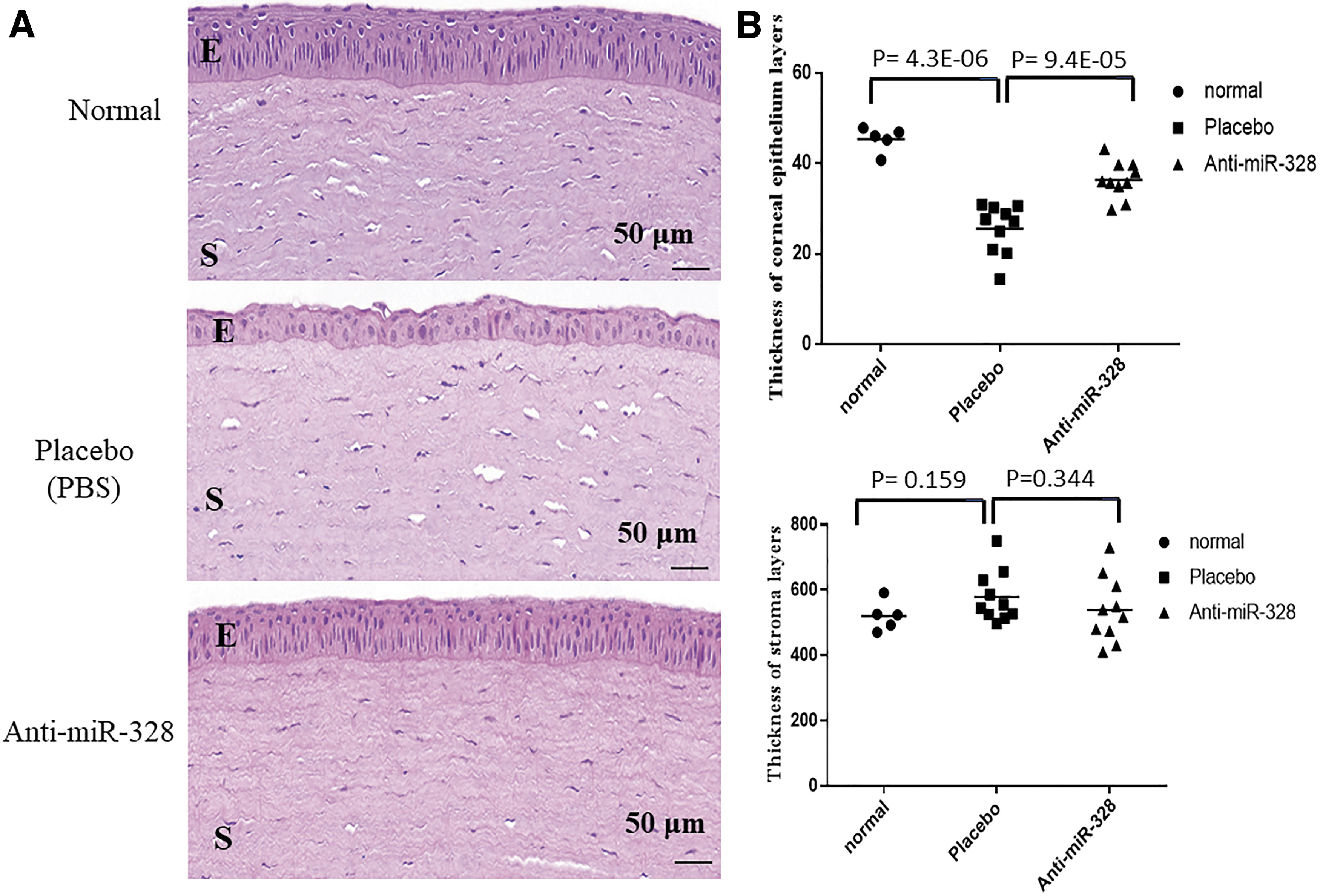
Representative images of H&E staining of corneal sections from the second batch of rabbits and normal control.
TUNEL is a common method to detect DNA fragmentation from apoptotic signaling cascades. In this assay, cells that had suffered severe DNA damage were stained dark brown. There were more apoptotic cells in the PBS group than in the anti-miR-328 group in both corneal epithelium (53 ± 3 cells vs. 39 ± 3 cells, P = 0.002) and stroma (84 ± 7 cells vs. 65 ± 5 cells, P = 0.029) (Fig. 3). Notably, there were only few apoptotic cells in the normal cornea (8 ± 1 cells in the epithelium and 10 ± 3 cells in the stroma) (Fig. 3).
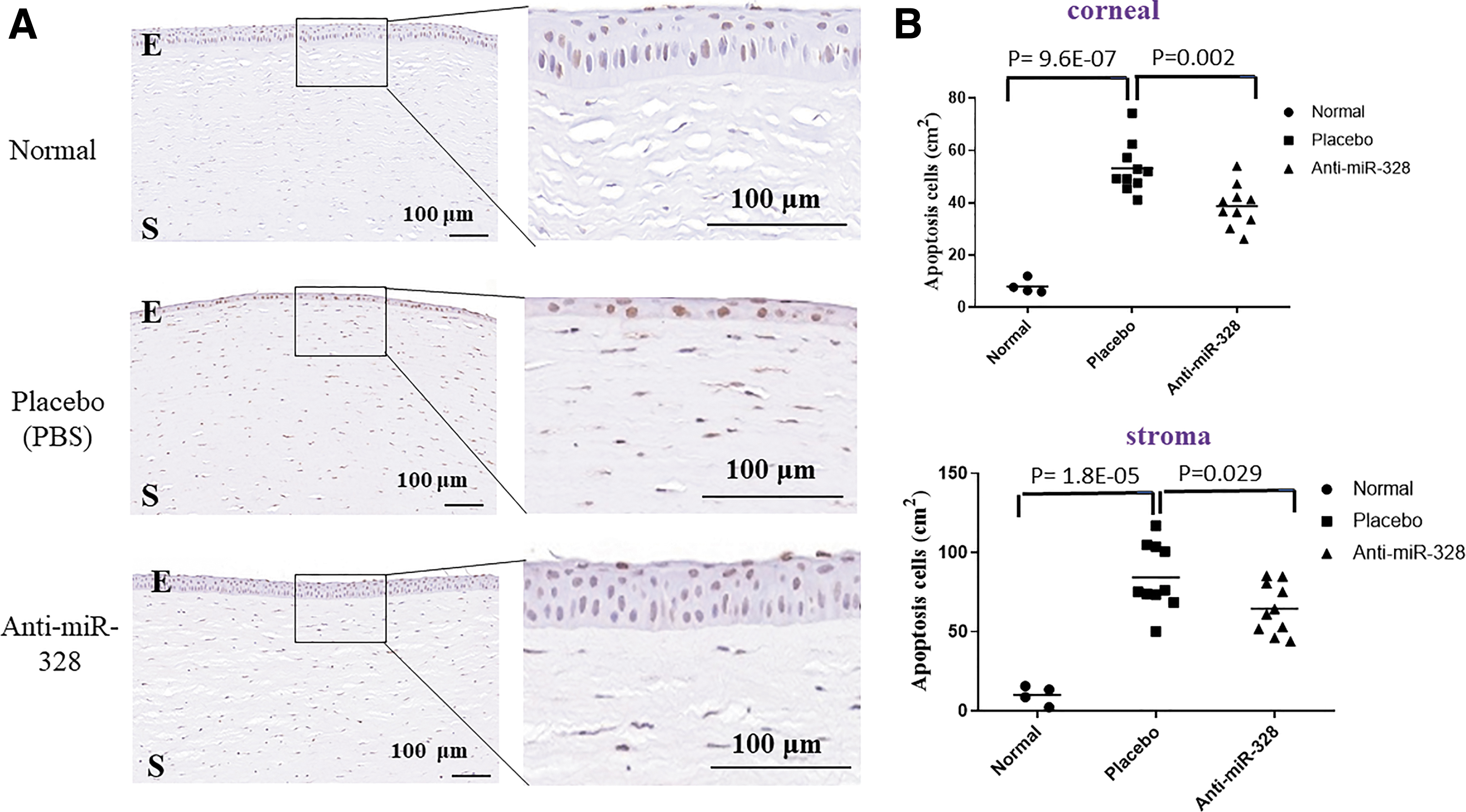
Representative images of TUNEL staining of corneal sections from the second batch of rabbits and normal control.
The histological examination of Meibomian glands also showed that the mean percentage of obstruction was lower in the anti-miR-328 group than the PBS group (56.4 ± 7.59 vs. 78.5 ± 8.17 vs. P = 0.059) (Fig. 4), although the difference did not reach a significant level of 0.05. However, the corneal cytokine data on IL-1, IL-6, and IL-8 revealed no difference between the PBS and anti-miR-328 groups (Supplementary Data), and the TNFα level was even higher in the anti-miR-328 group.
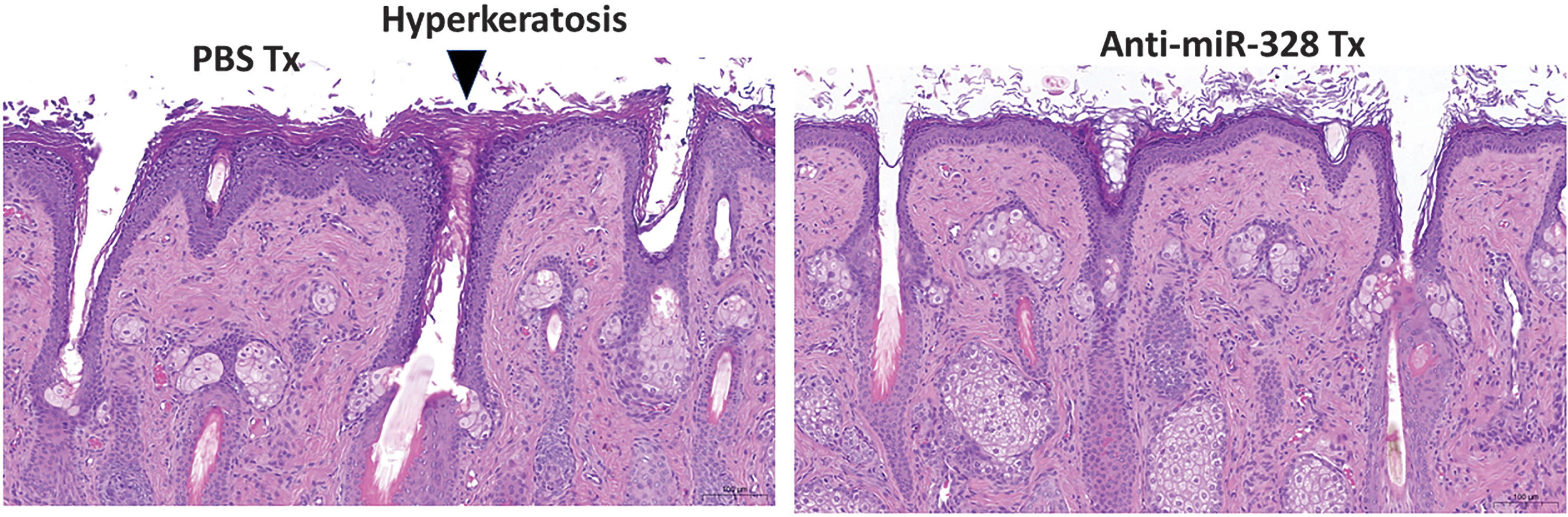
Representative images of the orifices of Meibomian glands from the second batch of rabbits. Hyperkeratosis (arrow) in the orifice of Meibomian glands of PBS-treated eye. Scale bar = 100 μm.
Dose dependency
To find the dose to reach the maximal therapeutic effect on DED, we tested the following anti-miR-328 doses: 10, 30, 60, 90, 120, and 160 μM in DED mice. The data indicated a dose-dependent effect on reducing corneal staining and the eyes treated with the dose of 160 μM of anti-miR-328 contained eye drop almost had no fluorescent staining on day 14 (Fig. 5). Accordingly, anti-miR-328 at 160 μM may be the optimal dose for treating DED in mice.
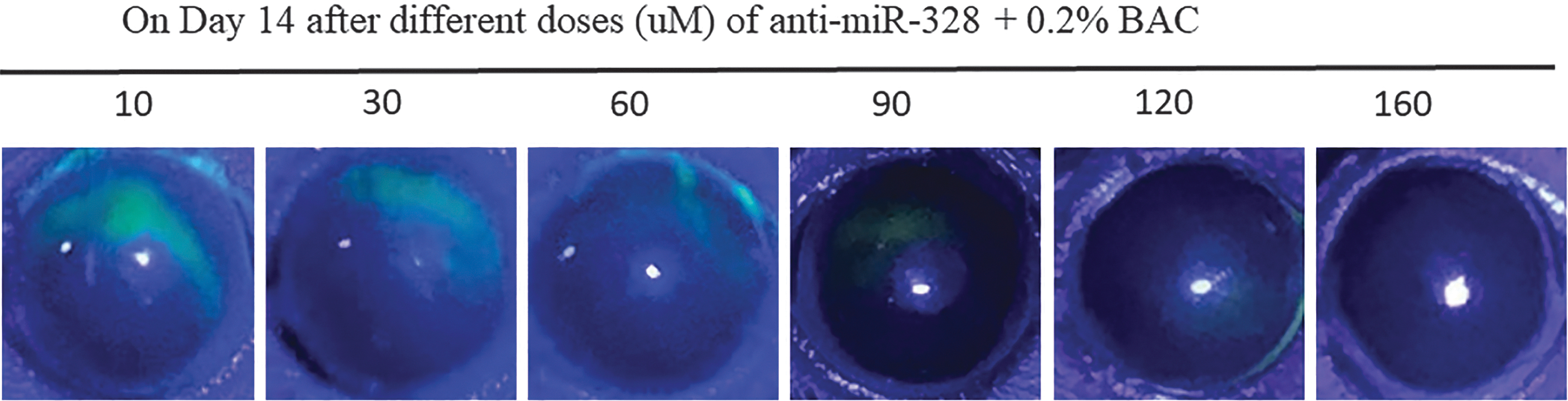
Representative pictures of dose-dependent effect of anti-miR-328. Mouse eyes were simultaneously treated with both anti-miR-328 and 0.2% BAC (with a 10-min interval) twice per day for 14 days. The representative pictures were taken on day 14. BAC, benzalkonium chloride.
Discussion
This study first demonstrated that BAC induced the miR-328 overexpression in SIRC cells, which might contribute to the development DED. To test this hypothesis, we instilled eye drops with an anti-miR-328 oligonucleotide in BAC-induced DED mice and rabbits. For the rabbits, the therapeutic effects of anti-miR-328 were demonstrated by (1) a reduction of corneal fluorescein staining, (2) decreased limbal and conjunctival congestion, (3) the restored thicknesses of corneal epithelial cells, (4) a decreased number of apoptotic cells in the corneal epithelium and stroma, and (5) less obstruction in Meibomian gland orifices.
For the mouse eyes, we only used the corneal fluorescein staining to assess the therapeutic effect, and the results also indicated that anti-miR-328 was better than the vehicle placebo. Accordingly, this study suggested that eye drops with anti-miR-328 oligonucleotide may be a novel agent to DED treatment.
Although dysregulation of miRNAs has been implicated in many complex diseases, the reports of miRNAs in commonly seen DED (i.e., not caused by Sjögren syndrome or other autoimmune diseases) were sparse.14,29,30 To our knowledge, this is the first report to demonstrate miR-328 as a risk factor for DED animal models. We first demonstrated that both BAC and hyperosmolarity induced overexpression of miR-328 in a corneal cell line (Supplementary Fig. S1a, b). miR-328 expression was also higher in the corneas of BAC-treated rabbits (Supplementary Fig. S1c).
Subsequent anti-miR-328 treatment indicated that DED could be attributed to overexpressed miR-328. Although transfection of miR-328 to THP-1 cells led to an increase IL-6 by 50% (data not shown), anti-miR-328 eye drops did not show anti-inflammatory effects in the BAC-induced DED animal models. Therefore, the detrimental effects of miR-328 on the ocular surface is probably not due to the inflammatory change. In contrast, previous studies have shown that overexpression of miR-328 induced cell apoptosis in cardiac myoblasts and hepatocellular carcinoma cells.21,31 Overexpression of miR-328 also increased intracellular Ca2+ concentration, which could be harmful to the cells. 31
In addition, an increase of miR-328 level inhibited cell proliferation and promoted cell apoptosis. 32 miRNA-328 has been reported to suppress the insulin growth factor 1 (IGF1) receptor, IGF-1 has a marked influence on the function of human Meibomian gland epithelial cells.33,34 Taken together, BAC-induced overexpression of miR-328 may cause ocular cell apoptosis and MGD, all of which eventually lead to DED development.
Two FDA-approved topical ophthalmic drugs for DED are cyclosporine and lifitegrast, both of which exert their therapeutic effects through anti-inflammation.35,36 Although the primary mechanism of action for anti-miR-328 therapy may not be anti-inflammatory, corneal protection may be a major effect of anti-miR-328 on BAC-induced DED animals. Further molecular studies are warranted to illustrate the detailed role of miR-328 in DED development. This study provided a proof of concept of miRNAs therapy for DED. The promising results from rabbit and mouse data warrant further exploration of detailed molecular mechanisms of anti-miR-328 therapy.
There are a couple of reasons supporting that the therapeutic effect is due to our anti-miR-328 oligonucleotide rather than random oligo effect. First, we have tested 49 antisense oligonucleotides with different lengths or modifications (see the details in US patent no. 101 79913B2), and only few antisenses reduced miR-328 levels in vitro and in vivo. Second, we showed dose-dependent effect, which is unlikely due to random oligo effects. Third, PBS solution is unlikely to contain sequence-specific antisense to generate therapeutic effect. The possibility of a sequence-specific antisense oligonucleotide is extremely small (∼1/421). Fourth, PBS or vehicle improved DED is commonly reported in animal studies or human clinical trials because of the wetting effect.
In conclusion, this study is the first report to indicate a potential of miRNAs therapy for DED. Although more molecular studies are needed to elaborate how miR-328 contributes to DED development, we showed that anti-miR-328 increased re-epithelialization of cornea, decreased apoptosis of corneal epithelial and stromal cells, and reduced obstruction of Meibomian gland orifice. This study may provide an alternative intervention for the DED patients who do not benefit from anti-inflammatory therapy.
Footnotes
Author Disclosure Statement
C-L.L. and S-H.H.J. are the authors for the patent of anti-miR-328 oligonucleotides. Other coauthors declare no competing interests.
Funding Information
This study was supported by China Medical University intramural grant (CMU-109-MF-29), Ministry of Science and Technology of Taiwan (MOST 108-2314-B-039-049-MY3), and the “Drug Development Center, China Medical University” from The Featured Areas Research Center Program within the framework of the Higher Education Sprout Project by the Ministry of Education in Taiwan.

References
Supplementary Material
Please find the following supplemental material available below.
For Open Access articles published under a Creative Commons License, all supplemental material carries the same license as the article it is associated with.
For non-Open Access articles published, all supplemental material carries a non-exclusive license, and permission requests for re-use of supplemental material or any part of supplemental material shall be sent directly to the copyright owner as specified in the copyright notice associated with the article.
