




Abstract
Purpose:
To compare a novel, sustained release formulation and a bolus injection of a targeted nanocarrier for the ability to specifically deplete cells responsible for the development of posterior capsule opacification (PCO) in week-long, dynamic cell cultures.
Methods:
A novel, injectable, thermosensitive poly(D,L-lactic-co-glycolic acid)-b-poly(ethylene glycol)-b-poly(D,L-lactic-co-glycolic acid) (PLGA-PEG-PLGA) triblock copolymer hydrogel was engineered for the sustained release of targeted, nucleic acid nanocarriers loaded with cytotoxic doxorubicin (G8:3DNA:Dox). Human rhabdomyosarcoma (RD) cells were used due to their expression of brain-specific angiogenesis inhibitor 1 (BAI1), a specific marker for the myofibroblasts responsible for PCO. Under constant media flow, nanocarriers were injected into cell cultures as either a bolus or within the hydrogel. Cells were fixed and stained every other day for 7 days to compare targeted depletion of BAI1+ cells.
Results:
The formulation transitions to a gel at physiological temperatures, is optically clear, noncytotoxic, and can release G8:3DNA:Dox nanocarriers for up to 4 weeks. In RD cell cultures, G8:3DNA:Dox nanocarriers specifically eliminated BAI1+ cells. The bolus nanocarrier dose showed significantly reduced cell depletion overtime, while the sustained release of nanocarriers showed increased cell depletion over time. By day 7, <2% of BAI1+ cells were depleted by the bolus injection and 74.2% BAI1+ cells were targeted by the sustained release of nanocarriers.
Conclusions:
The sustained release of nanocarriers from the hydrogel allows for improved therapeutic delivery in a dynamic system. This method can offer a more effective and efficient method of prophylactically treating PCO after cataract surgery.
Introduction
Cataracts are the leading cause of vision impairment and blindness worldwide. 1 Over 10 million cataract surgeries are performed annually with their rate expected to double over the next 10 years. 2 As a result, in 2013 the World Health Organization released a global action plan to increase access to cost-effective cataract surgeries.3–5 Cataract surgery involves removal of opacified tissue in the lens, replacing the lens with an artificial intraocular lens (IOL). Although surgery is safe and effective, over time the wound healing process initiated by surgical intervention can lead to a vision-impairing secondary cataract known as posterior capsule opacification (PCO). 6
PCO occurs in up to 40% of adult patients and nearly all children after cataract surgery. 7 Currently, the most effective treatment for PCO is neodymium-doped yttrium–aluminum–garnet (Nd:YAG) laser capsulotomy, which improves visual acuity by rupturing the opacified tissue with short, high-powered pulses of light. 8 However, Nd:YAG therapy is not available worldwide and there are often side effects from treatment, including intraocular pressure spikes, cystoid macular edema, retinal detachment, and IOL damage.9–12 Improvements in surgical techniques, IOL design, and understanding the influence of biomaterials on IOL performance have improved patient outcomes regarding PCO, but it still presents a significant burden on patients and the health care system.13–16
Cytotoxic drugs and chemicals, including anti-inflammatories, immunomodulating agents, anticell migration compounds, and cytotoxins, have been administered after cataract surgery to prevent the initial migratory cell response of PCO, but these drugs eliminate cells nonspecifically and drug diffusion to surrounding tissue can initiate an inflammatory response. 17 With the increase in cataract surgeries expected worldwide, there is an urgent, unmet need for more effective, prophylactic treatment strategies to prevent PCO.
The fibrotic cell response that leads to PCO is due to the migration of a specific subpopulation within the lens known as Myo/Nog cells.18–20 Myo/Nog cells were first identified in the chick embryo blastocyst by their expression of the skeletal muscle-specific transcription factor MyoD and the bone morphogenic protein inhibitor noggin.21,22 Previous experiments revealed the commitment of these cells to the skeletal muscle lineage regardless of their environment. 18 In cultures of human lens tissue, Myo/Nog cells differentiate into myofibroblasts in response to wound healing.18,19 Depletion of Myo/Nog cells in short-term and long-term cultures of human lens tissue prevented the accumulation of myofibroblasts.18,19 A third marker of Myo/Nog cells is brain-specific angiogenesis inhibitor 1 (BAI1) recognized by the G8 monoclonal antibody (mAb).21,23,24
The anti-BAI1 G8 mAb specifically targets Myo/Nog cells for depletion by complement-mediated cell lysis or when conjugated to dendritic DNA nanocarriers loaded with cytotoxic doxorubicin (G8:3DNA:Dox).18–21,23,25,26 Injections of G8:3DNA:Dox into the rabbit lens capsule during cataract surgery significantly reduced clinical signs of PCO after 28 days. 20 However, in humans, PCO develops more slowly than it does in rabbits. 27 Additionally, over time Myo/Nog cells from the ciliary body may traverse the zonules and repopulate the lens. 28 Therefore, a controlled release system for G8:3DNA:Dox would be more effective in reducing PCO over a longer period of time.
In this study, we tested a sustained delivery formulation for delivery of G8:3DNA:Dox in cultures of human rhabdomyosarcoma (RD) cells containing a subpopulation that expresses BAI1. 29 The formulation consists of a biodegradable, in-situ forming poly(D,L-lactic-co-glycolic acid)-b-poly(ethylene glycol)-b-poly(D,L-lactic-co-glycolic acid) (PLGA-PEG-PLGA) hydrogel that releases G8:3DNA:Dox for up to 4 weeks. 30 Herein, we compare a bolus dose versus hydrogel-mediated sustained delivery of G8:3DNA:Dox for cell targeting and depletion within a dynamic, microfluidic flow environment. The hypothesis underlying this work is that administration of the drug in a sustained delivery formulation will specifically eliminate the subpopulation of cells that express BAI1 more effectively than a bolus dose in long-term, dynamic systems.
Methods
Materials
Triblock copolymer, PLGA-PEG-PLGA, with d,l-LA/GA ratio of 15/1, PLGA/PEG ratio of 2/1, and PEG with a molecular weight 1,500 Da was purchased from PolySciTech, Inc., (West Lafayette, Indiana). Poly(ethylene glycol) Mn 400 (PEG400) was purchased from Alfa Aesar (Harverhill, MA). G8:3DNA:Dox nanocarriers were obtained from Genisphere, LLC. RD cells were purchased from the American Type Culture Collection (ATCC CCL-136) (Manassas, VA) and used in accordance with Rowan University Institutional Biosafety Committee (IBC) protocol 2018-14. Phosphate-buffered saline (PBS) was purchased from VWR (Radnor, PA).
3DNA nanocarrier synthesis and formulation
The term 3DNA refers to a novel, dendritic nanocarrier comprising nucleic acid monomers designed specifically for step-wise hybridization and layer-by-layer assembly, described previously.19,31,32 It consists of ∼3,000 DNA bases with 36 single-stranded peripheral regions. The approximate molecular weight, diameter, and zeta potential of 3DNA are 106 Da, 60 nm, and −28 meV, respectively.
Doxorubicin (Sigma-Aldrich, St. Louis, MO) was intercalated into double-stranded regions of 3DNA by incubating at room temperature at a ratio of 500:1 Dox:3DNA, resulting in >99% loading efficiency. The G8 mAb was conjugated to a DNA oligonucleotide through amine-to-sulfhydryl attachment using a heterobifunctional crosslinker (Pierce Crosslinking Kit; Thermo Fisher Scientific) and hybridized to 3DNA peripheries through complementary base pairing. The final 3DNA construct contained 4 mAb per particle and had a diameter of 120 nm.
Injectable hydrogel formulation and characterization
Solutions consisting of 10 (w/v)% PLGA-PEG-PLGA, 1.6 (v/v)% PEG400 were mixed on a tube rotator at 4°C for 24 h. This formulation was previously shown to transition into a hydrogel at 35°C with acceptable physical and optical properties. 30 Solutions were lyophilized and reconstituted with G8:3DNA:Dox to a final concentration of 0.7 ng 3DNA/μL. Solutions were kept at 4°C until ready for use. To confirm the formation of a physical hydrogel, the viscoelastic properties of the hydrogel were investigated using an ATS RheoSystem NOVA Rheometer (State College, Pennsylvania).
A stress-controlled temperature ramp was performed between 2 flat plates of diameter 25 mm with the gap between the plates set to 0.3 mm. The heating rate was 1°C/min and the stress was 4 dyn/cm2 at a frequency of 1.0 rad/s. Optical clarity was determined by measuring light transmittance on a 96-well plate at 35°C using a UV/Vis spectrophotometer (SpectraMax M3, Molecular Devices). The wavelength range was between 400 and 700 nm. The release of G8:3DNA:Dox from hydrogels was performed in a 400 μL chamber under physiological fluid flow (2.5 μL/min) and detected in the supernatant through a fluorophore (Alexa647) conjugated to the nanocarriers.
Cell culture and cytotoxicity tests
A Myo/Nog-like subpopulation was identified in cultures of RD cells by colocalization of antibodies to noggin and BAI1. 29 Therefore, this cell line was utilized to observe the targeting and depletion of BAI1+ cells with exposure to G8:3DNA:Dox. Cells were cultured in complete Dulbecco's modified Eagle's medium (10% fetal bovine serum, 1% antibiotic/antimycotic) in a 37°C cell incubator (VWR) with 5% CO2. Cell viability tests were performed on cells cultured on 96-well tissue culture plates (VWR). To assess cytotoxicity, these cells were incubated with an aliquot of either hydrogel, 42 ng of G8:3DNA or 42 ng of G8:3DNA mixed with hydrogel. Viability was determined through calorimetric (3-[4,5-dimethylthiazol-2-yl]-2,5 diphenyl tetrazolium bromide) cell viability assay (PromoCell).
Dynamic cell culture experiments
RD cells were cultured in a standard 24-well tissue culture plate, which has a similar diameter (15.62 mm) to the lens capsule (12.53 mm). 30 Each well was capped using polydimethylsiloxane lens molds of the anterior capsule to form chambers of 400 μL volume with inlet and outlet ports for fluid flow (Fig. 1). Cells were seeded into devices at a density of 2 × 10⁵ cells/mL and allowed to attach overnight. Then, 42 ng of G8:3DNA:Dox nanocarriers as a bolus or with hydrogel was injected into each device. A syringe pump was used to maintain a flow rate of fresh media at 2.5 μL/min for up to 7 days.
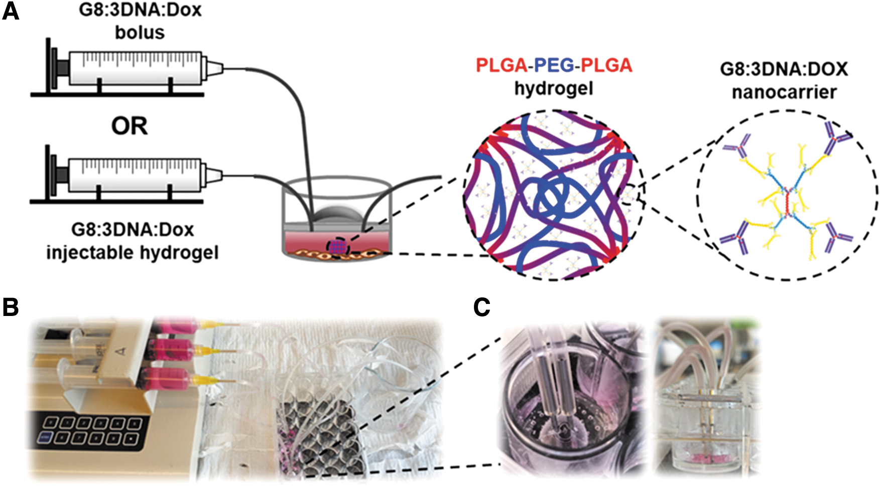
Cells were stained through the covalent, dead cell-specific Live-or-Dye NucFix™ Red Staining Kit (Biotium, Fremont, CA) and then immediately fixed with 2% paraformaldehyde followed by permeabilization with 0.5% Triton X-100. BAI1 was localized with the G8 IgM mAb. 24 The primary antibody was visualized using affinity-purified, F(ab’)2 goat anti-mouse IgM μ-chain conjugated with a fluorophore (Bio-techne, Minneapolis, MN). Finally, nuclei were stained with Hoechst dye (Bio-techne). Immunofluorescence was analyzed with an inverted fluorescent microscope (Zeiss) equipped with AxionCam ICm1 camera and Multi-Image-04 ZEN 2 lite image analysis software program. Cell counts and identification were performed using ImageJ.
Results
The RD human RD cell line was used to test the efficacy of the drug delivery system. About 12 (±9)% of the RD136 cell population is BAI1+. 29 A novel microfluidic setup was designed to culture the cells in a small volume and under dynamic fluid flow to mimic the lens capsule environment more closely (Fig. 1). Thus, G8:3DNA:Dox nanocarriers, either as a bolus or with a sustained release hydrogel, could be injected into each well and cell targeting could be compared over time.
This study utilized a PLGA-PEG-PLGA triblock copolymer hydrogel for its injectability, controlled gelation temperature, and ability to sustain the release of G8:3DNA:Dox. Rheological characterization is shown in Fig. 2A. The storage modulus of the hydrogel is near zero below 30°C, suggesting a Newtonian fluid with good injectability. Qualitatively, gelation can be confirmed through the vial-inversion method 33 and it was confirmed that the hydrogel transitions at a temperature of 35°C. Rheology revealed a storage modulus of 170.5 Pa at this temperature. The storage modulus reaches its peak at 37°C (474.6 Pa).
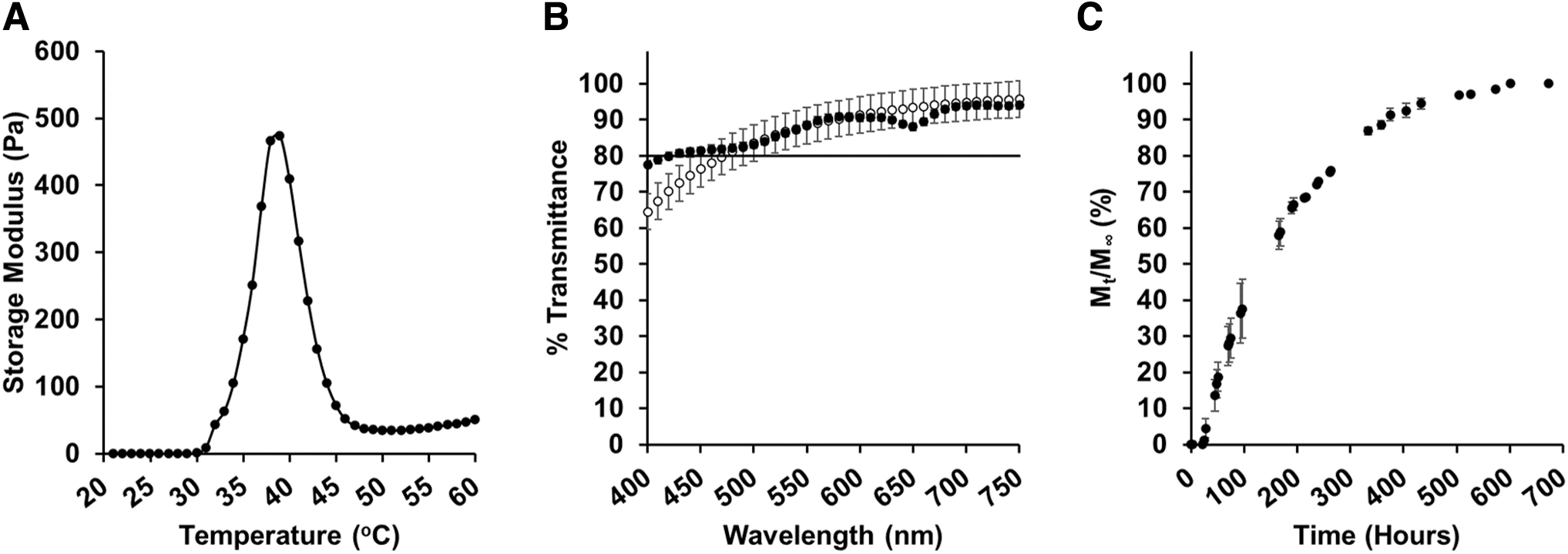
Characteristics of G8:3DNA:Dox/PLGA-PEG-PLGA hydrogel.
Furthermore, the hydrogel appears optically clear with over 80% light transmittance over the wavelengths 470–750 nm (Fig. 2B). After adding G8:3DNA:Dox, the light transmittance is slightly improved and retains over 80% transmittance over wavelengths 420–750 nm. Additionally, in Fig. 2C, we show the release of G8:3DNA:Dox from hydrogels under microfluidic conditions in PBS. In these conditions, the release of the nanocarrier is sustained for 4 weeks. After 24 h, only 1.2% of nanocarriers are released, indicating that no burst release is occurring and that the transport of the nanocarriers out of the gel is controlled.
Cell viability assays were used to determine the cytotoxicity of PLGA-PEG-PLGA hydrogel and G8:3DNA nanocarriers without intercalated doxorubicin. The results are shown in Fig. 3. Aliquots of hydrogel, G8:3DNA nanocarriers, and a combination of the 2 showed high cell viability indicating that the hydrogel and targeted nanocarrier are nontoxic.
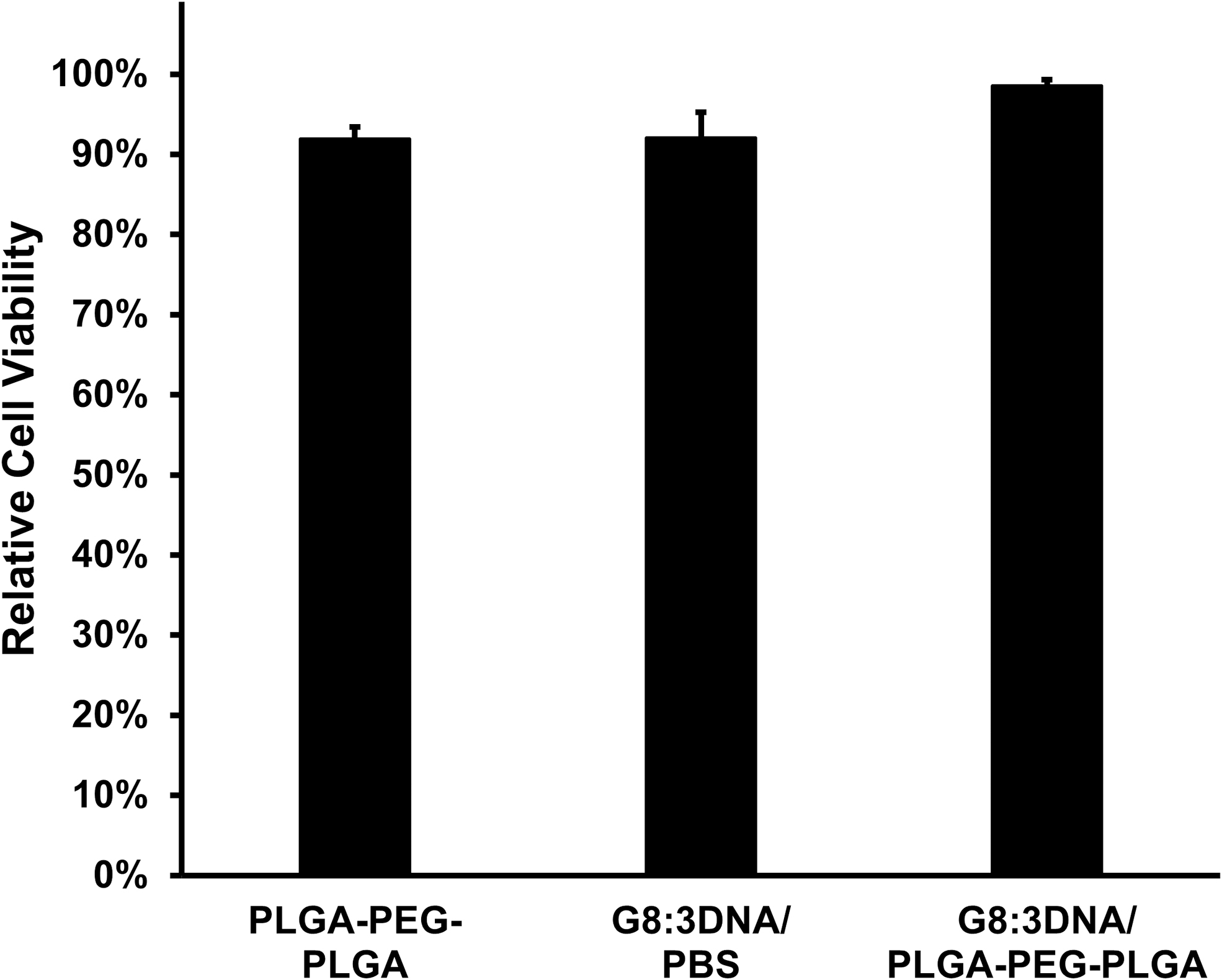
Results of MTT cell viability assay after treating cultures with PLGA-PEG-PLGA hydrogel, nanocarriers in PBS, and nanocarriers with hydrogel. Cell viability was measured without the presence of Dox. All components retain above 90% cell viability indicating that they are noncytotoxic (n = 3). MTT, (3-[4,5-dimethylthiazol-2-yl]-2,5 diphenyl tetrazolium bromide); PBS, phosphate-buffered saline.
We compared a bolus dose and sustained delivery of G8:3DNA:Dox for their ability to kill BAI1+ cells in long-term, dynamic cell cultures (Fig. 4). At 24 h, the bolus outperforms the hydrogel with 26% ± 15% and 10.4% ± 14.2% of BAI1+ cells nonviable, respectively, although the averages are statistically similar. By day 3, only 5.5% ± 2.3% of BAI1+ cells were nonviable in cultures treated with the bolus. The relative targeting of BAI1+ cells treated with bolus is reduced over time, with only <2% BAI1+ cells being nonviable on day 7.
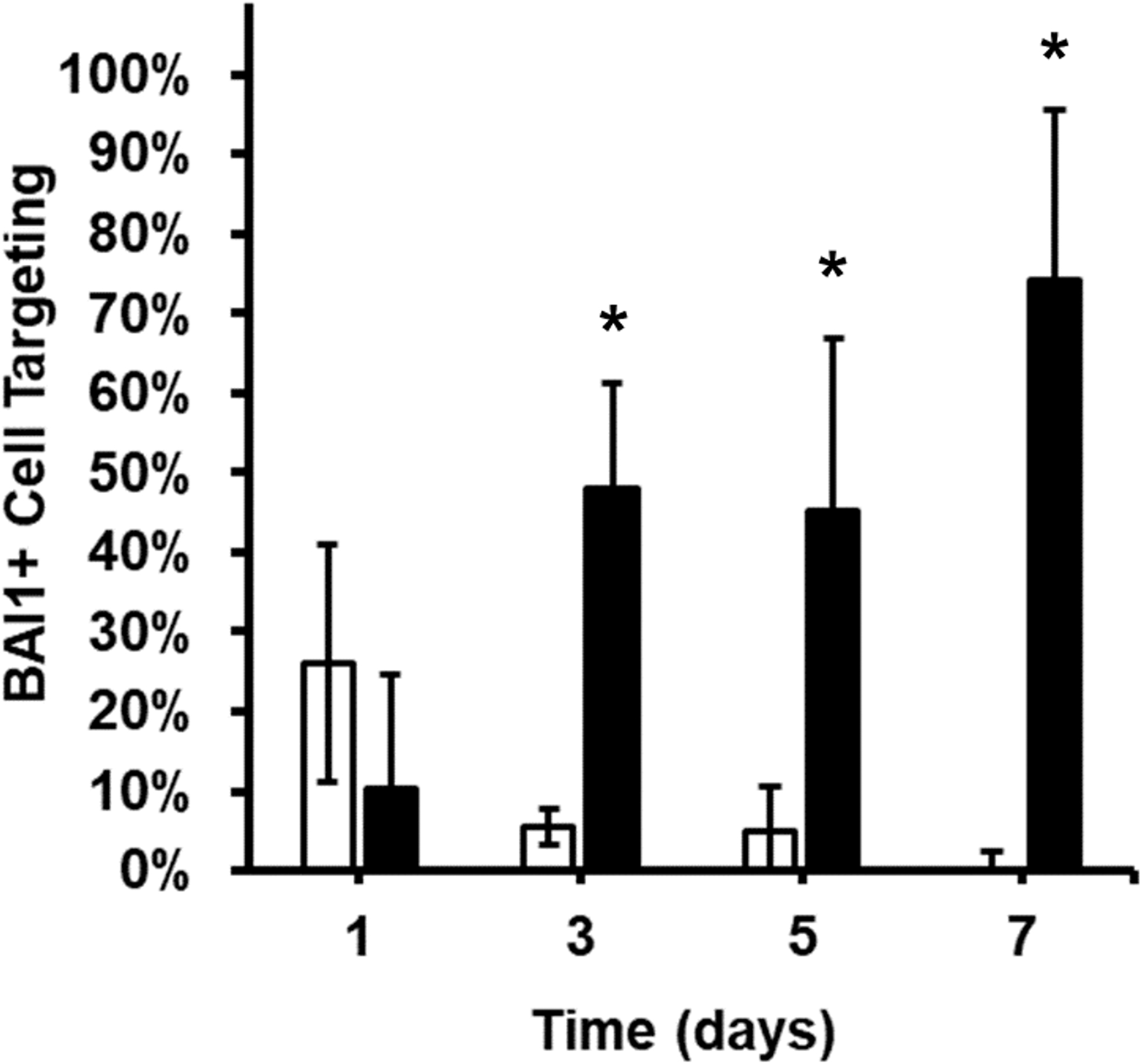
BAI1+ cell targeting in dynamic cell cultures. Cells were treated with G8:3DNA:Dox nanocarriers as a bolus (▪) or with hydrogel (□) at t0. The percentage of BAI1+ cells that were targeted were monitored every other day for 7 days. Significant differences are seen in targeting percentage between bolus and sustained release systems, denoted with an asterisk (one-tailed t-test, P < 0.05; n = 4). BAI1, brain-specific angiogenesis inhibitor 1.
In cultures treated with G8:3DNA:Dox nanocarriers and hydrogel, targeting of BAI1+ cells continued to increase. By day 3, 47.9% ± 13.4% of BAI1+ cells were nonviable. The result is similar at day 5, with 45.2% ± 21.5% BAI1+ cells targeted. At day 7, 74.2% ± 21.4% of BAI1+ cells were nonviable with release of G8:3DNA:Dox from the hydrogel.
Images of specific targeting of BAI1+ cells by G8:3DNA:DOX nanocarriers is shown in Fig. 5. In cultures treated with nanocarriers and hydrogel, the number of BAI1+ cells declined for the entire 7 days. In cultures treated with a bolus of the drug, almost all BAI1+ cells were viable at the final time point. Importantly, toxicity was specific to BAI1+ cells. Indeed, over the course of the experiment, an average of only 0.8% ± 1% of off-target (BAI1−) cell depletion was observed.
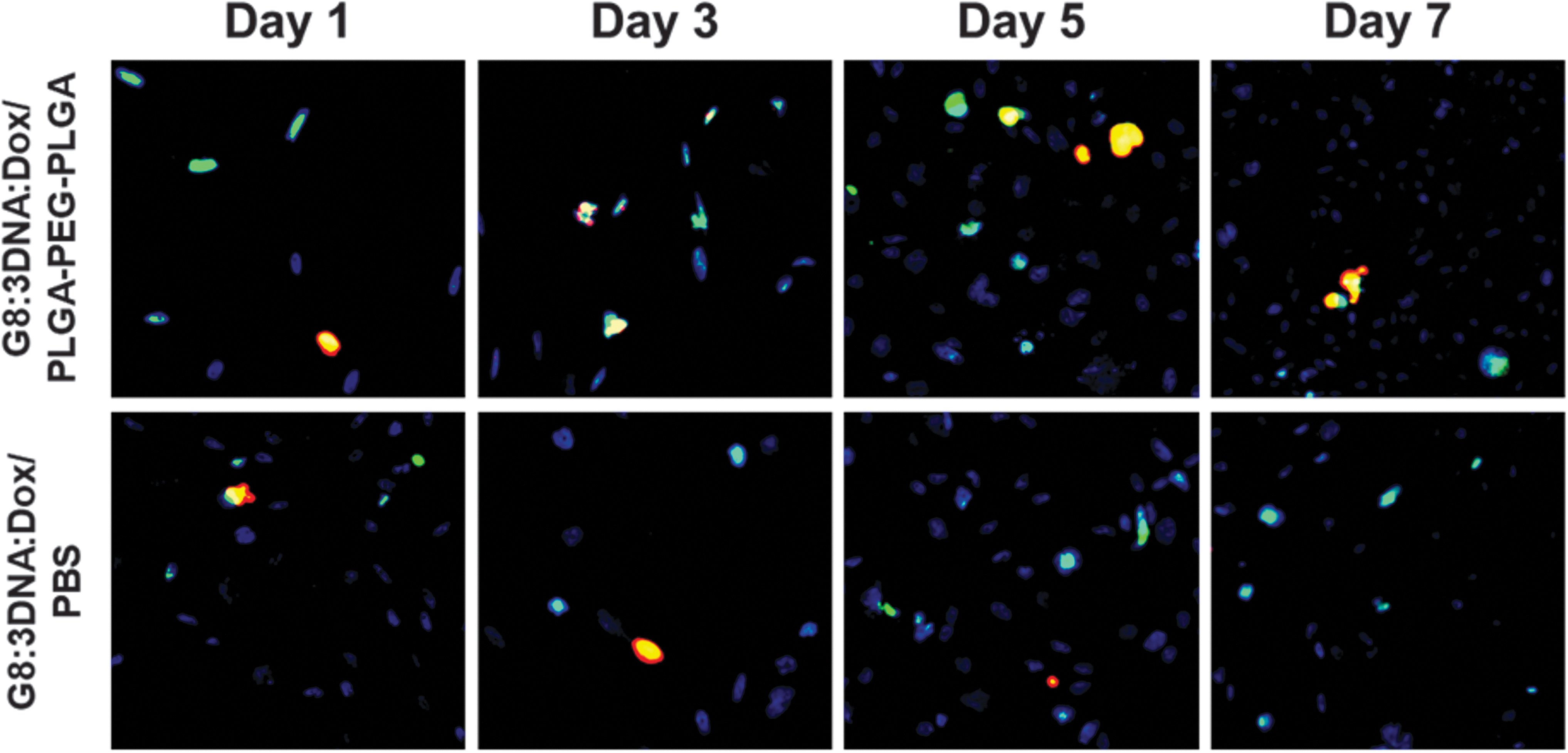
Specific targeting of BAI1+ cells over 7 days. Cells were treated with G8:3DNA:Dox nanocarriers in a sustained release hydrogel (G8:3DNA:Dox/PLGA-PEG-PLGA) or as a bolus injection (G8:3DNA:Dox/PBS). Cell nuclei (blue), dead cells (red), and BAI1+ cells (green) were stained. Targeted cells appear yellow due to colocalization of BAI1+ cells and dead cells (green+red). Nanocarriers given as a bolus show very little targeting after 7 days. Nanocarriers released from hydrogel continue to target BAI1+ cells at an increased rate over 7 days.
Discussion
PCO is expected to rise with the rate of cataract surgery over the next 10 years. There are no reliable indicators of vulnerability to developing PCO, and therefore, effective methods of preventing its development are imperative to preventing vision loss in patients, especially for those lacking access to Nd:YAG laser therapy.
A large barrier in this case is the challenge of effective drug delivery to treat ocular disorders due to the unique physiological barriers present within the eye, ocular fluid dynamics, difficulty in accessing posterior portions of the eye, and low drug bioavailability.34–36 The structure of the lens capsule and the fluid dynamics therein make intraocular drug delivery difficult.37,38 Due to these challenges, many effective ocular treatments are hindered by the need for multiple doses or invasive surgical interventions. As the healing process after cataract surgery can last for weeks, it is imperative that the release of a therapeutic is sustained.
At the forefront of ocular drug delivery are thermoresponsive hydrogels and nanoarchitectures that can achieve a sustained release of therapeutics within the eye, including hydrophilic, hydrophobic, and bioactive drugs.39–42 Injectable, in situ gelling systems in particular present an excellent area of focus due to the ability to provide minimally invasive, site-specific dosages of drugs for long-term administration.43,44 In particular, PLGA-PEG-PLGA triblock copolymer hydrogels have emerged as excellent vehicles for ocular therapeutics due to biocompatibility and sustained therapeutic release.45–47
When in contact with the aqueous humor within the eye, PLGA degrades into its monomers, lactic acid and glycolic acid, which are metabolized by natural biological processes. Additionally, PLGA-PEG-PLGA hydrogels have tailorable mechanical and optical properties and the capability to sustain therapeutic release. Similar polymers have been studied previously and show no sign of toxicity.48,49 We show similar observations for our hydrogel in Fig. 3.
In a previous study, a bolus injection of G8:3DNA:Dox administered in a static environment required 2 doses to deplete all BAI1+ cells in cultures of human lens tissue. 19 Therefore, we developed a microfluidic system within a 24-well tissue culture plate as a high-throughput method to investigate the long-term prophylactic capabilities of G8:3DNA:Dox in a dynamic environment. Microfluidic designs are able to simulate complex physiological processes and environments to better assess fluid dynamics and biochemical concentration gradients.50–52 This allowed us to compare a bolus dose of G8:3DNA:Dox and an injectable, sustained release formulation of G8:3DNA:Dox in a more physiologically relevant environment.
In dynamic cultures of RD136 cells in which a subpopulation expresses BAI1, we see that after 3 days the bolus injection of nanocarriers targets less than 10% of BAI1+ cells. This value continues to decrease for the 3DNA bolus injections until nearly no BAI1+ cells were targeted at 7 days. This is due to the high turnover of fluid, which occurs in 3–4 h within the chamber, which would rapidly reduce the nanocarrier concentration.
Conversely, G8:3DNA:Dox nanocarriers in hydrogel continue to deplete BAI1+ cells at an increasing rate over the course of 7 days. On day 7, over 70% of all BAI1+ cells were targeted and killed by nanocarriers released from the hydrogel. This significant difference in cell depletion is due to the transition at physiological temperatures to a nonflowing hydrogel that resides within the culture chamber. The hydrogel subsequently continues to release nanocarriers leading to extended exposure to cells over a longer time period.
In all cases, G8:3DNA:Dox specifically targeted the BAI1+ subpopulation through the conjugated G8 mAb, which is necessary to avoid nonspecific cell depletion. While it is unknown whether BAI1+ cells remaining in these cultures arose from the proliferation of untargeted BAI1+ cells or from de novo expression of this molecule, these results demonstrate that sustained delivery of the drug in the hydrogel formulation is more effective than bolus delivery for targeting this population.
This work can be compared with previous in vivo studies involving bolus injections of G8:3DNA:Dox in adult rabbits during cataract surgery. 20 Animals treated with G8:3DNA:Dox showed little to no clinical evidence of PCO (determined through slit lamp analysis and histology) 28 days after cataract surgery. However, BAI1+ Myo/Nog cells remained in the lens and more Myo/Nog cells may enter the lens by traversing the zonules from the ciliary body, 28 suggesting that vision impairment can still emerge over time. In the system used here, the ability of a bolus injection of G8:3DNA:Dox to deplete BAI1+ cells began to wane after only 3 days. The sustained hydrogel release, on the other hand, continues to specifically kill BAI1+ cells for at least 7 days.
Conclusions
Administration of a drug that prevents PCO would reduce vision loss in patients and significantly lessen the burden on the health care system. PCO can be reduced by injecting a drug that targets Myo/Nog cells during cataract surgery. However, the physiological conditions within the lens indicate that the concentration of a drug will decline over time. Herein, we showed that a biodegradable, in situ forming hydrogel loaded with G8:3DNA:Dox-targeted nanocarriers significantly outperformed a bolus injection in killing BAI1+ cells in dynamic cell cultures. The hydrogel is in situ forming, nontoxic and optically clear at 37°, and therefore, could be injected into the lens capsule during cataract surgery. Over time, the hydrogel would degrade into nontoxic components.
A microfluidic device utilizing cells cultured on 24-well plates under physiological fluid flow allowed for high-throughput comparisons of BAI1+ cell toxicity over 1 week through treatment with either a bolus injection of G8:3DNA:Dox or within our PLGA-PEG-PLGA hydrogel formulation.
Over time, BAI1+ cells were targeted at a much higher rate using the sustained release hydrogel. A bolus injection did kill BAI1+ cells within the first 24 h of treatment; however, nearly all remaining BAI1+ cells were viable after 7 days, whereas sustained release of the drug continued to kill cells over the course of a week. We expect that targeting would extend for longer than 7 days, as G8:3DNA:Dox was observed to release from the hydrogel for up to 4 weeks. This hydrogel formulation has potential for sustaining drug delivery to prevent PCO. Additionally, the versatility of the nucleic acid conjugate accentuates the platform nature of the 3DNA and the system as a whole. Therefore, we envision highly tailorable nanocarriers with controllable release profiles for the treatment of a wide variety of ocular disorders.
Footnotes
Authors' Contributions
M.E.B., M.G-W., and R.G. conceived the project, and L.L.O, R.J.M., and M.E.B. designed the experiments. L.L.O. formulated and characterized the hydrogel used in this study and performed cell viability and initial cell studies, G.G., J.G., and M.G-W. aided in cell culture and imaging, and R.J.M. and P.L.P performed cell studies and bolus and dynamic flow experiments. R.J.M., P.L.P., and L.L.O. performed cell imaging and analyzed the results of dynamic experiments. R.G. and J.B. synthesized and characterized the nanocarriers. L.L.O., R.J.M., M.E.B., and M.G-W. analyzed data and wrote the article. All authors were involved with reviewing and discussing the data.
Author Disclosure Statement
This work is the subject of pending U.S. patent 16/498,689 with M.E.B., L.L.O., and M.G-W. as inventors with financial interest. M.E.B. is founder with equity interest in OcuMedic, Inc. The remaining authors have no conflict of interests to report.
Funding Information
This work was funded by the Cooper Foundation (Myo/Nog cell program to M.B. and M.G-W.). PLP is an NSF REU Fellow supported by National Science Foundation Grant EEC 1757815.