




Abstract
Purpose:
To characterize the ocular toxicity of an antibody–drug conjugate (ADC), depatuxizumab mafodotin (Depatux-m), in nonclinical species and to evaluate the effects of drug–antibody ratios (DARs), variations of the ADC construct, and potential methods for mitigation of the corneal toxicity. Depatux-m contains the potent cytotoxic agent monomethyl auristatin F as the ADC payload.
Methods:
Depatux-m was administered intravenously to cynomolgus monkeys at doses up to 30 mg/kg and to mice up to 100 mg/kg. Ocular toxicity was evaluated by clinical ophthalmic examinations and histopathology. Potential mitigation was tested through agents to block target engagement and multiple topical ophthalmic treatments (antioxidant, vasoconstrictor, tear stimulant).
Results:
Effects primarily involved corneal epithelium and were dose-dependent with respect to onset, severity, and time to reversal in both monkeys and mice. On slit lamp biomicroscopy, the initial effect in monkeys was superficial multifocal punctate opacities (granularity), which migrated axially and were followed by pigmentation and multifocal punctate fluorescein staining. Microscopically, findings were characterized by single-cell necrosis, pigmentation, disordered basilar layer, and thinning of the corneal epithelium. Increased toxicity was associated with a higher DAR or more stably attached linker. Treatment with agents to block target engagement did not affect toxicity, and none of the topical treatments was successful.
Conclusions:
The corneal findings observed were similar to the effects described in clinical trials with Depatux-m and other ADCs. Collectively, these studies and available literature support the hypothesis that ADC-mediated toxicity is driven primarily by mechanism of action of the payload.
Introduction
Depatuxizumab mafodotin (depatux-m) is an antibody–drug conjugate (ADC) consisting of an antibody targeted against epidermal growth factor receptor (EGFR) conjugated through a noncleavable maleimidocaproyl (mc) linker to a potent cytotoxic agent, monomethyl auristatin F (MMAF). Ocular adverse events (AEs) were common in clinical trials with Depatux-m and were variably reported by affected patients as blurred vision, photophobia, eye pain, dry eye, and/or foreign body sensation. 1 Ocular AEs in human clinical trials with Depatux-m and other ADCs included the terms corneal epitheliopathy, keratopathy, microcystic keratopathy, keratitis, and/or corneal epithelial microcysts. 2
Despite clinical reports of ocular AEs with ADCs, these effects have not been well described or understood in terms of histopathologic effects or reversibility, both of which are better evaluated in nonclinical species.
The studies reported here characterized the ocular effects of Depatux-m in nonclinical toxicology studies. Nonhuman primates (cynomolgus monkeys) were administered Depatux-m followed by ophthalmic and histopathologic evaluations to better understand the onset and progression of ocular effects. Studies in monkeys were also conducted to evaluate methods to potentially mitigate ocular toxicity. In addition, reversibility was evaluated in subchronic (13-week) toxicity studies in monkeys and mice administered Depatux-m. Table 1 shows the different studies conducted, their objectives, and key points regarding the study design for each.
Study Designs, Objectives, and Key Points
DAR, drug–antibody ratio; Depatux-m, depatuxizumab mafodotin; EGFR, epidermal growth factor receptor; MMAF, monomethyl auristatin F; rhEGF, recombinant human epidermal growth factor.
Methods
Animals
Animal welfare for all studies was in compliance with the U.S. Department of Agriculture's (USDA) Animal Welfare Act (9 CFR Parts 1, 2, and 3). The Guide for the Care and Use of Laboratory Animals, Institute of Laboratory Animal Resources, National Academy Press, Washington, DC, 1996, was followed. All studies were approved by the appropriate Institutional Animal Care and Use Committees. In addition, animals were treated in accordance with the Association for Research in Vision and Ophthalmology Statement for the Use of Animals in Ophthalmic and Vision Research.
Naïve male and female cynomolgus monkeys of Chinese or Vietnamese origin were obtained from Covance Research Products, Inc. (Alice, TX). Monkeys were 2 to 6 years of age and weighed 2 to 4 kg at the start of the studies. Animals were pair or triple housed for 3 of the 4 studies and were individually housed with commingling enrichment in the fourth study.
CD-1 mice [CD® (SD); Charles River Laboratories, Inc.] were 9 or 10 weeks old and weighed 21 to 38 g at the start of dosing. Mice were housed individually in polycarbonate solid bottom ventilated cages equipped with feeders and automatic watering.
Test articles and dose administration
Depatux-m and variants of Depatux-m (ABT-414p, ABT-414-F2, and ABT-806-bac-MMAF) were formulated in 15 mM histidine, 7% sucrose, 0.01% Tween 80, pH 6.0 for intravenous (IV) injection by slow bolus (1–2 min) administration. The anti-EGFR monoclonal antibody, depatuxizumab, was formulated in 15 mM histidine, 7% sucrose, 0.01% polysorbate 80, pH 6.0, for IV slow bolus injection.
The topical ophthalmic treatments were the following: (1) ABT-806 for eye drops (depatuxizumab, 15 mM histidine, 70 mg/mL sucrose, 0.1% polysorbate 80, pH 6; AbbVie, Inc.), (2) Visine AC® (tetrahydrozoline HCl 0.05%, zinc sulfate 0.25%; Johnson & Johnson Consumer, Inc.), (3) BSS PLUS Part I® and BSS PLUS Part II® (combined as per manufacturer's instructions; Alcon Laboratories, Inc.), (4) recombinant human epidermal growth factor (rhEGF; Sigma-Aldrich, Inc.), and (5) Restasis® (Allergan, Inc.). Depatuxizumab was formulated at 151 mg/mL and was provided in standard eyedropper vials; at typical eye drop volume of 0.05 mL, the depatuxizumab dose was 7.55 mg per eye drop.
Lyophilized rhEGF was reconstituted per package insert instructions to 1 mg/mL using filtered 10 mM acetic acid in water for injection and was diluted to the final concentration of 10 μg/mL using 0.1% bovine serum albumin (Sigma-Aldrich, Inc.) in phosphate-buffered saline and adjusted to pH 7.4. Dose administered was 0.5 microgram rhEGF per eye drop. Eye drops were administered into the conjunctival sac. Animals were sedated with ketamine for administration of eye drops.
Studies and experimental design
Multiple studies were conducted in nonclinical species. A 3-week study was conducted in monkeys to characterize ocular toxicity of Depatux-m and variants of Depatux-m with different drug–antibody ratios (DARs) or linkers. Monkeys (3/sex/dose group) were administered 10 or 30 mg/kg up to 2 times (dosing days 1 and 22) followed by an observation period of up to 4 weeks. Necropsy was conducted on day 15 or 50 to perform histopathologic evaluation of the eyes. This study was conducted at Covance Laboratories, Inc. (Madison, WI) for AbbVie, Inc.
Two studies were conducted in monkeys to evaluate potential methods for mitigation of Depatux-m-induced corneal toxicity, either by systemic treatment with the anti-EGFR antibody or by topical administration of various eye drops. In the systemic pretreatment study, depatuxizumab (30 mg/kg) was administered intravenously at 4 or 24 h (n = 3/group) before a single IV dose of 30 mg/kg Depatux-m. In the topical administration study, monkeys (n = 3/group) received a single IV dose of 30 mg/kg Depatux-m.
The topical ophthalmic treatments were administered as eye drops twice daily to the right eyes of animals in each treatment group for 25 consecutive days (beginning 3 days before and continuing for 2 weeks following IV administration of Depatux-m). Purified water was applied to the left eyes of study animals as a control. Necropsy was not performed in these studies; all but 1 animal were returned to the stock colony at the termination of the studies. These studies were conducted at MPI Research (Mattawan, MI) for AbbVie, Inc.
A 13-week study in monkeys and a 13-week study in mice were conducted under Good Laboratory Practices to support clinical trials with Depatux-m; these studies were general toxicity studies with the objective to evaluate potential toxicity, toxicokinetics, and reversibility of Depatux-m. The 13-week studies were not dedicated ocular toxicity studies, and only the ocular toxicity and reversibility of ocular toxicity are discussed within this article. Monkeys (6/sex/group) were administered Depatux-m at dosages of 0, 2, 4, or 8 mg/kg/dose every other week for 13 weeks (7 doses). Two animals/sex/group were maintained as postdose recovery groups for an additional 12 weeks.
Mice (10/sex/group) were administered Depatux-m at dosages of 0, 10, 50, or 100 mg/kg/dose every other week for 13 weeks (7 doses). An additional 5 mice/sex/group were maintained as postdose recovery groups for an additional 16 weeks after cessation of dosing. Necropsy was performed at the end of the dosing and recovery periods in these studies. These studies were conducted at MPI Research for AbbVie, Inc. (monkeys) and at AbbVie, Inc. (mice).
Study procedures
In all studies, the minimal parameters evaluated included observation of clinical signs, body weights, food consumption, and evaluation of clinical pathology parameters (hematology, clinical chemistry, coagulation). Blood samples were collected for toxicokinetic analyses to confirm exposure to Depatux-m.
Ophthalmic examinations
Clinical ophthalmic examinations were performed in the monkey studies, but not in the mouse study. In the monkey studies, slit lamp biomicroscopy and indirect ophthalmoscopy were performed following pupillary dilation with 1% tropicamide once predose and then weekly or biweekly (13-week study) during the dosing phase on all surviving animals by a veterinary ophthalmologist.
Corneal fluorescein staining was performed utilizing 1 of 2 techniques. In the “saline flush” technique, a drop of diluted (0.33 mg/mL) fluorescein solution was applied to each eye, which was then irrigated from the eye with sterile 0.9% sodium chloride for injection before the ocular surface was examined. This method is widely used in a clinical setting and is optimized to identify areas of overt epithelial loss where the water-soluble fluorescein stain binds to the hydrophilic underlying stroma, but does not bind to the intact hydrophobic corneal epithelium. However, flushing the eye with saline can lead to diffusion of the dye into the surrounding structures, dilution of very small amounts of the dye to the point they are no longer visible, or potentially irrigation of the dye from within epithelial cells in the case of corneal microcysts.3,4
Therefore, a different “no saline flush” technique was utilized. With this technique, a cotton swab soaked in a 1 mg/mL solution of fluorescein stain was used to apply a very small amount of stain (a few microliters) to the ocular surface. The eyelids were closed to distribute the stain over the corneal surface and the eye immediately examined with the slit lamp biomicroscope as redistribution and quenching of superficial micropunctate fluorescein rapidly occur. The “no saline flush” method was used in the characterization study of Depatux-m and variants, whereas the “saline flush” method was used in the systemic and topical mitigation studies and in the 13-week study.
Histopathologic evaluation
When necropsy was performed, the eyes (with bulbar conjunctivae) were examined macroscopically; eyelids (with palpebral conjunctivae) were included in some but not all studies. Eyes, including the optic nerves, were fixed using a modified Davidson's fixative followed by placement into 10% neutral buffered formalin. Following fixation, ocular tissues were trimmed, paraffin embedded, sectioned, and stained with hematoxylin and eosin for microscopic examination by a veterinary pathologist.
Results
Depatux-m induces reversible corneal epithelial changes in monkeys
Monkeys administered Depatux-m manifested bilateral, usually symmetrical, ophthalmic abnormalities limited to the eyelids and anterior segment, especially the cornea. No posterior segment/fundus abnormalities were observed on either slit lamp biomicroscopy of the anterior vitreous or on indirect ophthalmoscopy. Investigative studies demonstrated reproducible Depatux-m-related corneal changes, noted first as multifocal punctate white perilimbal opacities involving the corneal epithelium or very superficial anterior stroma (corneal granularity, Fig. 1A).

Corneal observations in monkey on slit lamp biomicroscopy.
This initial lesion progressed axially toward the center of the cornea and was followed by pigmentation and superficial multifocal punctate corneal fluorescein stain retention without an epithelial defect and/or overt fluorescein stain uptake indicative of loss of the corneal epithelium (Fig. 1A–C). Variable effects on the eyelids such as epithelial degeneration and/or inflammation of the conjunctiva were observed at the highest doses administered (10 and 30 mg/kg).
Perilimbal corneal pigmentation initially trailed the leading edge of the granular changes, and, over time, pigment extended as far axial into the cornea as the granular changes. Pigmentation was generally observed ∼1 week later than the granularity (or concurrent with the onset of granularity at some lower doses). In some cases, the axial extent of corneal pigmentation was considered likely to diminish the animals' visual field.
Visualization of corneal microcystic changes was optimized with the “no flush” technique. With this technique, fluorescein stain retention was more readily observed in areas of punctate granular changes and was considered likely to represent corneal microcystic changes. Multifocal punctate corneal fluorescein retention was observed concurrent with the onset of pigment or 1 to 2 weeks after the granular change. This finding is consistent with previous reports3,4 where the superficial micropunctate corneal fluorescein staining profile has been associated with corneal epithelial cells undergoing apoptosis.
The corneal findings noted by ophthalmic examination correlated with microscopic findings of corneal epithelial cell degeneration/individual cell necrosis, increased mitosis, pigment accumulation, and minimal-to-severe loss of corneal epithelium and/or separation from underlying corneal stroma (Fig. 2). Separation/loss of epithelium was observed only at the higher doses and was attributed to antemortem loss of adherence of the basal layer of the epithelium to the stroma secondary to degeneration/necrosis; however, separation/loss was also partially attributed to processing artifact.

Microscopic findings in corneal epithelium of monkey.
Degeneration/individual cell necrosis was characterized by varying degrees of attenuation and disorganization of the epithelium with dissociation and necrosis of individual epithelial cells and globular eosinophilic intercellular debris. Increased mitosis was characterized by varying numbers of frequently bizarre mitotic figures predominantly in the basal layer of the epithelium. Pigment was characterized as granular brown intracytoplasmic pigment in all layers of the epithelium.
At 30 mg/kg, the highest dose administered to monkeys, effects on the eyelids such as swelling and erosion of the eyelid margins were observed. Microscopic findings in the eyelids were observed at both 10 and 30 mg/kg and consisted of epithelial cell degeneration/individual cell necrosis and increased mitosis along with increased thickness of the epidermis and mixed cell infiltrates. Similar findings, including mixed inflammatory cell infiltrates, were also present in the palpebral and bulbar conjunctival epithelium.
The incidence and severity of corneal effects in monkeys was dose-dependent (Table 2), with effects observed in 100% of animals administered 30 mg/kg and in 67% of animals administered 10 mg/kg. Likewise, time of onset was dose-dependent, with effects observed by day 7 after a single dose of 30 mg/kg, whereas animals administered 10 mg/kg had effects observed by day 28 (after 2 dose administrations on days 1 and 21).
Corneal Epithelial Dose-Dependent Effects in Cynomolgus Monkeys
EPI: Corneal epithelial findings described as degeneration/single-cell necrosis/atrophy.
PGM: Increased pigment observed in corneal epithelium.
Monkeys were administered doses of 2, 4, and 8 mg/kg every other week for 13 weeks (total of 7 doses administered), 10 mg/kg once every 3 weeks (total of 2 doses), or 30 mg/kg as a single dose.
Earliest onset of finding in weeks after first dose. Number indicates maximum incidence of animals with finding/total number of animals evaluated.
Necropsy performed at end of 13-week dosing period (day 92) for 2, 4, 8 mg/kg dosage levels. Necropsy performed on day 50 for 10 mg/kg dose level. Necropsy performed on days 15 or 50 for 30 mg/kg dose level (incidences shown were the same at both time points).
Recovery assessed after a 12-week dose-free recovery period for 2, 4, 8 mg/kg dosage levels. Recovery not assessed at 10 and 30 mg/kg dose levels.
Diffuse corneal granularity and pigmentation on ophthalmic examination. Pigment (minimal to mild) within corneal epithelium on microscopic evaluation in 3 of 4 animals.
N/A, not assessed.
At lower dosages in a 13-week study, corneal epithelial changes occurred in a dose-dependent manner (Table 2), with no ophthalmic effects at 2 mg/kg and corneal changes at 4 and 8 mg/kg. At doses of 4 and 8 mg/kg, there were dose-related corneal and eyelid findings that progressed in a similar manner to that described for previous studies, that is, dose-related corneal granularity, pigmented opacities, and fluorescein-positive lesions that developed after the appearance of granularity, eyelid swelling (blepharitis), and mucoid discharge on the eyelid margins; fluorescein uptake and mucoid discharge were only observed at 8 mg/kg. In animals administered 8 mg/kg, overt corneal ulceration indicative of loss of the corneal epithelium was occasionally observed.
Microscopic findings were also dose-dependent (Fig. 3), with minimal nonadverse effects at a low incidence at 2 mg/kg and adverse corneal epithelial degeneration/atrophy at 4 and 8 mg/kg.
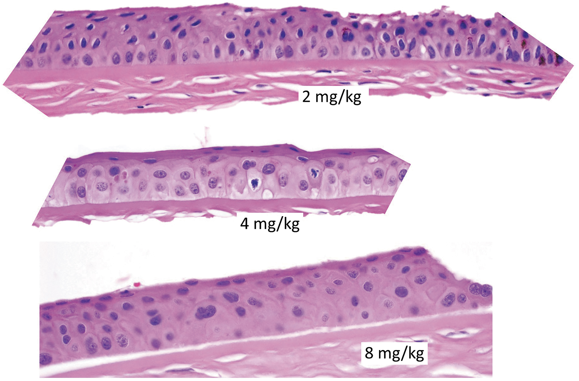
Dose–response relationship of microscopic findings in the corneal epithelium of monkey. Animals received 2, 4, or 8 mg/kg Depatux-m every other week for 13 weeks. Magnification 12.6 × .
Depatux-m-related corneal effects in monkeys were reversible in a dose-related manner, with complete resolution at 2 and 4 mg/kg and partial resolution at higher doses with more severe effects (≥8 mg/kg, Table 2). The observation of multifocal punctate corneal granularity resolved at the earliest. As expected, corneal pigmentation was slower to resolve. Subjective severity scores on ophthalmic evaluation indicated that the ocular lesions appeared to progress during the dosing period, particularly in the longer 13-week study, and improved during the recovery period. Histopathology evaluation demonstrated partial to complete resolution of corneal effects. In recovery animals, there were no ocular findings at 2 and 4 mg/kg, a finding consistent with complete recovery.
Microscopic changes present in recovery animals at 8 mg/kg were limited to minimal to mild pigment within the corneal epithelium, consistent with partial recovery. In animals administered 30 mg/kg, the severity of microscopic changes was generally decreased at day 50 compared with changes at day 15 or in unscheduled necropsies, consistent with partial reversibility.
Higher DAR elicited a higher incidence and earlier onset of corneal toxicity
The effect of DAR on corneal toxicity was evaluated by comparing Depatux-m with 2 variants of Depatux-m that differed by manufacturing process. Depatux-m has a broad distribution with a DAR of 4. ABT-414p and ABT-414-F2 both incorporated the same antibody, linker, and payload as Depatux-m, and had a DAR of 3.0 and 2.0, respectively.
Monkeys were dosed at 0 (vehicle control), 10, or 30 mg/kg. The high dose, 30 mg/kg, was not tolerated and therefore only a single dose was administered. The vehicle control and 10 mg/kg doses were administered on days 1 and 22. No ocular abnormalities were noted for animals receiving vehicle control. For the 10 mg/kg groups, all animals (6/group) were euthanized for necropsy on day 50 (28 days after the second dose). For the 30 mg/kg groups, 3 animals per group were euthanized on days 15 or 50, respectively.
In general, a higher DAR elicited a higher incidence and/or earlier onset of corneal toxicity. At 10 mg/kg (Table 3), although the earliest onset of granularity was earlier for DAR2 and DAR3 compared with DAR4, the peak incidence was lower for DAR2. The timing of peak incidence was comparable for all 3 DARs. At 30 mg/kg (data not shown), the onset of corneal toxicity was at the first ophthalmologic examination 1 week after the dose for all DARs tested. For DAR4 and DAR3, all animals were affected starting from week 2 and continuing through the end of the study period. For DAR2, the incidence was lower. Overall, taking both the 10 and 30 mg/kg dose levels into consideration, the onset and severity of corneal findings were equivalent for Depatux-m (DAR4) and ABT-414p (DAR3), and less with ABT-414-F2 (DAR2).
Summary of Corneal Epithelial Effects
Cynomolgus monkeys were administered 10 mg/kg once every 3 weeks (total of 2 doses).
Ophthalmic examinations were conducted once weekly for a total of 7 weeks. Earliest onset of finding in weeks after first dose. Number indicates maximum incidence of animals with finding/number in group.
An incidence of 6/6 was recorded once during the study; on all other examinations the incidence was ≤3/6.
ABT-806-BAC-MMAF was not tolerated after a single dose of 10 mg/kg. After the week 2 ophthalmoscopic examinations, 3 of the 6 animals were sent to necropsy.
Bromoacetamide linker increased corneal toxicity compared with mc linker
ABT-806-bac-MMAF is an ADC made with the same antibody and MMAF payload as Depatux-m, but with the stable attachment noncleavable bromoacetamide (bac) linker instead of the mc linker of Depatux-m. Repeated doses of ABT-806-bac-MMAF were not tolerated; therefore, animals received a single administration of vehicle control, 10 or 30 mg/kg ABT-806-bac-MMAF (6/group). No ocular abnormalities were noted in animals receiving vehicle control.
For the group that received 10 mg/kg ABT-806-bac-MMAF, an interim necropsy on day 15 (3/group) was performed to characterize the corneal findings present at ophthalmic examinations on day 14, and a necropsy was also performed on day 50 (3/group, 28 days after the second dose). Five of 6 animals administered 30 mg/kg ABT-806-bac-MMAF were euthanized between days 11 to 21 due to severe ocular toxicity, and the sixth animal was euthanized at the end of the observation period on day 50.
Compared with Depatux-m, corneal effects were observed earlier in animals administered ABT-806-bac-MMAF. At a dose of 10 mg/kg, corneal granularity was observed as early as day 7 for ABT-806-bac-MMAF, whereas for Depatux-m, corneal granularity was typically not observed until after 2 administrations at 10 mg/kg (Table 3). Increased corneal toxicity, as well as decreased tolerability, was associated with the bac linker compared with the mc linker.
Systemic pretreatment with anti-EGFR antibody did not mitigate Depatux-m corneal toxicity in cynomolgus monkeys
This study investigated whether the Depatux-m-induced corneal toxicity could be mitigated by systemic pretreatment with the anti-EGFR monoclonal antibody, depatuxizumab (ABT-806), to prevent binding of Depatux-m to EGFR on the cornea where the EGFR is known to be expressed. The dose of depatuxizumab (30 mg/kg) was selected to saturate EGFR binding (data not shown).
Ophthalmic examination findings, including blepharitis/eyelid swelling, keratitis, and corneal granularity, were observed across both groups and started on day 14 and persisted through the day 21 and 28 examinations. In addition, on days 21 and 28, all animals in both groups were observed with corneal pigmentation. One animal in the 4-h group had positive fluorescein stain uptake on day 28 and was subsequently treated with topical nonsteroidal anti-inflammatory drugs and antibiotics on days 28 to 32 for a presumed corneal ulceration. The animal had no further veterinary observations on ophthalmic examination and was returned to the stock colony on day 32.
Overall, no significant difference was noted in the corneal effects between the 2 treatment groups, demonstrating no clear advantage to a single treatment with depatuxizumab either 4 or 24 h before a single administration of Depatux-m.
Topical administration of various eye drops did not mitigate Depatux-m-related corneal toxicity
This study investigated the effects of 5 different topical ophthalmic treatments in monkeys administered a single IV dose of 30 mg/kg Depatux-m. The topical ophthalmic treatments, administered as eye drops, were the following. (1) Depatuxizumab (potentially blocking the receptor target expressed on the corneal epithelium), (2) Visine AC (vasoconstrictor), (3) BSS PLUS® (irrigating solution including glutathione, antioxidative), (4) rhEGF (potentially blocking the receptor target), and (5) Restasis (cyclosporine, immunomodulator/tear stimulant). Dose levels of depatuxizumab and rhEGF were selected to saturate their respective receptors although receptor saturation was not directly measured.
The clinical signs and ophthalmic effects that developed among animals were similar among all treatment groups. Effects in eyes were similar to those in previous studies and included periocular redness, swelling, and partial/complete closure of the eyes. In most cases, the ocular abnormalities observed were seen bilaterally, and there was no clear trend or distinction between the eye receiving treatment eye drops (right) or the control eye (left). On slit lamp examination, all animals in all groups had adnexal and anterior segment findings of both eyes by the 3-week (day 21) time point. Onset of findings was typically between the 1- and 3-week time points. The most common findings observed included eyelid erythema and/or swelling, ocular discharge, corneal granularity, and corneal pigmentation.
These findings were noted in similar frequency and severity among all 5 dose groups, and additionally within each group between both the treatment eye drop (right) and control (left) eyes. Fluorescein stain uptake was noted for 1 animal on day 14, and 3 (of 15) animals on day 21.
To optimize visualization of corneal effects, on day 28, the “no saline flush” fluorescein method was used on the left (control) eye of each animal; this method resulted in higher incidence of staining in all groups compared with the “saline flush” method used on the right (eye drop) eye, although the incidences of corneal granularity and pigmentation were identical. Fluorescein stain uptake was observed in 1 or 2 animals in all groups except the ABT-806 group on day 28.
Progressive Depatux-m-related corneal findings were observed and did not appear to be mitigated by any of the treatments with twice-daily topically administered eye drops; however, optimal administration of the treatments may not have been achieved.
Depatux-m induces reversible corneal epithelial changes in mice
There were no clinical ophthalmic examinations in the mouse study; eyes were evaluated postmortem for histopathologic changes. There were no ocular effects in the 10 mg/kg dose group at the end of the 13-week dosing period. The majority of animals at 50 or 100 mg/kg had bilateral corneal epithelial atrophy, with dose-dependent severity, variably characterized as unevenly spaced and/or variably sized cells resulting in disordered arrangement, variable thickness (as little as 2–3 cells thick), occasional single-cell necrosis and/or mitotic figures, and/or locally extensive separation (possibly artifactual) of corneal epithelium from the underlying stroma. Eyes from mice maintained for 17 weeks after the last dose had no findings related to the cornea, demonstrating reversal of the effect (Table 4).
Corneal Epithelial Effects in Mouse Were Dose-Dependent and Reversible
Findings were variably characterized by occasional single-cell necrosis and/or mitotic figures, unevenly spaced and/or variably-sized cells resulting in disordered arrangement, variable thickness (as little as 2–3 cells thick), and/or locally extensive separation (possibly artifactual) of corneal epithelium from underlying stroma.
Discussion
In nonclinical studies, the corneal effects of Depatux-m were observed in both nonhuman primates (cynomolgus monkey) and mice. The studies conducted in monkeys characterized the nature and temporal progression of the toxicity; reversibility was demonstrated in both monkeys and mice at 12 and 18 weeks, respectively (Table 1).
Corneal epithelial effects were dose-dependent with respect to onset, severity, and/or incidence; in both species, complete resolution of effects occurred at lower dosages and partial resolution occurred at higher dosages. Depatux-m administered to monkeys produced corneal epithelial effects that were detected by an ophthalmic examination and a histopathologic evaluation. There were no effects in the fundus observed with indirect ophthalmoscopy, and no effects on the retina upon histopathologic evaluation. Using a slit lamp biomicroscope to evaluate the cornea, the time course of effects in monkeys was characterized as multifocal punctate granularity to the perilimbal cornea, followed by corneal pigmentation and superficial micropunctate fluorescein retention.
These findings correlated with histopathologic observations in eyes collected at the end of the dosing and recovery periods. The ophthalmic and histopathologic observations are described in further detail as follows:
Multifocal punctate granularity was the initial ophthalmic observation. This granular change in the corneal epithelium was noted first in the perilimbal region and progressed axially toward the center of the cornea. Pigmentation initially trailed the leading edge of the granular changes and over time extended as far axial into the cornea as the granular changes. Pigmentation was characterized histologically as granular brown intracytoplasmic pigment in all layers of the epithelium. Corneal fluorescein stain retention occurred in 2 patterns. Most commonly it manifested as a superficial multifocal punctate staining profile of the corneal epithelium, which correlated with the corneal granularity changes observed on slit lamp biomicroscopy and the epithelial cell degeneration/individual cell necrosis on histopathology.
4
With this pattern, however, the corneal epithelium remained intact. The second staining pattern was observed in animals administered 8 mg/kg in the 13-week repeat-dose toxicity study in monkeys and represented a continuation of the epithelial cell damage to the point that portions of the corneal epithelium were lost thereby exposing the underlying stroma. Animals occasionally presented with marginal eyelid swelling and erythema, bilateral blepharitis, and/or corneal edema. Ophthalmic examination findings correlated with histopathologic observations of single-cell necrosis of the corneal epithelium, pigmentation, disordered basilar epithelium, and thinning of corneal epithelium. The effects appeared to originate in the perilimbal area and migrate axially toward the center of the cornea and were eventually reversible.
In a 13-week GLP toxicity study with Depatux-m, the NOAEL (no-observed adverse effect level) for corneal effects in monkeys was determined to be 2 mg/kg, and the lowest dose at which adverse corneal effects were observed was 4 mg/kg. Exposures to Depatux-m at these doses were similar to the exposures achieved in human clinical trial subjects at a therapeutic dose, indicating that corneal effects in monkeys were observed and were reversible at clinically relevant exposures.
In a 13-week GLP toxicity study in mice, Depatux-m at 50 and 100 mg/kg was associated with microscopic findings similar to those observed in monkeys. These effects were not observed after a 16-week dose-free recovery period, indicating reversibility in this species. There were no effects on the corneal epithelium in mice administered 10 mg/kg, either at the end of the dosing or recovery periods.
Investigative studies were conducted to evaluate the effects of different DARs, variations of the ADC construct (different linkers), and/or potential methods for mitigation of the corneal toxicity (Table 1). In a direct comparison of DAR, there was a general trend for higher DAR to be associated with increased toxicity, such that the corneal effects were similar with DAR4 and DAR3, and slightly less with DAR2. A variation of Depatux-m containing a stable attachment noncleavable linker, bac, was associated with increased ocular toxicity as well as decreased tolerability.
Potential mitigation for the Depatux-m-related corneal toxicity was tested in monkeys. Interference with EGFR engagement through topical or systemic administration of ABT-806 antibody did not mitigate corneal toxicity, nor did the following topical ophthalmic treatments: Visine AC (vasoconstrictor), BSS PLUS (irrigating solution including glutathione, antioxidative), rhEGF (as a decoy to block the potential binding to the receptor target expressed on the corneal epithelium), and Restasis (cyclosporine, anti-inflammatory). None of the eye drop treatments tested was successful in ameliorating the corneal toxicity in monkeys.
While suggestive that these approaches were not successful, the studies were limited in study design, such as administration schedule of systemic ABT-806 antibody or the number of times per day that eye drops could be administered.
The corneal findings observed by ophthalmic examination in monkeys were similar to descriptions of microcystic corneal changes in patients administered cytarabine,5–7 SGN-75, 8 or Depatux-m.1,9 In monkeys, the single-cell necrosis observed histologically and the multifocal punctate granularity with a superficial micropunctate corneal fluorescein staining profile observed ophthalmically appeared to mimic the microcystic keratopathy described in humans. The distribution pattern differed for cytarabine compared with SGN-75, with cytarabine-associated corneal changes located primarily in the central cornea, whereas SGN-75-associated corneal epithelial changes occurred in a ring pattern starting in the periphery and with migration toward the center.
In both cases, the corneal epithelial changes distributed in a pattern consistent with corneal epithelial cell turnover, either from basal to apical and superficial layers 8 or centripetal movement from the periphery toward the corneal center. A hypothesis for the development of microcystic corneal changes associated with Depatux-m is that the corneal changes occur in reaction to MMAF-induced microtubule inhibition in rapidly dividing transient amplifying cells that arise in the limbus of the cornea and migrate axially to populate the cornea. As a result of this damage, cells become necrotic or apoptotic, producing microcyst-like keratopathy in the basal corneal layers. Corneal epithelial cells undergoing apoptosis have also been previously reported to exhibit the superficial micropunctate corneal fluorescein staining profile observed here. 4 Apoptotic cells shrink, lose contact with adjacent cells, and/or are phagocytized, resulting in a spatial defect in the epithelium; these defects are visualized as “microcysts” on ophthalmic examination.
As the cornea regenerates, the microcysts traverse across the cornea, causing a variety of symptoms (such as dry eyes, blurred vision, or photophobia), and ultimately are sloughed off as the corneal epithelial cells regenerate. Although described as microcysts or microcystic-like keratopathy, no true cysts were observed either in patients administered high-dose cytarabine 8 or in animals administered Depatux-m.
Resolution of corneal epithelial effects is thought to occur as the corneal epithelium regenerates. Complete resolution has been observed in nonclinical species and in humans. In nonclinical species administered Depatux-m, recovery varied with dose level and duration of dosing; after 13 weeks of dosing, complete resolution of corneal effects occurred after ∼12 or 17 weeks in monkeys or mice, respectively. Resolution of ocular side effects in patients administered Depatux-m varied over several months. 1
The corneal effects observed with Depatux-m are associated with the cytotoxic effects of MMAF and occur with other ADCs regardless of target expression in the eye. EGFR is expressed in the cornea and ocular AEs occur with other EGFR targeting agents, for example, cetuximab, panitumumab. 2 However, depatuxizumab is a monoclonal antibody that targets a unique conformation of EGFR that is exposed due to overexpression, gene amplification, or a mutant form of EGFR that occurs in tumors and is largely inaccessible when the EGFR is expressed normally.10–12 EGFR is known to be expressed in the corneal epithelium during proliferation and wound healing, but it is not known whether the specific conformation of EGFR that depatuxizumab binds is accessible under these conditions.
Clinical studies of other ADCs have demonstrated ocular side effects similar to those observed in patients treated with Depatux-m, including ADCs with mafodotin and DM4 payloads.2,13,14 Notably, some of these ADCs have targets that are not expressed in ocular tissue structures, suggesting that the major component of the mechanism of action is not target mediated but is due to the cytotoxic activity of the payload. 2 Studies that attempted to block EGFR target expression on the cornea with systemic or topical administration of depatuxizumab or rhEGF demonstrated no amelioration of corneal effects, also suggesting that the mechanism of toxicity is not EGFR-mediated, although the constraints of the experimental designs for these blocking studies must be taken into consideration with the outcomes of the investigation. In addition, although the doses of depatuxizumab and rhEGF were selected with the intention to saturate EGFRs on the corneal epithelium, saturation was not confirmed in the studies.
In conclusion, studies investigating the corneal toxicity associated with Depatux-m in nonclinical species characterized the onset, progression, and resolution of corneal epithelial effects. The corneal epithelial effects occurred primarily as a result of the cytotoxic activity of the MMAF payload of the ADC.
Footnotes
Acknowledgments
The authors thank Dr. Katharine Whitney (AbbVie, retired) and Dr. Joshua Bartoe (Ophthalmology Services) for their expert assistance.
Authors' Contributions
L.I.L., T.A.H., J.K.J., P.E.M., and S.L.R. wrote the article. T.A.H., J.K.J., P.E.M., and S.L.R. designed the research; T.A.H., J.K.J., and P.E.M. performed the research; and T.A.H., J.K.J., and P.E.M. analyzed the data.
Author Disclosure Statement
Dr. Paul Miller is a Clinical Professor of Comparative Ophthalmology at the School of Veterinary Medicine, University of Wisconsin-Madison, and a consultant for Covance. Dr. Miller has served as a consultant for AbbVie and has received research funding and speaker fees from AbbVie. Drs. Lise Loberg, Sherry Ralston, Julie K. Johnson, and Tracy Henriques are employees of AbbVie and own AbbVie stock.
Funding Information
AbbVie sponsored the studies; contributed to the design; participated in the collection, analysis, and interpretation of data; and in writing, reviewing, and approval of the final version.