




Abstract
Purpose:
To investigate the efficacy and safety of scleral cross-linking (CXL) using Genipin in the treatment of juvenile guinea pigs with high myopia.
Methods:
Twenty-four 4-week-old tricolor guinea pigs with high myopia of diopter ≤ −6.0 DS in the right eye were randomly divided into two groups: Genipin CXL group and control group (n = 12 for each group). They received separately form-deprivation (FD) combined with sub-tenon injection, and the former was 0.5% Genipin solution, while the latter was 0.9% saline solution. Refractive error, axial length (AL), intraocular pressure (IOP), and structural and vasculature optic disc changes in optical coherence tomography (OCT) and OCT angiography (OCTA) were analyzed at baseline and at 3 weeks after injection.
Results:
Baseline parameters were similar between the two groups (P > 0.05). After 3 weeks of the intervention, the difference of AL between the two groups was statistically significant (t = −11.28, P < 0.001). Besides, IOP increased in both groups, and the changes of IOP between the two groups were statistically significant (t = 2.80, P = 0.01). The average cup-disc ratio (C/D) (t = 3.11, P = 0.006) and the vertical C/D (t = 2.96, P = 0.009) of OCT-related optic disc parameters in the Genipin CXL group increased, and the differences were statistically significant compared with the control group.
Conclusion:
The CXL method of sub-tenon injection of Genipin solution could effectively inhibit the progression of myopia in juvenile guinea pigs with highly myopic eyes combined with FD. The slightly elevated IOP and increased C/D of some fundus optic discs should be further assessed.
Introduction
Myopia is a global public health problem. In recent years, the incidence rates of myopia and high myopia have noticeably increased, and it has shown an upward trend in young adults.1–6 It was estimated that the number of cases with myopia will globally reach 5 billion by 2050 and there will be as many as 1 billion cases with high myopia. 7 High myopia and its further pathological development may cause retinal detachment, myopic macular degeneration, myopic optic neuropathy, and so on. These complications seriously affect the visual function of the human eyes and they may even cause blindness. In the East Asia, the disease has become the most common cause of irreversible visual impairment and blindness.8–11
High myopia is generally considered to have a refraction power ≤ −6.0 DS, and pathological myopia typically refers to high myopia with additional fundus complications, such as posterior scleral staphyloma. The pathogenesis of myopia has still remained elusive. The mainstream view is that it is the biomechanical mechanism of the eyeball,12,13 which is manifested as scleral thinning, eyeball expansion, ocular axial elongation, and formation and development of myopia. As mentioned previously, the control of myopia to enhance scleral stiffness and limit scleral expansion is effective, and posterior scleral reinforcement is based on this principle. 14 In recent years, the sub-tenon injection of scleral cross-linking (CXL) agent in the treatment of experimental eye form-deprivation (FD) and lens-induced myopia has gradually attracted clinicians' attention. The results of previous studies showed that the mentioned method is effective in enhancing the scleral hardness and controlling the progression of myopia.15–17
However, CXL can increase the risk of elevated intraocular pressure (IOP) in glaucoma-induced conditions.18,19 A previous study reported glaucomatous changes in juvenile guinea pigs with FD myopia that were treated with CXL. 20 Therefore, the safety of application of CXL in the treatment of myopia is worthy of further promotion. In the study of the treatment of FD myopia in young guinea pigs using CXL method, the experimental eyes read “hyperopic.” However, in clinical practice, patients who are expected to be treated with CXL are high myopia or pathological myopia. Therefore, the existing research on the CXL method in hyperopic eyes complicated with FD is methodologically deviated from the clinical application of the CXL method in the treatment of pathological myopia. Therefore, theoretically, it may be biased to use the results of such studies as a reference for the clinical treatment of pathological myopia in humans by the CXL method.
In this study, the CXL method was used to treat juvenile guinea pigs with high myopia combined with FD to assess its effectiveness and safety. The experiment is closer to the clinical indications of the posterior scleral reinforcement that is currently carried out in human eyes from the design level. Using Genipin as a CXL agent has less toxicity compared with other CXL agents such as glyceraldehyde. Guinea pigs are commonly used models for myopia. The results may provide experimental evidence for the future application of the CXL method in the treatment of high myopia in human eyes.
Methods
Experimental animals and grouping
Experimental animals
The right eyes (refraction power ≤ −6.00 DS) of twenty-four 4-week-old tricolor guinea pigs, weighing 200–240 g, were selected as the study eyes. Animals with congenital cataracts, corneal abnormalities, or other ocular diseases that might affect vision were excluded at baseline. Animals were raised at room temperature (20°C–25°C) and humidity of 40%–70%. The cage was 23 cm × 26 cm × 35 cm, and 5–6 animals were kept in one cage. Guinea pigs had free access to food and water, and vitamin C was added to the diet. Fluorescent lamps with timing devices were used for illumination, the illuminance was 450–500 LEX, and the illumination period was 12/12 h.
Animals were purchased from Tianjin Yuda Laboratory Animal Breeding Co., Ltd. (Tianjin, China), and the breeding and handling were approved by the Animal Ethics Committee of China Medical University (China). All animal experiments were conducted in compliance with the ARVO Statement for the Use of Animals in Ophthalmic and Vision Research.
Grouping
Totally, 24 tricolor guinea pigs were randomly divided into 2 groups (n = 12 eyes in each group). Pigs in the Genipin CXL group received FD combined with sub-tenon injection of 0.5% Genipin solution, whereas those in the control group received FD combined with sub-tenon injection of 0.9% saline solution. Each injection dose was 0.1 mL, and the injection time points were 0, 7, and 14 days. Refractive error, axial length (AL), IOP, and structural and vasculature optic disc changes in optical coherence tomography (OCT) and OCT angiography (OCTA) were measured at baseline and at 3 weeks after injection. After 3 weeks, 3 right eyes from each group were randomly selected for histological examination of the retina, choroid, and sclera.
Experimental method
An animal model of FD myopia
A size-6, 0.02 ± 0.01 mm thick, translucent nontoxic latex balloon (Qujie Co., Ltd, Suzhou, China) with a light transmittance of 7%–9% was used to cut a headgear exposing the left eye, ears, and mouth, and nose according to the animal's head. The right eye was deprived of form by the facemask, and the animal model of FD myopia was established. The facemask was used for the first 2 weeks with a size-6 balloon, and after 2 weeks, it was replaced with a size-8 balloon. The facemask position was checked daily in real-time to ensure that it completely covered the experimental eyes and it might not cause stress or injury to the animal. If the facemask was fallen off, it should be replaced timely, the deprivation included all right eyes, and the deprivation cycle was 3 weeks. During deprivation, periocular secretions were scrubbed with 0.9% saline daily. The facemask was daily replaced with a new one daily in a dark room (Fig. 1).
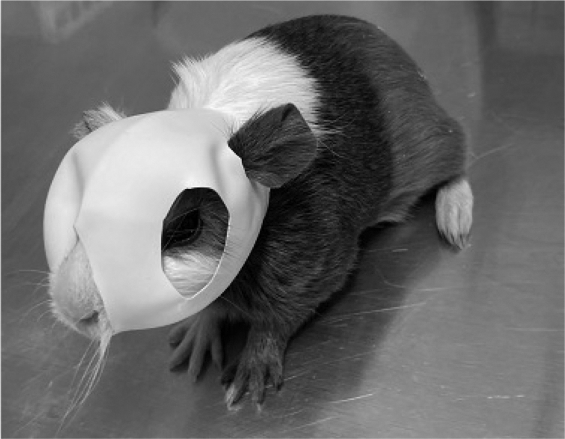
Single-eye facemask form deprivation-induced myopia in guinea pigs.
Refraction assessment
Before and after 3 weeks of intervention, the guinea pigs underwent mydriasis and retinoscopy (YZ-24 Streat Retinoscape; Suzhou Liuliu Vision Technology Co., Ltd., Suzhou, China) in an awake state. In this study, one drop of 0.5% tropicamide was instilled into eyes for 3 times, with an interval of 10 min/session. Retinoscopy was performed at 30 min after the last instillation. Retinoscopy was conducted by the same experienced optometrist. The results were recorded after retinoscopy, and the astigmatism was included in the spherical equivalent.
AL measurement
The guinea pigs were awake with 0.5% proparacaine hydrochloride eye drops for topical anesthesia. Measurement of the AL of guinea pig was performed using the manual A-mode ultrasound (Aviso, Quantel Medical, Inc., Paris, France). The diameter of the probe was 5 mm, the resolution was 0.01 mm, the transmission frequency was 11 Hz, and the pulse was received. The measured value represented the linear distance from the anterior apex of the cornea to the anterior surface of the retina at the posterior pole. Measurement parameters, including anterior chamber depth, lens thickness, vitreous depth, and overall AL were measured for 10 times, and their average values were calculated.
IOP measurement
When the guinea pig was awake, the IOP was measured using a rebound tonometer (iCare, Vantaa, Finland). Each measurement was carried out for 6 times, and the average value was obtained by 3 measurements. The measurement was started at 5 s after the guinea pig was taken or the headgear was removed. Equipped with a timer, the examination time points were at 0, 7, 14, and 21 days. The measurement time period was from 9:00 AM to 12:00 PM, and no surface anesthetic was used during the measurement.
OCT measurement
The optic disc was measured by spectral domain OCT (Cirrus HD 5000; Zeiss, Munich, Germany) and OCTA under general anesthesia, and 0.5% compound tropicamide was used to dilate pupils. The guinea pig was placed on a self-made guinea pig bracket, which was fixed on the jaw rest of the OCT apparatus and could be moved forward, backward, up, and down. The optic disc cube 200 × 200 protocol was utilized for scanning. The system automatically identified the center of the optic disc, automatically determined the position of the calculation ring, and selected the retinal nerve fiber layer (RNFL) and optic nerve head (ONH) programs to analyze the optic disc–related parameters, such as the area of the disc edge, the area of the optic disc, the average cup-to-plate ratio, the vertical cup-to-plate ratio, and the cup volume.
A 6 mm × 6 mm area of the retina at the posterior pole was scanned by a macular cube 512 × 128 protocol. The horizontal midline of the scanning area was connected to the midline of the optic disc. The left scan line was tangent to the temporal side of the optic disc. Retinal thickness at 3 mm from the optic disc and retinal volume over the entire scanned area were measured. Retinal thickness was the distance from the inner limiting membrane to the retinal pigment epithelium. OCT examination was repeated for >3 times. In addition to the analysis of RNFL and ONH, as part of the OCT examination, 3D-OCT images of the optic disc were captured, along with a map of avascular blood flow. The intraperitoneal anesthesia was induced, and the drug was 1% pentobarbital sodium at a dose of 0.33 mL/100 g (Fig. 2).
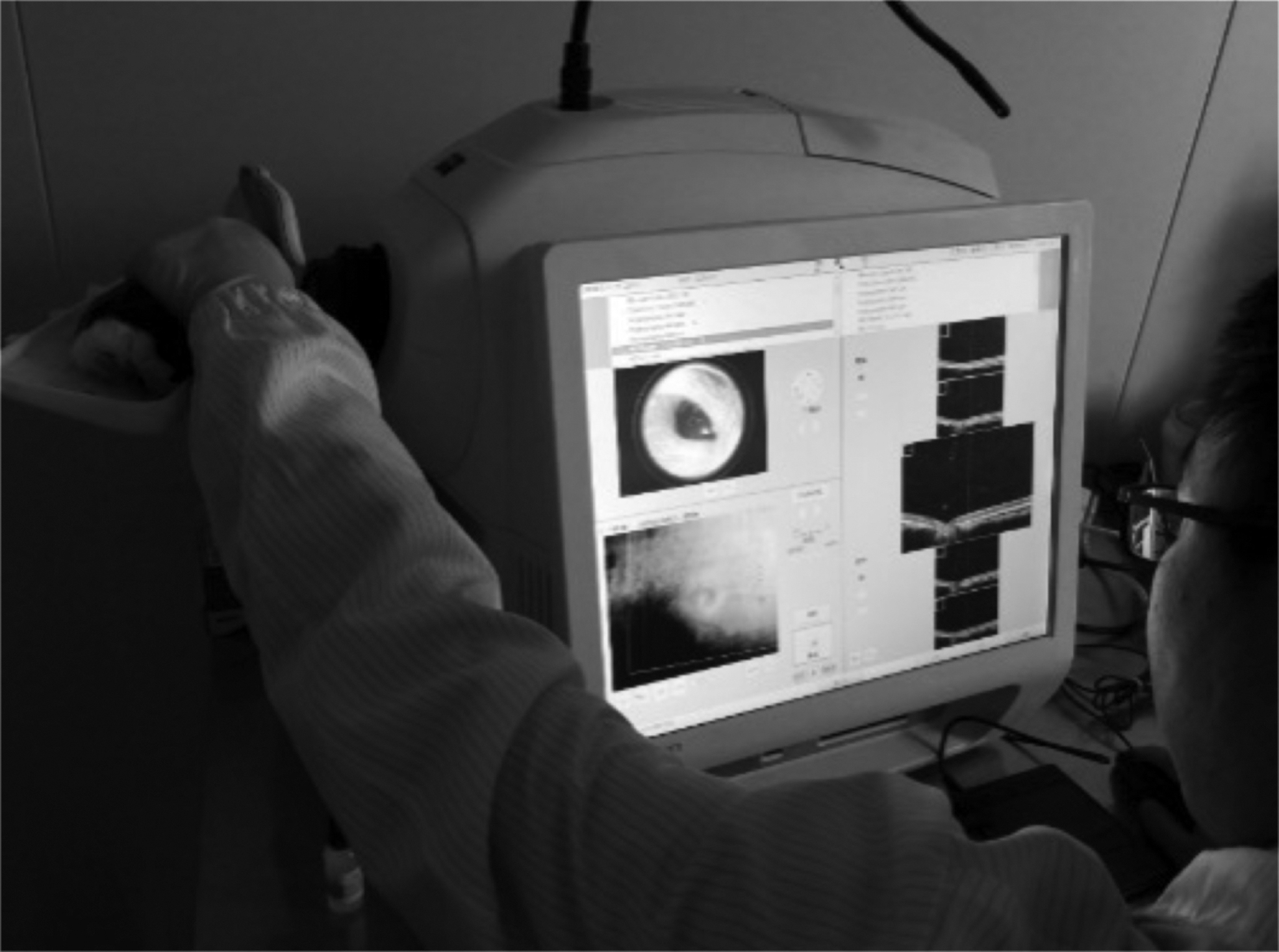
Optical coherence tomography examination of eyes of guinea pig under general anesthesia.
Procedure of CXL
For details of the CXL procedure, refer to Wang et al. 15 and Guo et al. 20 Guinea pigs were under general anesthesia. Under a 10 × microscope, a 29 G insulin injection needle was used, 0.5% Genipin (FUJIFILM Wako Pure Chemical Industries, Osaka, Japan) 0.1 mL was inserted at 3 mm behind the limbus cornea of the right eye, and sub-tenon injection was then performed. The injection was recorded at 0, 7, and 14 days, respectively. The points were located infratemporal, supranasal, and supratemporal, respectively. After injection, the micro-strabismus hook was used to push gently back and sides to make it even. It is essential to ensure that the conjunctival vesicles were intact and the drug could not leak. The ocular surface was rinsed with normal saline, and the eyelid was disinfected with iodophor. Guinea pigs wore a facemask after waking up, the eye area was washed with 0.9% normal saline every day after the surgery, and a new facemask was replaced. The method of 0.9% saline injection in the control group was carried out the same as mentioned previously (Fig. 3).

Sub-tenon injection.
Histological examination
After 3 weeks of intervention, 3 right eyeballs were randomly selected from the Genipin CXL group and control group, and they were placed in a fixative solution to prevent retinal detachment. They were fixed with 10% neutral formaldehyde for 24 h, embedded into paraffin, and 2 pieces of tissue were continuously cut from the temporal side of the optic nerve with a thickness of 0.5 mm for hematoxylin and eosin staining. The thicknesses of retina, choroid, and sclera, as well as choroidal blood supply were observed under a light microscope.
Type of outcome measures
The primary efficacy measure of this experiment is the control of myopia degree, and the secondary measure is the changes in AL. The primary safety measure is changes in IOP, and the secondary measure is the changes in optic disc OCT parameters.
Statistical analysis
Statistical analysis was performed using SPSS 18.0 software (IBM, Armonk, NY, USA). Normally distributed data with a normal distribution were expressed as mean ± standard deviation. The independent-samples t-test was used to compare the differences in refraction, IOP, AL, and OCT-related optic disc parameters between the 2 groups of guinea pigs before the intervention and at 3 weeks after the intervention. Differences in IOP, AL, and OCT-related optic disc parameters were evaluated using paired t-test to compare the intragroup differences at different time points. Descriptive statistics were utilized to analyze the histological imaging features of retina, choroid, and sclera, and 3D-OCT was used to assess the morphological changes of the optic disc. The blood flow at the optic disc was analyzed by the OCT avascular flow map. P < 0.05 was considered statistically significant.
Results
In total, 36 myopic guinea pigs of 200 three-week-old guinea pigs were screened and underwent FD for 1 week. At the age of 4 weeks, 24 eyes of guinea pigs with refraction power ≤ −6.0 DS were obtained and randomly divided into 2 groups, including 12 eyes in each group.
Baseline data
At baseline, there was no statistically significant difference in IOP, AL, refraction, and OCT-related optic disc parameters between Genipin CXL group and control group (P > 0.05; Table 1).
Baseline Comparison of Axial Length, Refraction, Intraocular Pressure, and Optical Coherence Tomography Parameters
CXL, cross-linking.
Results after 3 weeks of intervention
Comparison of AL, refraction, and IOP
Comparison of AL changes
According to the baseline comparison, there was no statistically significant difference in AL between the 2 groups. After 3 weeks of the intervention, AL in the Genipin CXL group was 7.95 ± 0.18 mm, and it was 9.03 ± 0.22 mm in the control group. There was a statistically significant difference between the 2 groups (t = −11.99, P < 0.001). Compared with the baseline, AL in the Genipin CXL group was significantly shortened by 0.34 ± 0.21 mm (t = −5.13, P = 0.001), whereas AL in the control group was significantly elongated by 0.64 ± 0.18 mm (t = 11.48, P < 0.001). There was a significant difference between the 2 groups in the changes of AL before intervention and at 3 weeks after the intervention (t = −11.28, P < 0.001) (Fig. 4).
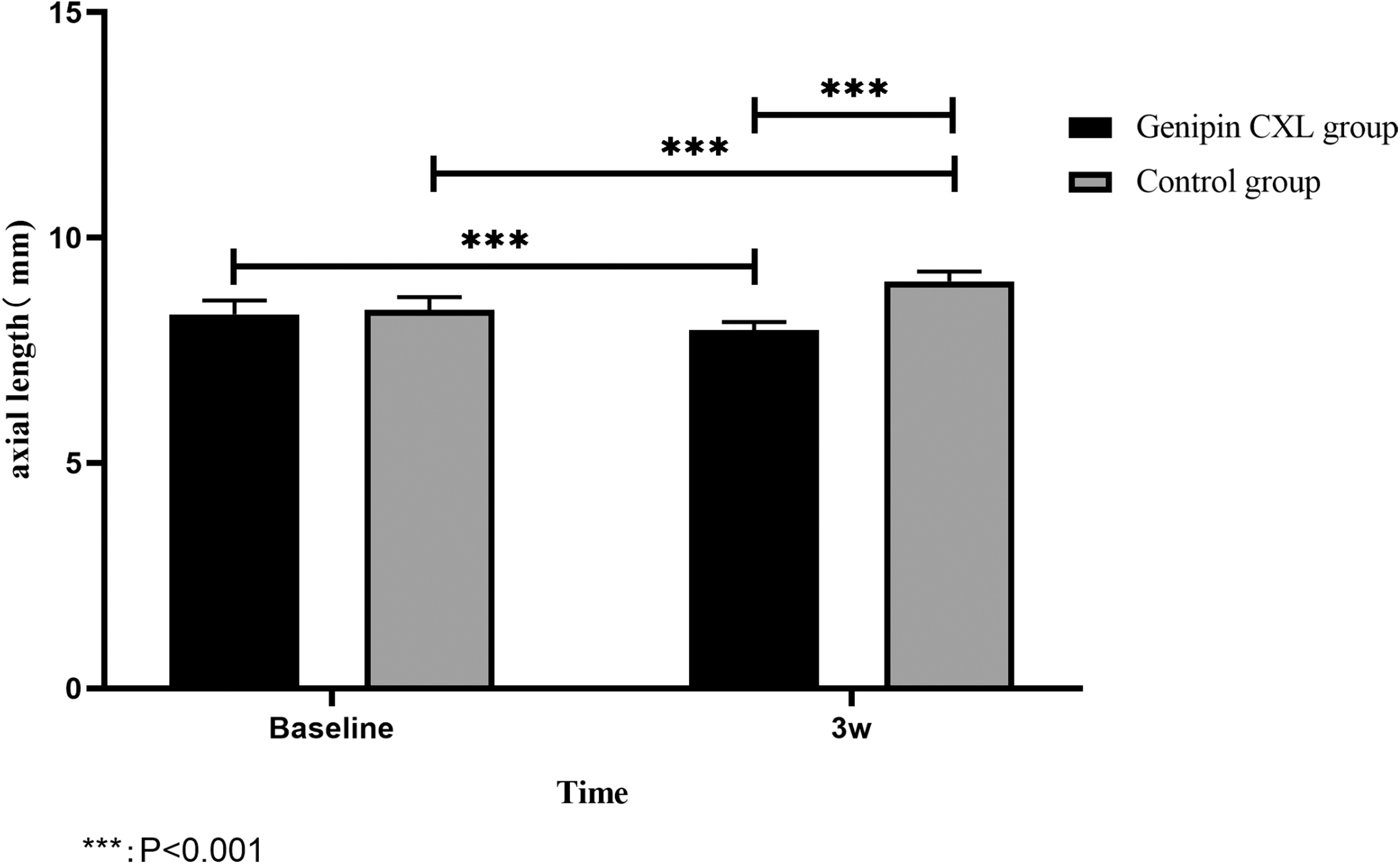
Comparison of changes of axial length before and after intervention (***P < 0.001).
Comparison of refraction changes
According to the baseline comparison, there was no statistically significant difference in refraction between the 2 groups. After 3 weeks of the intervention, refraction in the Genipin CXL group was −4.10 ± 1.60 DS, and it was −11.10 ± 1.78 DS in the control group. There was a significant difference between the 2 groups (t = 9.27, P < 0.001). Compared with the baseline data, refraction in the Genipin CXL group was significantly reduced by −2.75 ± 0.98 DS (t = 8.88, P < 0.001), whereas refraction in the control group significantly increased by −3.6 ± 0.74 DS (t = −15.43, P < 0.001). There was a significant difference between the 2 groups in the changes of refraction before intervention and at 3 weeks after the intervention (t = 16.38, P < 0.001) (Fig. 5).
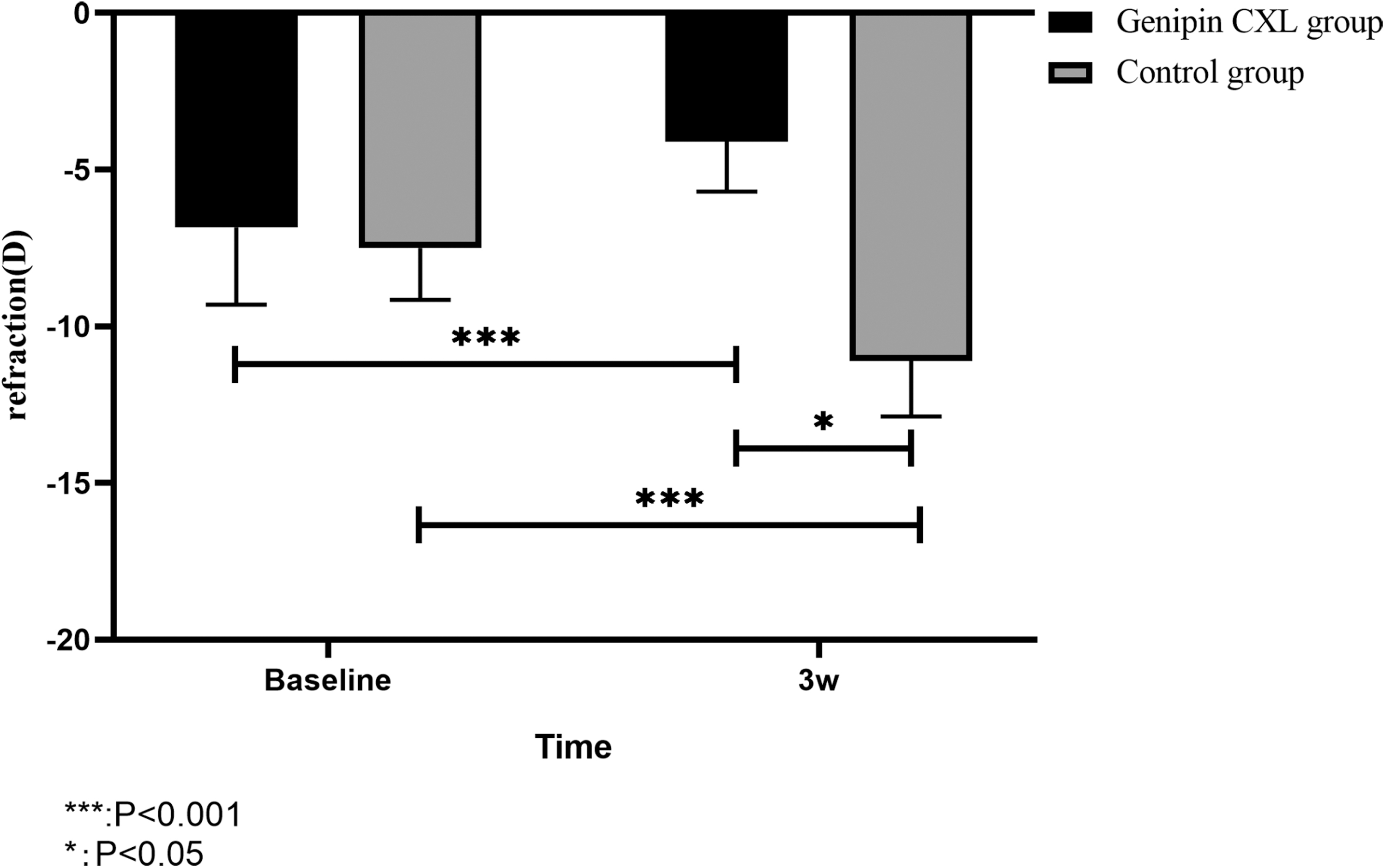
Comparison of changes of refraction before and after intervention (***P < 0.001).
Comparison of IOP changes
Over time, IOP increased in the Genipin CXL group and control group. At different time points, no statistically significant difference was found between the 2 groups at baseline. In the third week, IOP in the Genipin CXL group was 22.2 ± 2.35 mmHg, it was 19.5 ± 2.51 mmHg in the control group, and IOP in the Genipin CXL group was higher than that in the control group (t = 2.49, P = 0.02). Compared with the baseline data, IOP in the Genipin CXL group was elevated by 4.5 ± 2.59 mmHg after 3 weeks of intervention, IOP in the control group increased by 1.9 ± 1.37 mmHg, and the changes of IOP between the 2 groups were statistically significant (t = 2.80, P = 0.01) (Fig. 6).
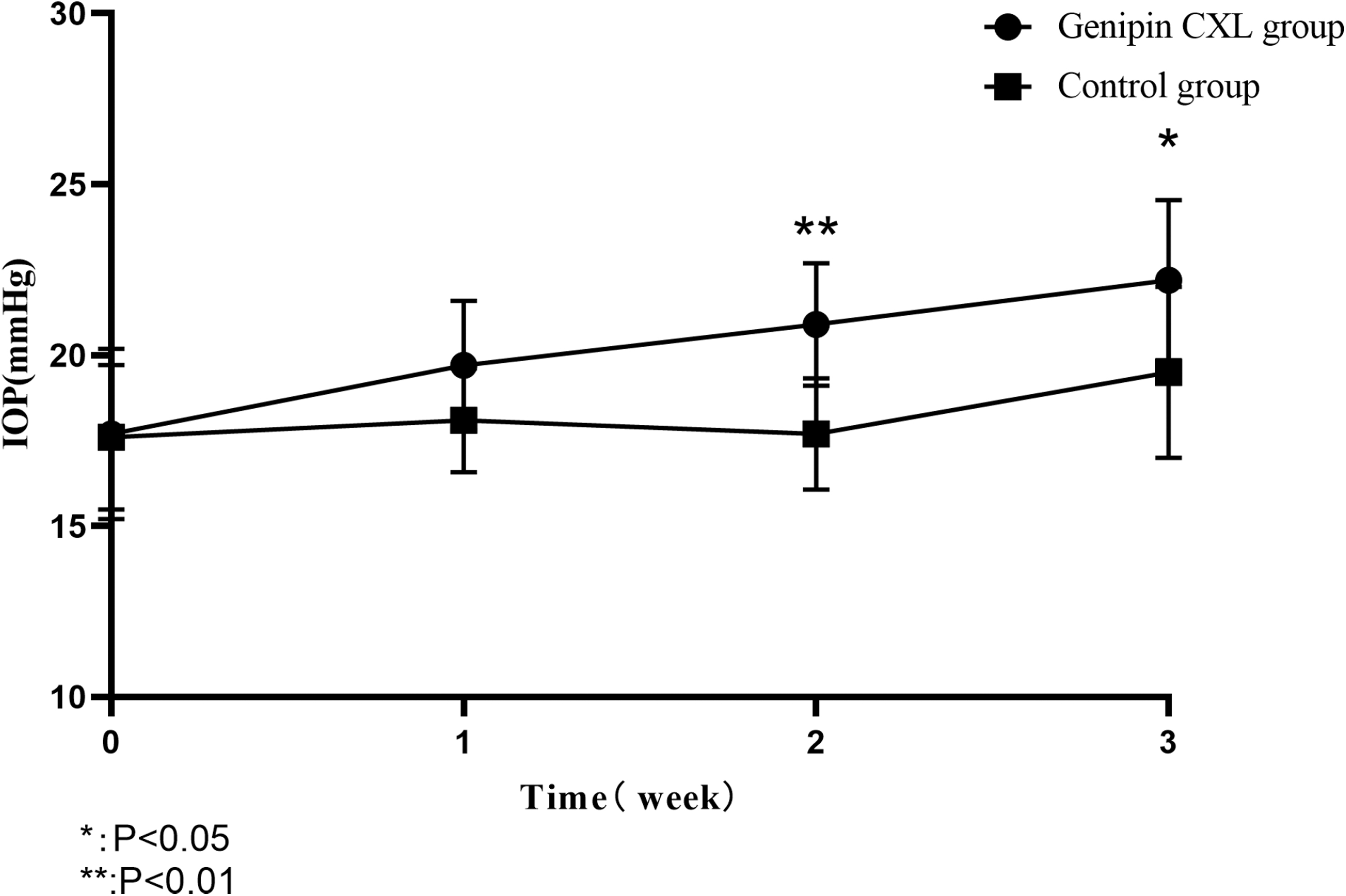
Comparison of changes of intraocular pressure before and after intervention (**P < 0.01, *P < 0.05).
Comparison of changes in OCT-related optic disc parameters
There were no significant differences in the average cup–disc ratio (C/D), vertical C/D, rim area, and cup volume between the Genipin CXL group and control group at baseline (P > 0.05). When CXL was combined with FD for 3 weeks, the average C/D and vertical C/D increased, and disc edge area was reduced, in which the difference between the 2 groups was statistically significant, whereas the difference in cup volume was not statistically significant (Table 2). When the guinea pig eyes of the Genipin CXL group were intervened for 3 weeks, compared with the baseline data, the average C/D and the vertical C/D were elevated, the rim area was reduced, and the cup volume was elongated (Table 3). These differences were statistically significant. However, in the control group, the changes of these parameters were not statistically significant compared with the baseline data (Table 4).
Comparison of Optical Coherence Tomography Parameter Values of Guinea Pig Optic Disc Between Genipin Cross-Linking Group and Control Group After 3 Weeks of Intervention
Self-Comparison of Optic Disc Optical Coherence Tomography Parameters in Genipin Cross-Linking Group Before and After 3 Weeks of Intervention
Self-Comparison of Optic Disc Optical Coherence Tomography Parameters in Control Group Before and After 3 Weeks of Intervention
Analysis of blood flow at the optic disc by the OCT avascular flow map and 3D-OCT
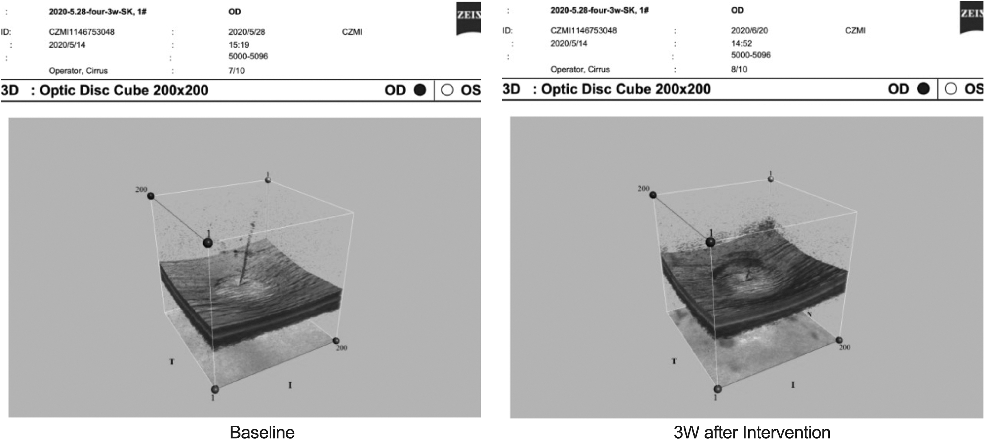
In the Genipin CXL group, at 3 weeks after intervention, the optic disc blood flow was significantly reduced compared with the OCT avascular flow map at baseline, the annular elevation of the optic disc decreased, the blood vessel peak disappeared, and the optic disc depression was significantly deepened compared with 3D-OCT graphs at baseline (Figs. 7 and 8). However, there were no significant changes in the control group after the intervention (Figs. 9 and 10).
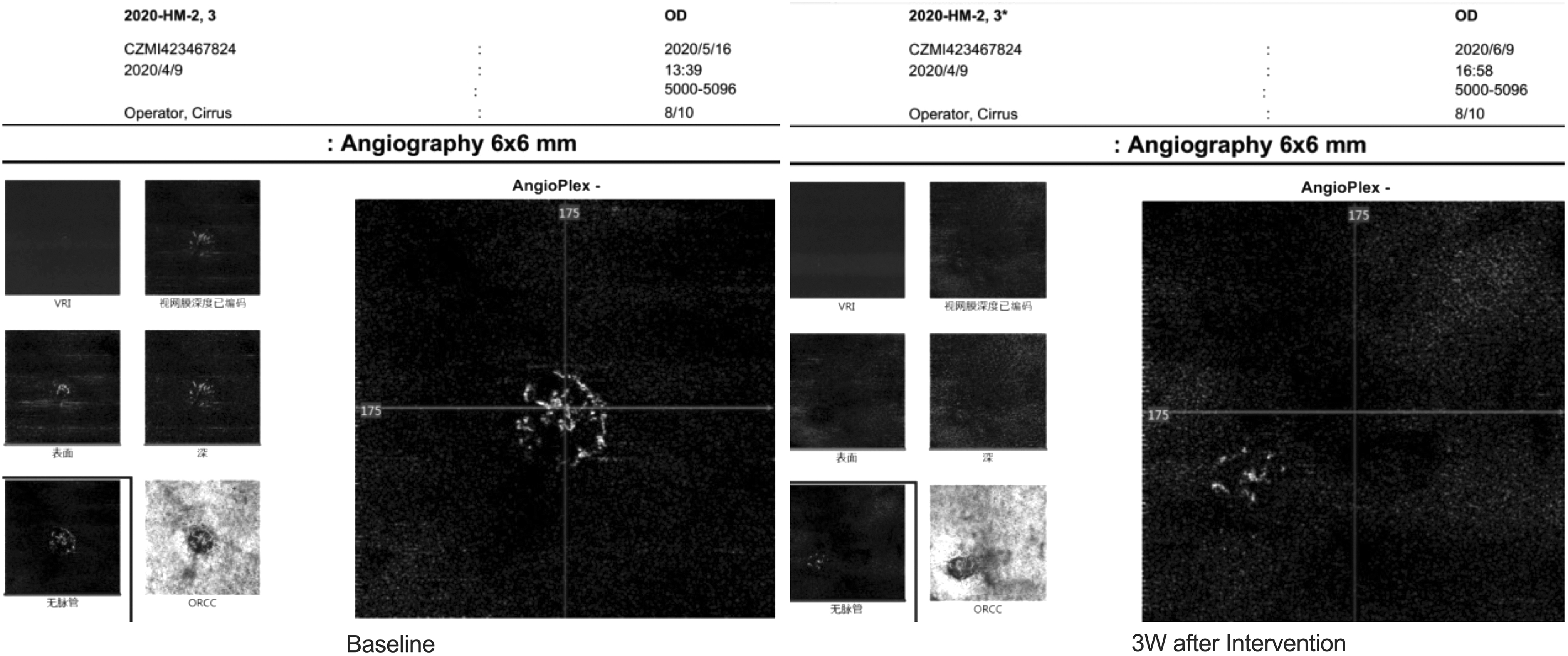
Comparison of OCT avascular blood flow map in the Genipin CXL group at baseline and after 3 weeks of intervention, and the optic disc blood flow was significantly reduced after 3 weeks. CXL, cross-linking; OCT, optical coherence tomography.
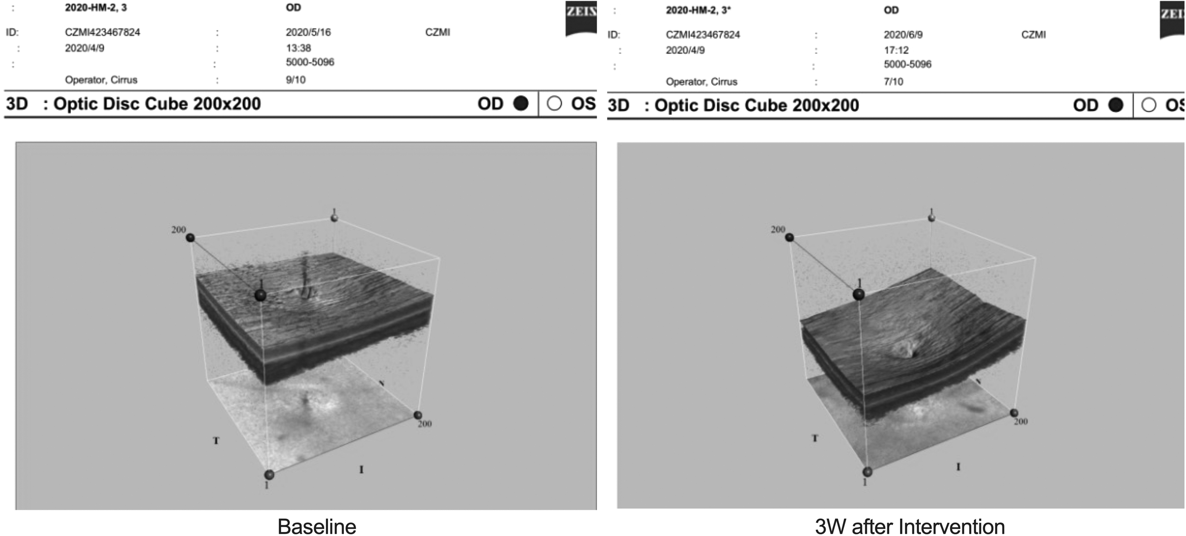
Comparison of 3D-OCT graphs in the Genipin CXL group at baseline and at 3 weeks after intervention. After 3 weeks, the annular drop of the optic disc decreased, the blood vessel peak disappeared, and the optic disc depression was significantly deepened.
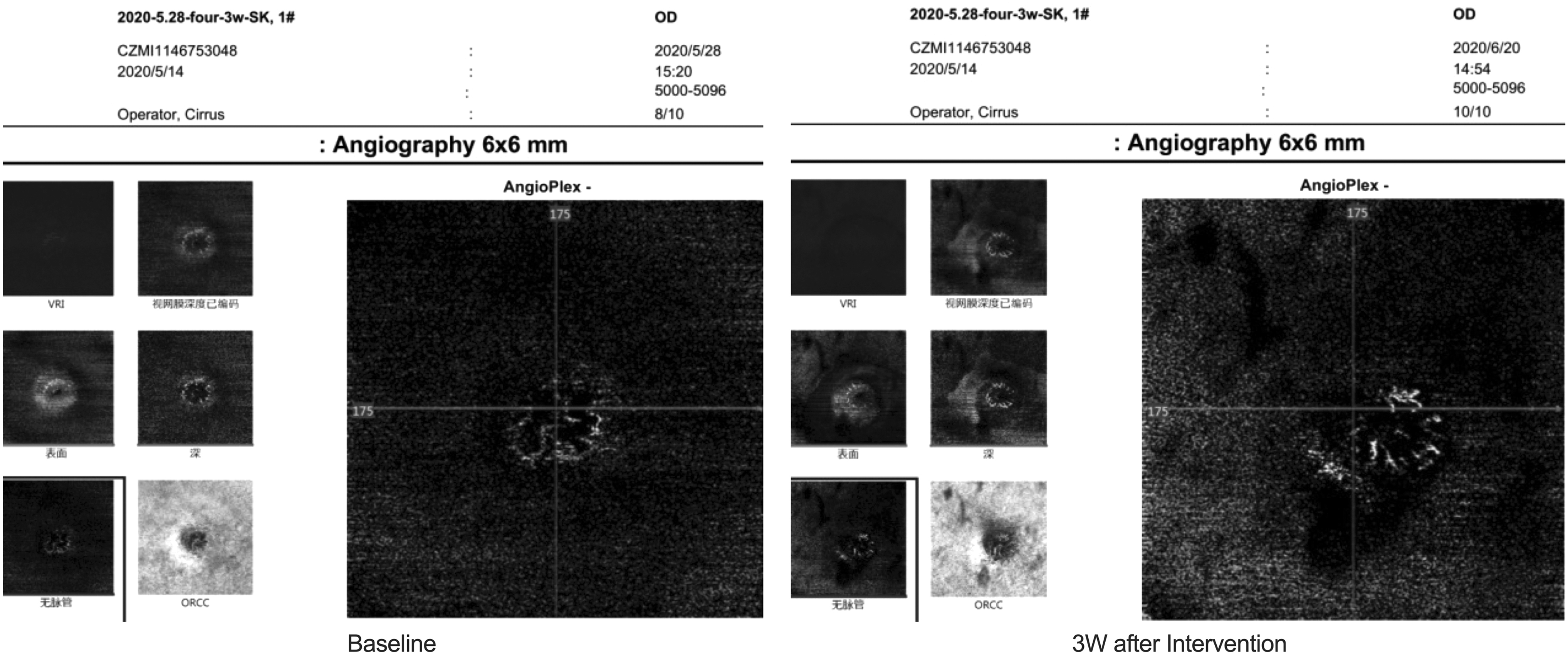
OCT avascular flow map showed no significant change in optic disc blood flow in the control group at baseline and after 3 weeks of intervention.
Comparison of 3D-OCT images in the control group at baseline and at 3 weeks after intervention, and there was no significant change in optic disc morphology after 3 weeks.
Histological examination
After 3 weeks of intervention in the Genipin CXL group, the choroidal blood perfusion was obvious. The control group exhibited a significant reduction in the choroidal blood flow (Fig. 11).

Left: Under a light microscope, the choroidal blood perfusion was obvious when the Genipin CXL group was intervened for 3 weeks. Right: Under a light microscope, the choroidal blood flow significantly decreased after 3 weeks of intervention in the control group (as indicated by the arrow).
Discussion
The results of this study showed that 0.10 mL sub-tenon injection of 0.5% Genipin solution could effectively inhibit development of high myopia in guinea pig eyes in the state of FD, which was more effective than sub-tenon injection of 0.9% saline on days 0, 7, and 14. Moreover, it was found that IOP of guinea pigs was slightly elevated after scleral CXL, and the OCT imaging of optic nerve showed glaucomatous changes, which were manifested by the increase of the average C/D and vertical C/D, as well as deepening of the cup volume. The 3D-OCT indicated that the annular bulge of the optic disc disappeared, the blood vessel peak disappeared, and the optic disc depression occurred. The optic disc avascular flow map showed that the optic disc blood flow was significantly reduced, which is similar to our previous findings of CXL in the FD myopia group induced by hyperopia. 20
However, there were some differences between the 2 models. From the perspective of the degree of control of myopia, the efficacy of CXL in the treatment of high myopia combined with FD was significantly higher than that of hyperopia combined with FD. It was revealed that AL in the high myopia group was shortened, and refraction was reduced, whereas the hyperopic group had different degrees of increase. In terms of safety, the incidence and degree of glaucomatous in the high myopia group were lower than those in the hyperopia group in our previous study.
The high myopia group had an IOP of 22.2 ± 2.35 mmHg at 3 weeks after intervention, whereas the hyperopia group reached 31.5 ± 3.6 mmHg; the OCT-related optic disc parameters changed, in which the high myopia group had an average C/D of 0.56 ± 0.08 mm and a cup volume of 0.05 ± 0.01 mm3 at 3 weeks after intervention, whereas the hyperopia group had an average C/D of 0.65 ± 0.06 mm and a cup volume of 0.65 ± 0.06 mm3. 20 It is noteworthy that the baseline cup volume for hyperopic eyes was 0.03 ± 0.01 mm3, whereas it was 0.04 ± 0.02 mm3 for highly myopic eyes. The depth of cup volume in the hyperopic group was significantly greater than that in the high myopia group after CXL and FD.
Therefore, according to the results of this study, the efficacy and safety of CXL in the treatment of juvenile guinea pigs with high myopia were higher than the effect of the treatment of FD myopia on hyperopic eyes. The results suggest that the next experimental research on CXL treatment of myopia should take high myopia as the experimental eye into account. Such experimental results are precious for the future clinical trials on CXL in the treatment of pathological myopia, because the posterior sclera reinforcement of human eyes is performed in highly myopic eyes. The efficacy and safety of CXL in highly myopic guinea pigs were reported for the first time in this study.
The visual development of the guinea pig eye undergoes the same process of hyperopia formalization as the human eye. Of note, myopia occurs in different proportions in childhood. The incidence of myopia in juvenile guinea pigs that were 3–4 weeks old was ∼18% in this study. Meanwhile, high myopia can also be acquired in juvenile guinea pigs after induction by FD or lenses.21,22 Therefore, it is feasible to select high myopia for experimental eyes from the perspective of experimental enrollment. The sclera with high myopia is thinner and less rigid. Therefore, the hardness of scleral tissue hardness after CXL should theoretically be lower than that after CXL in hyperopic eyes. Regarding the relationship between sclera biomechanics and IOP, the stronger the scleral tissue hardness, the higher the induction of IOP.19,23
Therefore, the increase in IOP after scleral CXL in highly myopic eyes is relatively small, and the pressure on the fundus optic nerve may also be reduced. The effect of sub-tenon injection of CXL agent on enhancing scleral tissue hardness is exact, and it is concentration and frequency dependent. Previous studies have clearly demonstrated this effect.14,16,19,24,25 According to the previously published findings, 20 no further measurement of scleral biomechanics was performed after CXL in this study. The selection of Genipin for CXL can be related to the fact that Genipin CXL agent is extracted from pure Chinese medicine, the CXL effect is exact, and its side effects are relatively limited. It has been clinically and experimentally proven to be effective, safe, and histocompatible. It is a CXL agent with satisfactory properties and has an anti-inflammatory effect.26,27
The efficacy of scleral CXL in the treatment of experimental eye FD myopia has noticeably attracted scholars' attention. However, several issues, including determination of the most appropriate CXL agent, reasonable injection frequency, and optimal CXL position and range remain elusive. More research is needed to determine the safe and effective dose–effect standard in the future. The in vivo OCT of optic disc in guinea pig eyes is an effective method to indicate whether optic disc changes in glaucoma occur after the treatment of myopia using CXL. 28 The inspection images are clear, the indicators can be quantified, and the measurement can be repeated in situ. Moreover, 3D-OCT and OCTA measure the optic disc from different angles, which can visualize the imaging changes of the optic disc and changes of blood flow.
The OCT reference value of optic disc of normal guinea pigs has been studied by scholars, and we have also clearly explained it in previous studies.29–31 Therefore, it is recommended to apply it to similar experiments. In addition, the standard values of OCT of optic disc of guinea pigs were sorted out under the condition of high IOP caused by CXL, providing a theoretical reference for the future experimental studies. In similar previous studies, histological sections have not shown abnormal changes in glaucoma. We will also carry out electron microscopy in the next studies to observe the changes of the optic disc cribriform plate after CXL treatment of guinea pigs with high myopia, to confirm the results of this study from different prospects, such as imaging and pathological features.
This study had some limitations. First, although subconjunctival vacuoles were intact after the sub-tenon injection, the cornea and ocular surface were flushed with saline after injection. However, it was infeasible to fully confirm whether cornea is cross-linked and whether it has an effect on IOP. Second, owing to limitations related to the inspection equipment, no corneal curvature was assessed, and it was impossible to determine whether the CXL could affect corneal curvature and refraction. Moreover, the elastic modulus of the sclera should be measured, and it is necessary to assess IOP and imaging changes of the optic nerve under different scleral biomechanical states. In addition, the duration of our experiment is relatively short, so further observation is needed to evaluate potential side effects of long-term application.
Conclusions
In summary, sub-tenon injection of 0.5% Genipin solution could effectively inhibit development of myopia in the state of FD in guinea pigs with high myopia. However, glaucomatous imaging changes were found in the optic disc. Therefore, the safety of the mentioned method still needs further research, and highly myopic guinea pigs are appropriate candidates for such experiments.
Ethics Approval
This study was carried out in accordance with the principles of the Basel Declaration and recommendations of the Animal Ethics Committee of China Medical University (China). All animal experiments were conducted in compliance with the ARVO Statement for the Use of Animals in Ophthalmic and Vision Research.
Availability of Data and Materials
The datasets used and/or analyzed during this study are available from the corresponding author on reasonable request.
Footnotes
Authors' Contributions
L.G. and L.F. conceived and supervised the study and designed the experiments. L.G., Z.W., and Z.G. participated in data collection, as well as drafting the article. L.G., Y.T., S.C., and X.Z. collected data and performed the experiments. L.G. and B.M. performed the statistical analysis. L.G., L.F., and Z.G. participated in the data analysis and revising the article. All the authors read and approved the final version of the article.
Author Disclosure Statement
The authors declare that they have no competing interests.
Funding Information
This study was funded by the Fund for Science and Technology Program of Shenyang City (No. 21-173-9-24) and the Fund for Heath Commission of Shenyang (No. 2022108).