




Abstract
Purpose:
This study aimed to evaluate the effects of a new treatment—conditioned medium from human orbital adipose-derived stem cells (OASC-CM)—on corneal recovery after alkali burns in a rabbit model.
Methods:
The corneal alkali burn rabbit model was established and treated with OASC-CM, conditioned medium from human abdominal subcutaneous adipose-derived stem cells (ABASC-CM), and fresh control culture medium (con-CM) three times a day for 7 days, respectively. Subsequently, the treatment effects were evaluated and compared through clinical, histological, immunohistochemical, and cytokine evaluations.
Results:
Clinically, OASC-CM alleviated corneal opacity and edema and promoted recovery of corneal epithelium defect. Histologically and immunohistochemically, OASC-CM inhibited neovascularization, conjunctivalization, and immuno-inflammatory reaction, while promoting corneal regeneration and rearrangement. Increased secretion of interleukin-10 and inhibited protein levels of cluster of differentiation 45, interferon-γ, and tumor necrosis factor-α were observed in the alkali-burned cornea after OASC-CM treatment, which might be the relevant molecular mechanism.
Conclusions:
OASC-CM showed significant effects on the recovery of rabbit corneal alkali burns and eliminated immunological and ethical limitations, representing a new option for corneal wound treatment.
Introduction
Corneal alkali burn is an important cause of blindness, accounting for 60% of ocular chemical injuries. 1 Alkalis cause more serious damages to stem cells from corneal limbus than acids—the compounds alkalis forming with corneal tissues can penetrate not only lipophilic conjunctiva and corneal epithelium but also the hydrophilic corneal stroma and endothelium. 2 Various conventional methods are effective in treating mild corneal alkali burns, such as agents that promote epithelialization and inhibit inflammation and biological extracts containing growth factors. 1 For severe corneal alkali burns, emerging stem cell therapies using limbal stem cells 3 and stem cells from other tissues (e.g., embryo, 4 adipose, 5 and bone marrow 6 ) have been proved to be effective as well, but still have immunological and ethical limitations. 7 Thus, more feasible treatments for corneal alkali burns are still desperately needed.
Adipose-derived stem cell (ADSC)-free derivatives, including ADSC exosomes, ADSC-conditioned medium (ADSC-CM), and cell-free adipose tissue extracts, can eliminate limitations of rejection and ethics and are widely used to promote tissue regeneration due to their abundance and accessibility. 8 Orbital fat (OF) is a highly specialized adipose tissue depot that fills most of the orbital cavity. Like most ocular and orbital tissues, OF is from neural crest origin. 9 ADSCs vary in biological characteristics based on different anatomic regions of adipose tissue, 10 and depot-dependent biological functions of ADSCs have been found in recent studies. 11 Therefore, we hypothesized that orbital adipose-derived stem cells (OASCs) obtained from OF could be a better treatment option for corneal regeneration among ADSCs from different sources, including the most widely used abdominal subcutaneous adipose-derived stem cells (ABASCs). However, few studies have evaluated the effects of OASCs on treating corneal chemical defects, let alone those of CM harvested from OASCs.
In this study, we evaluated the effects of OASC-CM on corneal recovery and the relevant molecular mechanism for corneal regeneration in a corneal alkali burn rabbit model.
Materials and Methods
Animals
Nine healthy and purebred New Zealand white rabbits of both sexes, 4–6 months old, and weighing 2.0–2.5 kg were used in this study, which were provided by the Animal Experimental Center of Shanghai Tenth People's Hospital. All procedures involving animals strictly followed the Association for Research in Vision and Ophthalmology (ARVO) Statement and were ethically approved by the Animal Care and Use Committee of Shanghai Tenth People's Hospital (Approval No. SHDSYY-2020-0083).
Fat tissue samples
Fat tissue samples used in this study were surgically obtained from different cosmetic surgery patients in the plastic surgery department of Shanghai Tenth People's Hospital. OF tissue samples were harvested from OF pads in lower eyelid blepharoplasty, and abdominal fat tissue samples were from liposuction of the waist and abdomen. Donors were divided into two groups: The OF group (from young females 20–35 years of age, n = 6) and the abdominal subcutaneous fat (AF) group (from young females 29–44 years of age, n = 6).
Patients with obesity (body mass index >24 kg/m2), endocrine diseases, eye diseases, and other organic diseases were excluded. All participants provided their written informed consent to participate in this study and publish their case details. Experimental methods involving patients followed the tenets of the Declaration of Helsinki and were approved by the Medical Ethics Committee of Shanghai Tenth People's Hospital (Approval No. SHDSYY-2020-0084).
Isolation and Culture of OASCs/ABASCs
OASCs and ABASCs were isolated, respectively, from OF and AF tissue samples, as previously reported, with minor modification. 12 Briefly, OF/AF tissues were washed with phosphate-buffered saline (PBS; pH = 7.4, Sigma, Sigma-Aldrich Corp., Shanghai, China), and then further digested with 1% type-I collagenase solution (Worthington Biochemical Corp., Lakewood, NJ, USA) for 1 h at 37°C. After the addition and incubation with the culture medium [containing low-glucose Dulbecco's modified Eagle's medium (LG-DMEM; Gibco, Grand Island, NY, USA) with 10% fetal bovine serum (FBS, HyClone, Logan, UT, USA)], the centrifugation was performed and then the supernatant was discarded by aspiration.
The cellular pellet was suspended, and single-cell suspension was plated into 100-mm culture dishes (Falcon, B&D Bioscience, San Jose, CA, USA) at a density of 5,000 cells/cm2 and cultured at 37°C. The culture medium was replaced in the first 48 h after plating and changed every 3 days thereafter. Cells adherent on the plastic substrate were regarded as OASCs/ABASCs of Passage 0 (P0). Upon reaching ∼80%–90% confluence, OASCs/ABASCs were detached with 0.05% trypsin/0.5 mM ethylenediaminetetraacetic acid (Sigma, Sigma-Aldrich Corp.), replated into new culture dishes at a density of 20,000 cells/cm2, and regarded as OASCs/ABASCs of Passage 1 (P1).
Characterization of OASCs with induced differentiation and flow cytometry
Adipogenic and osteogenic differentiation of OASCs were performed as previously reported. 13 After 2 weeks of differentiation, Oil Red and von Kossa staining were performed and evaluated under an optical microscope (IX70; Olympus, Tokyo, Japan).
Flow cytometry was performed to detect typical surface marker expressions of OASCs. 14 Briefly, cell aliquots (2 × 10⁶/mL) were incubated with monoclonal antibodies CD90, CD105, CD73, CD45, CD34, CD19, CD14, HLA-ABC, and HLA-DR (all from Santa Cruz Biotechnology, Dallas, TX, USA). Results were obtained on a FACScan cytometer (Coulter Epics Altra, Becton Dickson, San Jose, CA, USA) and analyzed with FlowJo software.
Preparation of OASC-CM/ABASC-CM
Preparation of OASC-CM/ABASC-CM was performed as previously reported, with minor modification. 11 Briefly, the culture medium was discarded when OASCs/ABASCs of P2 grew to about 85% confluence, and then cells were cultured with fresh LG-DMEM (without FBS or antibiotics) for 48 h. After 3 days, the culture supernatant was collected and filtered through a 0.22-μm sterile filter (Millipore, Millipore Corp., CA, USA). Finally, OASC-CM/ABASC-CM was concentrated (30-fold) using ultrafiltration with a cutoff of 10 kDa (Millipore, Millipore Corp.) and stored at −80°C for future use. LG-DMEM without cells, FBS, or antibiotics was used as a control conditioned medium (con-CM).
Establishment of the corneal alkali burn rabbit model
Rabbits were anesthetized by intramuscular injection with xylazine (5 mg/kg) and ketamine (45 mg/kg), and proparacaine hydrochloride drops (5 g/L) were used to provide topical anesthesia.
The corneal alkali burn rabbit model was established as previously described. 15 Briefly, a single-layer circular filter paper (10-mm in diameter) was soaked in 1 N sodium hydroxide solution for 60 s, and then placed on the cornea of rabbits for another 60 s. Eighteen eyes of the nine rabbits were randomly divided into three equal groups: (1) the OASC-CM group; (2) the ABASC-CM group; and (3) the con-CM group (six eyes per group). For each group, 0.3 mL of OASC-CM, ABASC-CM, and con-CM was applied, respectively, and topically three times a day for 7 days since the day of the injury (D0); 0.3% ofloxacin eye drops (Santen, Santen Pharmaceutical Co., Ltd., Osaka, Japan) was used twice a day for 7 days to prevent eye infections.
Result Evaluation
Clinical evaluation
Slit-lamp microscopy photographs were taken to evaluate opacity, conjunctivalization, and neovascularization of the cornea every two days. On the sixth day after the surgery (D6), corneal turbidity was scored with the protocol of Gupta et al. 16 (Table 1). The area of corneal neovascularization (CNV) was calculated on the same day by measuring the radius of the cornea (r), the reticule vessel length (L) from the limbus, and the number of clock hours (C) of the limbus involved. A formula of D'Amato et al. 17 was used to determine the area of CNV: C/12 × 3.1416 [r 2 –(r–L) 2 ]. On D6, epithelial defect photographs were taken under cobalt blue light after the cornea was stained with sodium fluorescein and scored with the criterion (Table 2).
Scoring Criteria of Corneal Turbidity
Scoring Criteria of Corneal Sodium Fluorescein
Histological and immunohistochemical evaluation
All experimental rabbits were killed by injecting air through the auricular vein on the seventh day after the operation (D7). For histopathological observation, half of each group's corneas (n = 3), including limbus and conjunctival region, were collected, and then cut into several 5-μm paraffin-embedded sections. Specifically, for histological examination, part of the tissue sections from each cornea was stained with the hematoxylin and eosin method. For immunohistochemical (IHC) examination, mouse anti-rabbit monoclonal primary antibodies [vascular endothelial growth factor (VEGF; dilution factor, 1:500), cytokeratin 19 (CK19; dilution factor, 1:500), and p63 (dilution factor, 1:500)] were applied separately to the other tissue sections from each cornea and incubated overnight at 4°C. Furthermore, sections were incubated with goat anti-rabbit secondary horseradish peroxidase (HRP) antibodies. 3,3′-diamino benzidine solution (AR1022; dilution factor, 1:500) was used as a chromogen and Mayer's hematoxylin was used for counterstaining.
Cytokine evaluation
The other half of each group's corneas (the other three corneas per group) was used for cytokine evaluation. Briefly, corneal tissues of the same weight were collected on D7 and ground with 500 μL of precooled PBS solution, followed by centrifugation (1,000 rpm) for 10 min. Levels of interferon interleukin-10 (IL-10), cluster of differentiation 45 (CD45), interferon-γ (IFN-γ), and tumor necrosis factor-α (TNF-α) in corneal samples were determined by enzyme-linked immunosorbent assay (ELISA; Elabscience Biotechnology Co., Wuhan, China).
Statistical analysis
Data obtained from three independent groups (n = 6) were expressed as mean ± standard deviation. Shapiro–Wilk and Levene analysis were performed to test the normality and homogeneity of the variance, respectively. One-way analysis of variance and the least significant difference t-test were used to determine possible significant differences between groups. Statistical analysis was done with the SPSS software package version 26.0 (IBM Corp., Armonk, NY, USA), with statistical significance set at P < 0.05.
Results
Morphological observation of OF and characterization of OASCs
Figure 1A showed a baggy appearance of a 55-year-old female patient preparing for lower eyelid blepharoplasty. After surgery, three fat pads (medial, central, and lateral) were taken from each side of the lower eyelids (Fig. 1B). Histologically, orbital adipocytes were separated by dense septum fibers and arrayed tightly, and the size of adipocytes was slightly irregular (Fig. 1C).
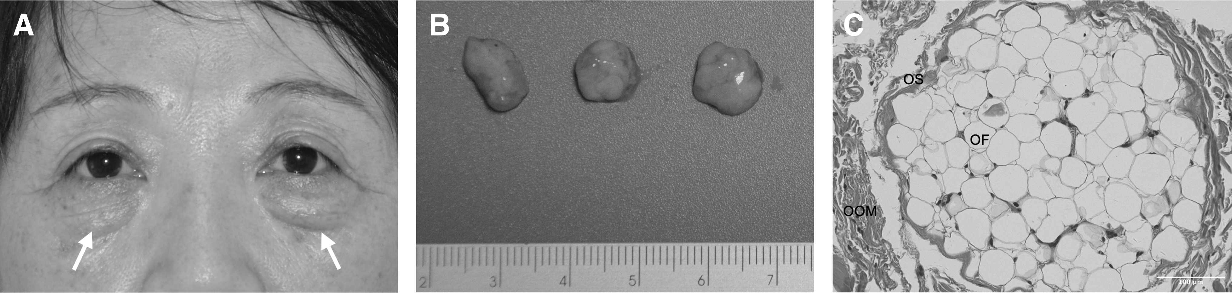
Morphological observation of OF.
OASCs of P1 extracted from OF tissue were spindle shaped (Fig. 2A). Osteogenic (Fig. 2B) and lipogenic differentiation (Fig. 2C) results were verified by von Kossa and Oil Red staining, respectively. Flow cytometry analysis revealed that OASCs were positive for CD90, CD105, CD73, and HLA-ABC, and negative for CD45, CD34, CD19, CD14, and HLA-DR (Fig. 2D).

Characterization of OASCs.
Establishment of the corneal alkali burn rabbit model
The whole study design is shown in Fig. 3A. Corneal alkali burn was induced in the rabbit model (Fig. 3B), with the change of corneal turbidity from clear to dense opacity (Fig. 3C, D).
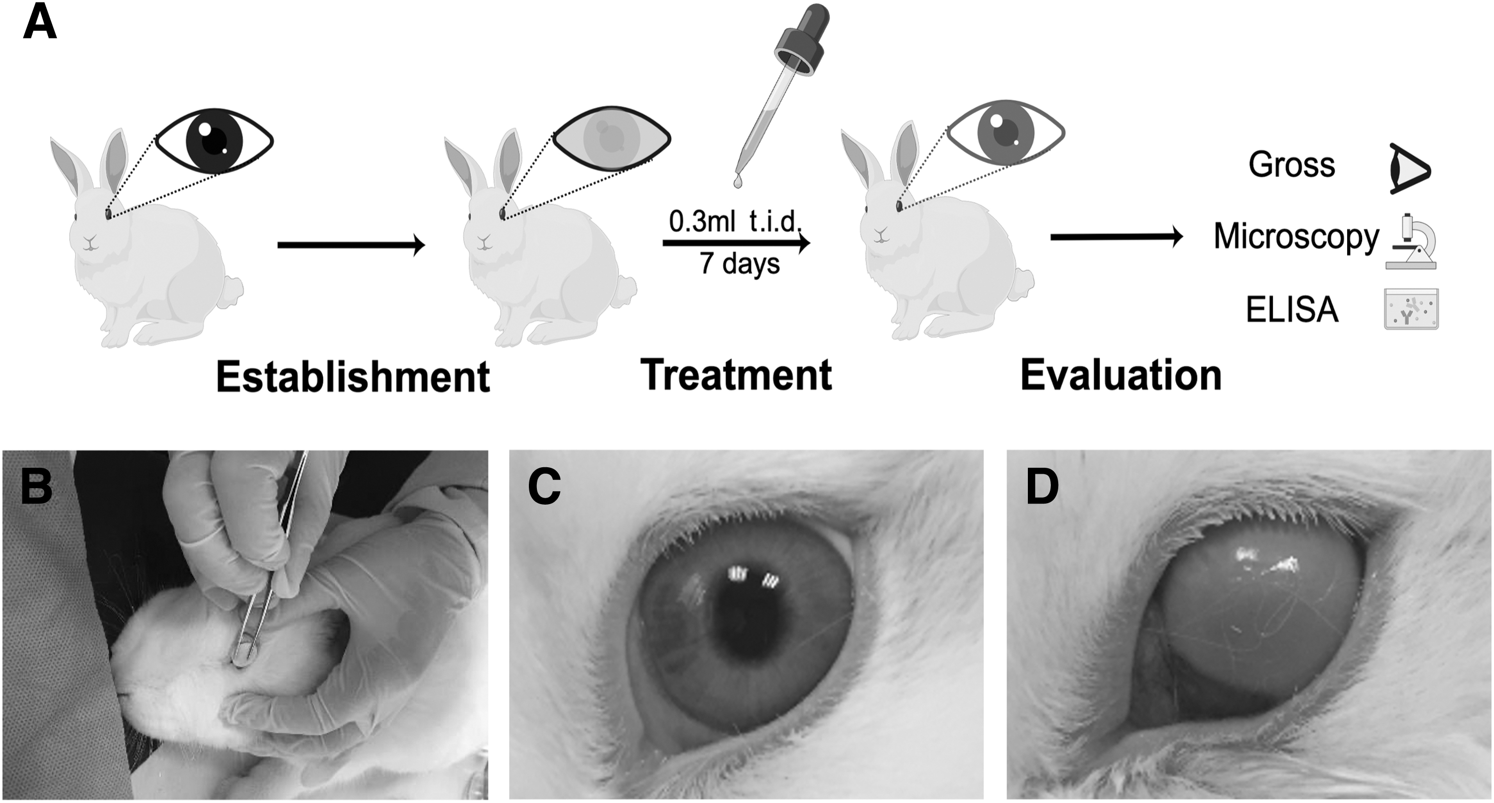
Establishment of the corneal alkali burn rabbit model.
Clinical evaluation
A round, white, and turbid area over the corneal limbus could be observed immediately in each eye after alkali burns (Fig. 4A). In the OASC-CM group (Fig. 4A), on the second day after the surgery (D2), the white haze on the corneal surface dispersed evenly, and the edema level alleviated. No new capillary grew into the corneal limbus. On the fourth day after the surgery (D4), the grade of corneal turbidity declined significantly, and there was no obvious hyperemia or edema. On the sixth day after the surgery (D6), the turbid area of the cornea was far smaller than the other two groups, with significant improvement in corneal clarity.

Clinical evaluation by eye.
In control groups (ABASC-CM and con-CM; Fig. 4A), on D2, large ulcers on the corneal surface of rabbits could be observed and corneal transparency decreased. On D4, new capillaries grew into the limbus of the cornea, and corneal edema could be observed as well. On D6, new capillaries gradually grew toward the center of the cornea, and conjunctivalization, edema, as well as hyperemia further led to poorer transparency in control groups. Corneal turbidity score (Fig. 4B) on D6 quantitatively showed that compared to control groups, the cornea was significantly clearer in the OASC-CM group (2.18 ± 0.51; n = 6, P < 0.05). Measurement of the neovascularization area (Fig. 4C) quantitatively showed neovascularization was significantly less in the OASC-CM group (10.59 ± 1.78 mm2; n = 6, P < 0.05).
The image panel (Fig. 5A) showed that all groups were positive for sodium fluorescein staining on D6 under the cobalt blue light of a slit-lamp microscope. In the OASC-CM group (Fig. 5A), the area of corneal epithelium defect was limited and significantly smaller than that in control groups, with less stained color. In control groups (Fig. 5A), a continuous corneal defect area could be observed, which was much larger than the OASC-CM group. Besides, the area was stained unevenly, and the rim was unclear. Intergroup differences of sodium fluorescein staining score quantitatively showed OASC-CM could promote regeneration of corneal epithelium (n = 6, P < 0.05; Fig. 5B).
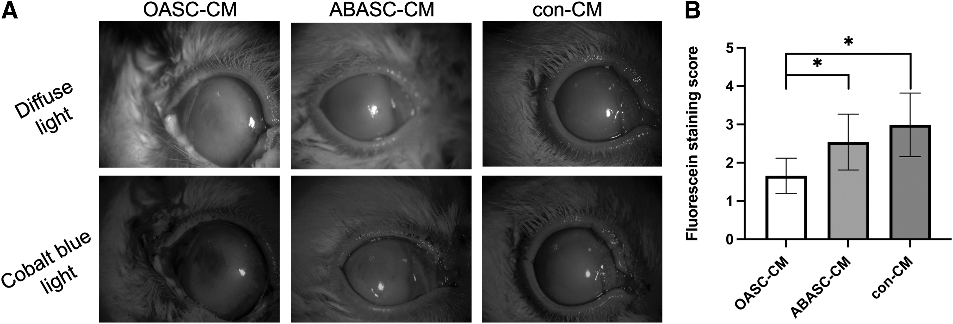
Clinical evaluation by sodium fluorescein staining.
Histological and IHC evaluation
Histological results (Fig. 6A) showed that in the OASC-CM group (n = 3), corneal epithelium showed integrity, and fibrous tissue in the stroma layer was arrayed in order. No obvious inflammatory cell infiltration and vascularization was found. The structure of the elastic layer and endothelial cell layer of the cornea was relatively intact. In the ABASC-CM group (n = 3), corneal epithelium did not show continuity and was partially detached from other layers of the cornea. The structure of the upper half of the stroma layer was loose, while the other half of the stroma layer was arrayed closely. In the con-CM group (n = 3), the posterior elastic layer and the endothelial cell layer were detached. The whole stroma layer was loose, and gaps between fibrous tissues were large. The number and diameter of new capillaries increased in both control groups.
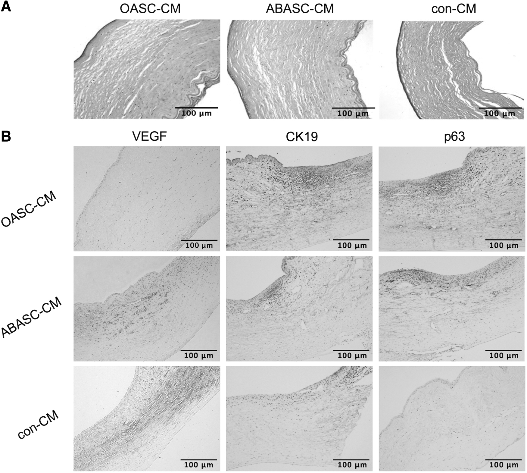
Histological and IHC evaluation.
IHC results (Fig. 6B) showed that in the OASC-CM group (n = 3), the cytoplasm of neovascular endothelial cells and inflammatory cells was stained with a color tan from the center of the cornea to the upper half of the stroma layer of the corneal limbus, showing a slight positivity of VEGF expression, while control groups (n = 3) exhibited a strong positivity. Besides, increased expressions of CK19 and p63 in the OASC-CM group were observed.
Cytokine evaluation
Both anti-inflammatory and proinflammatory cytokines were measured by the concentrations of protein per sample using ELISA. In the OASC-CM group, the mean CD45 (0.41 ± 0.30 pg/mL), IFN-γ (9.36 ± 8.29 pg/mL), and TNF-α (97.95 ± 85.19 pg/mL) levels were significantly lower than the other two groups, while the mean concentration of inflammatory inhibitory factor IL-10 (240.32 ± 210.05 pg/mL) was higher (n = 3, P < 0.01; Fig. 7A–D).
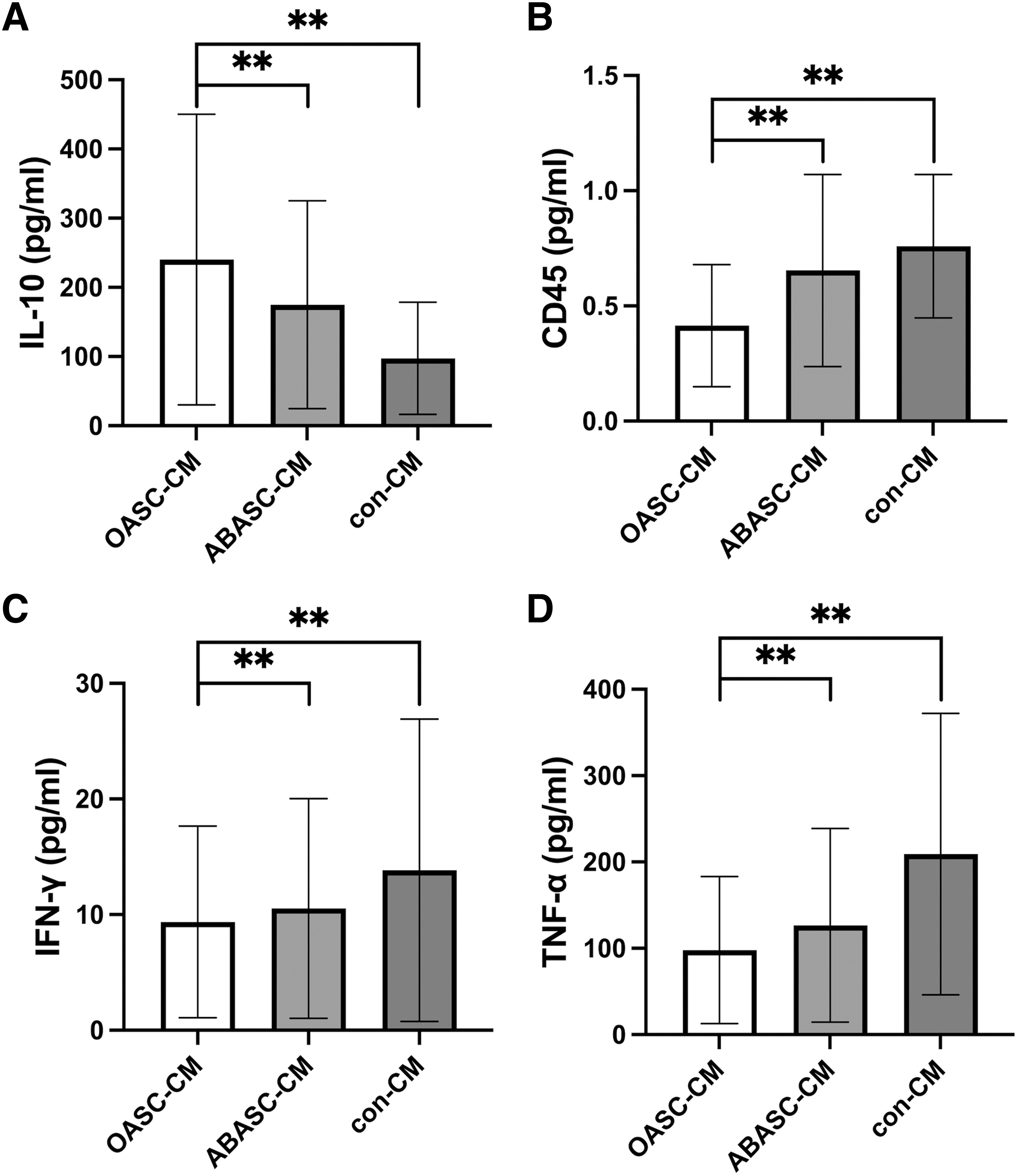
Cytokine evaluation. Levels of IL-10
Discussion
Current treatment methods of corneal alkali burn, due to the immunological and ethical limitations, still need to be improved. 7 This study proved that OASC-CM, as a simple nonsurgical and rejection-free method, could treat corneal alkali-induced injuries in a rabbit model. OASC-CM improved corneal transparency and regeneration of the alkali-burned cornea by inhibiting neovascularization, conjunctivalization, and immuno-inflammatory reaction.
Corneal epithelium plays a significant role in corneal transparency, 18 and precise arrangement of corneal stroma also contributes to optical clarity.19,20 Damage to any corneal element will result in a loss of corneal transparency; on the contrary, improvements on these elements can promote recovery of corneal clarity.19,20 Our histological and IHC evaluation results (Fig. 6) directly showed that OASC-CM promoted reepithelialization of corneal epithelium and rearrangement of stroma after alkali burns, which could improve corneal transparency.
In addition, the expression levels of CK19, a cornea-specific epithelial marker, 21 and p63, a proliferative corneal epithelial marker, 22 were significantly elevated after OASC-CM treatment (Fig. 6B), indicating its effects on corneal epithelium regeneration from the aspect of molecular biological mechanism. Furthermore, sodium fluorescein staining results (Fig. 5) visually and quantitatively showed that OASC-CM promoted corneal epithelium regeneration. Finally, our gross observation (Fig. 4A, B) confirmed the most opacity and edema regression in the OASC-CM group. Lin et al. 23 reported that eye drops containing OASCs could promote corneal epithelialization after alkali-induced burns. Compared to using OASCs directly, we found OASC-CM also had similar effects on corneal epithelium regeneration, but with elimination of immunological and ethical issues.
Expansion of vascular capillaries inside and into formerly avascular parts of the cornea is known as CNV, which can cause corneal edema, lipid deposition, fibrosis, and finally visual impairment. 24 VEGF plays a crucial role in this process. 25 As we found, expression of VEGF was significantly reduced in the OASC-CM group (Fig. 6B), leading to a significantly inhibited CNV clinically (Fig. 4C).
Inflammatory cells will be attracted to wounded regions to clear components released from damaged keratocytes, which is crucial to wound healing. However, excessive inflammatory infiltration decelerates the healing process. 26 As shown in our histological and IHC results (Fig. 6), OASC-CM reduced inflammation significantly. Mechanistically, inflammatory cytokines mediate immunological changes after corneal alkali burns, and various molecules, such as CD45, IFN-γ, CXC chemokines, IL-1, and TNF-α, participate in early immune response and affect corneal wound healing negatively.27–30
Overexpression of these inflammatory factors promotes the migration of inflammatory cells to the wound, induces the accumulation of collagen, and then leads to scar fibrosis. 28 In this study, significantly lower protein levels of CD45, IFN-γ, and TNF-α in the experimental group showed the anti-inflammatory capacity of OASC-CM (Fig. 7B–D). CD45 is also a fibrocyte marker indicating fibrocyte infiltration into the corneal stroma, 31 so we assumed that OASC-CM helped suppress corneal fibrosis formation by reducing the secretion of CD45. IL-10 is an anti-inflammatory factor that increases the wound healing rate in a corneal wound healing model in vitro. 32 The decreased IL-10 levels after OASC-CM treatment confirmed its effects on inflammatory inhibition (Fig. 7A).
In this study, OASC-CM showed superiority over ABASC-CM in clinical, histological, IHC, and cytokine evaluations. Studies have shown biological differences among ADSCs from different anatomic locations on differentiating potential, stem cell surface markers, and cytokine secretion. 33 Embryologically, adipose tissues of the eyelid originate from neural crest, 9 while most of the adipose tissues, including abdominal subcutaneous fat, derive from mesoderm. 34 Based on our hypothetical connection between embryological origins and biological characteristics, we proved OASCs—which have the same embryological origin with most of the ocular and orbital tissues—were more effective than ABASCs in repairing corneal defects. Our origin-dependent hypothesis on ADSC therapy has been confirmed in other studies: In a retinal lineage therapy, OASCs also showed better effects on the regeneration of retinal pigment epithelium compared to ABASCs. 35 In a cardiomyogenic therapy, epicardial ADSCs revealed a higher cardiomyogenic potential compared with omental ADSCs. 36
ADSC-CM eliminates immunological and ethical limitations of stem cell therapy. However, few studies proved its treatment effects on corneal wound healing treatment, except ours and another one. 37 What make ours unique are as follows: (1) We used ADSC-CM from OF rather than from epididymal and subcutaneous fat in that study. More innovatively, based on our origin-dependent hypothesis, we proved its better treatment effects than those of ABASC-CM. (2) Instead of harvesting fat tissue from rats, we used human adipose tissue from cosmetic surgeries, whose results were more persuasive for future clinical use. (3) We used alkali, but not 100% alcohol, to build a corneal wound model, which was more destructive and fatal in corneal chemical burns. To our knowledge, this study is not only the first to demonstrate the significant treatment effects of OASC-CM on corneal alkali-induced injuries but also the first to compare them with those of ABASC-CM.
Our study also had some limitations: (1) The immunosuppression mechanism mediated by OASC-CM has not been fully explained, since only four inflammatory factors responded to OASC-CM in corneal tissue, as was detected in this study. (2) Further investigation of potential functional molecules to treat corneal alkali burns in OASC-CM is needed. (3) The mechanism resulting in the different treatment effects between OASC-CM and ABASC-CM should be demonstrated in the future. (4) To achieve better treatment effects, the most optimal concentration of OASC-CM needs to be investigated. (5) Despite referring to the designs of other studies about corneal wound healing to set our sample size,35,38–41 a larger scale study may still be needed to add more statistical robustness to our findings.
Still, we demonstrated that OASC-CM could reduce the CD45, IFN-γ, and TNF-α levels, and increase the IL-10 expression level to regulate inflammatory reactions in the alkali-burned cornea. Through inhibition of CNV, conjunctivalization, and inflammation, OASC-CM promoted corneal tissue regeneration and rearrangement, showing significant treatment effects on corneal alkali burns. OASC-CM could be applied more widely for corneal wound healing and even for other tissue regeneration in the future.
Footnotes
Acknowledgment
The authors are grateful to Dr. Caihe Liao and Dr. Xiaoqiang Liu for their technical support.
Authors' Contributions
Y.C. designed and conducted the whole experiments, collected relevant data, as well as wrote the first draft of the article. G.L. analyzed and interpreted the data, and modified and confirmed the final version. Both authors read and approved the final article.
Author Disclosure Statement
No competing financial interests exist.
Funding Information
This work was supported financially by grants from the National Natural Science Foundation of China (Grant No. 31870974 and 51873101).