




Abstract
Purpose:
This study aimed to investigate the relationship between diclofenac sodium ophthalmic solution (DFNa) and corneal epithelial cell damage and to evaluate the preventive effect of rebamipide (RBM) on it.
Methods:
DFNa, DFNa/preservative-free (PF), or 0.5% chlorobutanol (CB) solution was instilled into the conjunctival sac of a normal rabbit eye, and corneal resistance measurement (using a corneal resistance device [CRD]) was performed 120 min after the end of instillation. Then, fluorescent staining (FL), corneal tissue staining (hematoxylin and eosin [H&E]), and immunostaining (zona occlusion-1) were performed (RBM-untreated group). However, RBM was instilled into the eyes of another group of normal rabbits, followed by each of the solutions; 120 min after the end of instillation, all evaluations were performed for this group (RBM treatment group).
Results:
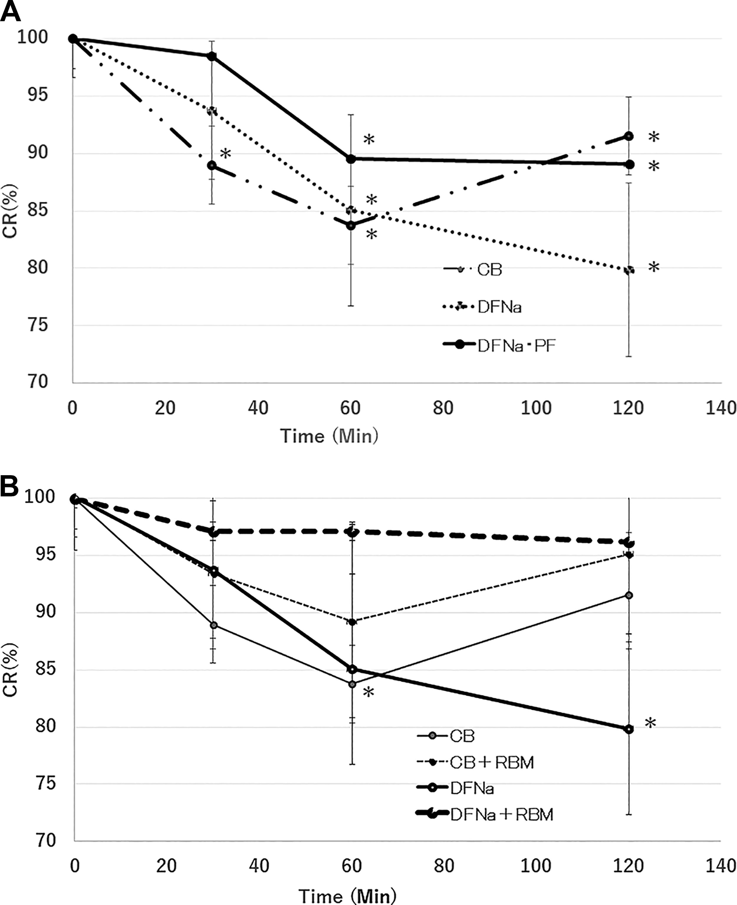
Using the CRD method, in the RBM-untreated group, corneal resistance (CR; %) was found to be significantly reduced in DFNa (79.9 ± 19.4%), DFNa/PF (89.1 ± 17.3%), and 0.5% CB (83.8 ± 10.6%). In addition, DFNa and 0.5% CB solutions showed positive staining in the FL staining method. In the H&E staining method, some clear voids were observed in the outermost layer of the cornea using DFNa and 0.5% CB solutions. However, corneal epithelial damage was suppressed in the RBM treatment group. ZO-1 immunostaining in DFNa and 0.5% CB solutions revealed discontinuous localization of ZO-1 at the cell periphery.
Conclusions:
RBM eye drops were effective in preventing corneal epithelial damage caused by DFNa eye drops, and CB was considered to be the main causative agent of this damage.
Introduction
The main pharmacological actions of nonsteroidal anti-inflammatory drugs (NSAIDs) are diverse and include anti-inflammatory, analgesic, and antipyretic actions. Generally, NSAIDs inhibit cyclooxygenase, thereby decreasing peripheral and central prostaglandin production1,2 and making them ideal agents for avoiding steroid-induced side effects after ophthalmic surgery. Diclofenac sodium (DFNa) is an anti-inflammatory analgesic derived from phenylacetic acid. As an oral drug, it has several antipyretic and analgesic effects in rheumatoid arthritis and osteoarthritis. 3 DFNa eye drops were first released in 1989 and are primarily used to prevent postoperative inflammatory symptoms during cataract surgery. However, the onset of corneal epithelial damage caused by this drug is regarded as an issue.4–6
Previous clinical reports7,8 have confirmed the development of adverse side effects, such as impaired corneal sensation, persistent epithelial defects, superficial punctate keratitis, corneal infiltration, and corneal lysis. In addition, some basic studies have confirmed that DFNa causes delays in corneal epithelial healing. 9 The authors investigated corneal epithelial damage in cultured rabbit corneal cells. 10 Herein, we investigated the effects on corneal epithelial cells by dividing the main agent (DFNa) and preservative chlorobutanol (CB) of the ophthalmic solution to elucidate the factors leading to corneal epithelial damage induced by DFNa ophthalmic solution. The preventive effect of rebamipide (RBM) on disability was evaluated in vivo.
Materials
Animals
Adult male albino New Zealand white rabbits (n = 15; body weight: 3.0–3.5 kg) were purchased from Sankyo Labo Service Co., Inc. (Toyama, Japan), and both eyes of each rabbit were included in the study. Animals were kept under a natural light–dark cycle in cages with ad libitum access to food and water in a room maintained at 23°C. This study was conducted in compliance with the standards of the Institutional Review Board of Kanazawa Medical University on the Use of Animals (protocol number: 2020-50) and the Association for Research in Vision and Ophthalmology (ARVO) statement on the Use of Animals in Ophthalmic and Vision Research. Somnolent intravenous injection of sodium pentobarbital at 64.8 mg/kg body weight (Kyoritsu Seiyaku Corporation, Tokyo, Japan) was used for euthanizing the animals in all subgroups.
Reagents
Ophthalmic Solution: Ophthalmic solution containing 0.1% diclofenac sodium (Wakamoto Pharmaceutical Co., Ltd., Tokyo, Japan; hereinafter referred to as “DFNa”): 60 μL per one droplet. Diclod ophthalmic solution 0.1% (original drug). Diclofenac sodium ophthalmic solution (0.1%; DFNa/preservative-free [PF]; Nitten Co. Ltd., Nagoya, Japan); hereinafter referred to as “DFNa/PF”): 60 μL per droplet. Diclofenac sodium PF ophthalmic solution 0.1% (Nitten; generic drugs) (Table 1). A 2% rebamipide ophthalmic solution (Mucosta® ophthalmic suspension UD 2%; Otsuka Pharmaceutical Co., Ltd., Tokyo, Japan; hereinafter referred to as “RBM”): 60 μL per droplet. Chlorobutanol (hereinafter referred to as “CB”; FUJIFILM Wako Pure Chemical Co., Ltd. Tokyo, Japan) adjusted to 0.5% with a normal saline solution. Mouse monoclonal anti-ZO-1 antibody (Invitrogen 33-9100; 1:100 dilution; Invitrogen ZO-1 Monoclonal Antibody) ZO1-1A12 (Invitrogen Co., Ltd.). Alexa 488 Goat anti-Mouse IgG antibody (Invitrogen A11001; 1:500 dilution; Alexa Fluor™ 488 Goat anti-Mouse IgG [H+L] Cross-Adsorbed Secondary Antibody; Invitrogen Co., Ltd.).
Comparison of the Original and Generic Drugs in Terms of Composition and Additives
Corneal epithelial injury assessment methods
Fluorescein staining method (120 min after using the eye drops)
Fluorescein (FL) staining was used to assess corneal epithelial injury using a Zeiss slit-lamp microscope 0320 w25 (Carl Zeiss Co., Ltd., Tokyo, Japan), with the degree of superficial punctate keratopathy (SPK) classified according to the system devised by Miyata et al. 10 Briefly, after FL corneal staining, the sum of the SPK area was graded from A0 to A3 and the density was graded from D0 to D3 according to the area and density (AD) classification. At baseline, the absence (A0D0) of corneal epithelial injury was confirmed in all rabbit eyes before the animals were assigned to treatment groups.
Corneal resistance device
Corneal resistance (CR) was used to quantify corneal epithelial injury measured using a newly improved electrical CRD (Fukuda Model 2015), which incorporates a vaginal impedance reader (Model MK-1°C; Muromachi Kikai Co., Ltd., Osaka, Japan) and corneal contact lens electrode (Mayo Corporation, Aichi, Japan).8,11–13 The ratio of posttreatment CR to the baseline CR was defined as the CR ratio (%) and was measured using CRD immediately before and 10, 30, and 60 min after the initial administration. CR ratio (%) = CR after administration × 100/baseline CR before initial administration.
Histological examination
Corneal tissue isolated from the harvested eye was fixed for 24 h in 4% paraformaldehyde, embedded in paraffin, and sectioned at approximately 4 mm. Paraffin-embedded sections were stained with hematoxylin and eosin (H&E) and processed for histopathological examination.
Immunofluorescence analysis
The corneal tissue from each group was collected 120 min after completing instillation and fixed with 4% PFA for 5 min. This was followed by a 5-min treatment with cold acetone, followed by three 5-min permeabilizations with 1% Triton X-100 and 1% DMSO in PBS (TD buffer). After treatment with 5% normal goat serum for 1 h, treatment with the primary antibody was performed overnight at 4°C. Thereafter, the corneal tissue was treated with a secondary antibody at room temperature for 2 h, washed, cut into four sections, and stretched. The samples were mounted with a fluorescent antifading agent, Fluor Keeper (Nacalai Tesque), and images were acquired and evaluated using a confocal microscope (ZEISS LSM710).
Statistical analysis
Quantitative data among the treatment groups were analyzed using Student’s t-test, with results presented as means ± standard deviation (SD) and any between-group differences calculated. Statistical significance was set at P < 0.05.
Methods
All rabbits used were subjected to a rabbit eye FL test and a CR measurement test before initiating the experiment to confirm no abnormalities and were subsequently used for the experiment.
Experiment 1: Evaluating corneal epithelial cell damage caused by DFNa, DFNa/PF, and 0.5% CB
In the RBM-untreated group, DFNa, DFNa/PF, or CB solution (containing 0.5%) was instilled into rabbits 10 times every 5 min, and the FL staining test was performed 120 min after completing the instillation. The CR value was measured 30 min after completing the instillation. The evaluations were made after 120 min. In addition, the H&E staining of the corneal tissue was performed 120 min after completing the instillation. Immunostaining was performed using a ZO-1 antibody 120 min after completing instillation (RBM-untreated group).
Experiment 2: Preventive effect on RBM against corneal epithelial cell damage by DFNa and 0.5% CB
After instilling RBM or saline into the normal rabbit eye once, 10 instillations of DFNa or CB solution were performed every 5 min. FL staining test was performed 120 min after completing the instillation, and the CR value was measured from 30 min to 120 min after completing the instillation. In addition, the corneal tissues were subjected to H&E staining 120 min after completing the instillation. Immunostaining was performed with the ZO-1 antibody 120 min after completing the instillation (RBM-treated group).
Results
Using the CRD method, the CR (%) after 120 min was significantly reduced to 79.9 ± 19.4% for DFNa, 89.1 ± 17.3% for DFNa/PF, and 83.8 ± 10.6% for the 0.5% CB solution (RBM-untreated group) (P < 0.05) (Fig. 1-a). Contrastingly, DFNa increased to 96.2 ± 3.3% in the RBM-treated group, which was significantly higher than that in the RBM-untreated group (P < 0.05) (Fig. 1-b).
In FL staining, both DFNa and 0.5% CB solutions showed corneal epithelial damage in the RBM nontreated group. The AD classification for participants in both groups was A2D2. However, in the RBM treatment group, corneal epithelial damage was reduced to A0D0 or A1D1 (after 120 min) (Fig. 2). In H&E staining, several clear voids were observed in the outermost superficial cells of the cornea in the DFNa, RBM-untreated, and 0.5% CB solution groups; however, corneal epithelial damage was markedly suppressed in the DFNa/RBM-treated group (after 120 min) (Fig. 3).
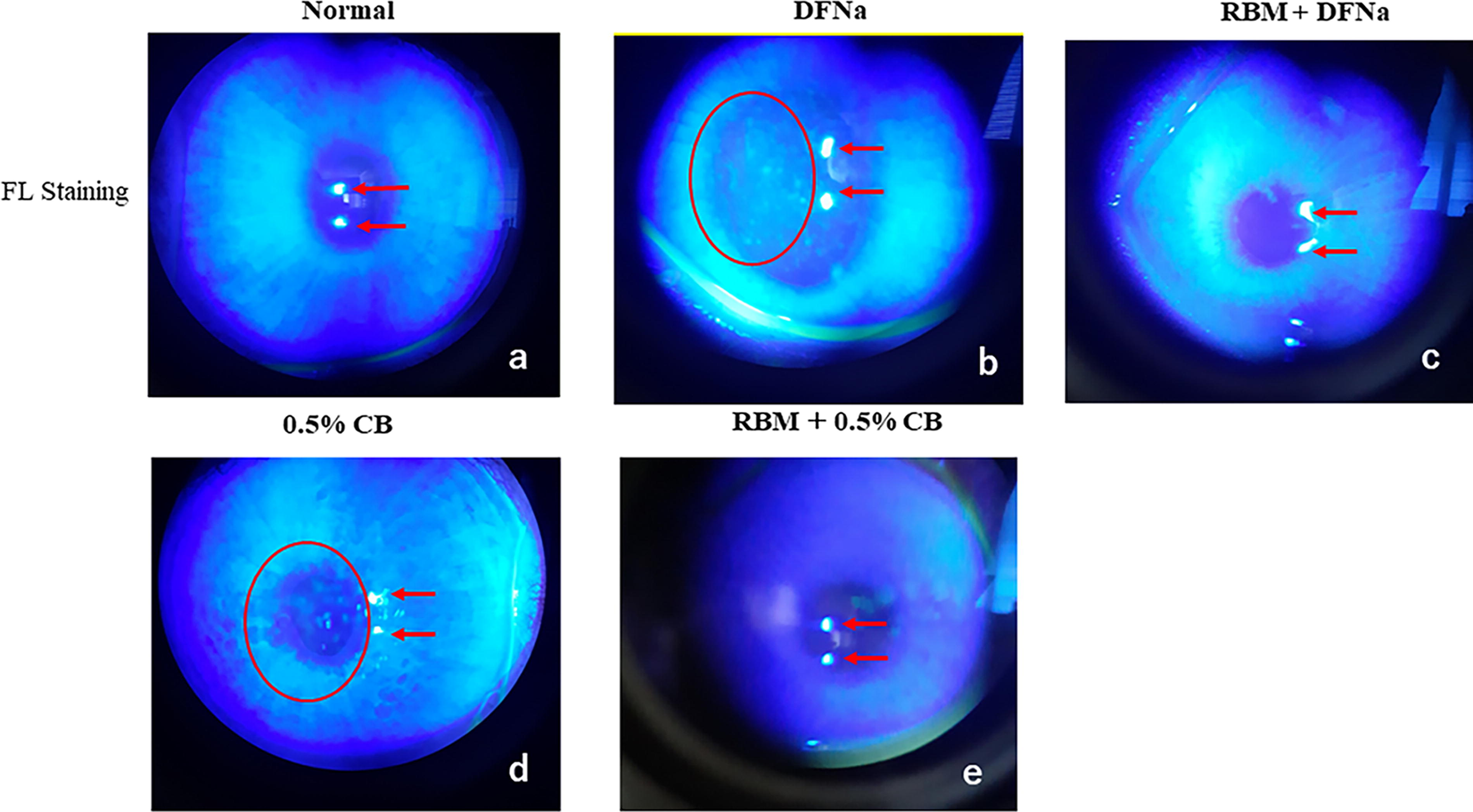
FL staining-based evaluation of corneal epithelial damage by DFNa and 0.5% CB solutions:

Hematoxylin and eosin staining-based evaluation of corneal epithelial damage by DFNa and 0.5% CB:
Immunostaining showed that in the RBM-untreated group containing DFNa and 0.5% CB solutions, the localization of ZO-1—a tight-junction constituent protein in the corneal epithelium, was discontinuous at the cell periphery. In contrast, with the use of the DFNa solution, ZO-1 localization in the RBM treatment group was not absent at the cell periphery. No obvious RBM effect was observed in the 0.5% CB solution. In the DFNa group, no lack of ZO-1 localization was observed at the cell periphery; however, in the 0.5% CB solution group, ZO-1 localization was discontinuous at the cell periphery after 120 min (Fig. 4) (Table 2).

Evaluation of the corneal epithelial damage by 0.5% CB using confocal laser microscope findings:
Results of Each Evaluation Method (120 m). Suppressive Effect of Rebamipide (RBM) Installation on Corneal Epithelial Damage
In each evaluation method, corneal epithelial damage was significantly reduced in the RBM group compared with the No RBM group.
Discussion
In this study, we investigated the effects of DFNa on the corneal epithelial cells by dividing the main agent (DFNa) and preservative (CB) of the ophthalmic solution to clarify the cause of corneal epithelial damage induced by the DFNa ophthalmic solution. Corneal epithelial damage caused by DFNa was confirmed, and the main cause was considered to be the preservative CB. In addition, corneal epithelial damage was confirmed, even with CB-free DFNa/PF. Our results are concordant with that of DFNa damage to the corneal epithelium obtained from cultured corneal cell lines (in vitro). 10
In this study, we assessed the damage to the corneal epithelium using a CRD in vivo.11–16 The principle of the CRD method is as follows: the cell membrane has high electrical resistance, the current applied between the tissues at a low frequency of 1,000 Hz mainly passes through the extracellular fluid, and the corneal epithelium has tight junctions (TJs), creating electrical resistance. If the corneal epithelium is physically or chemically damaged, these structures break, the flow of electrons through the cornea increases, and the electrical resistance of the cornea decreases depending on the degree of damage.
Contrastingly, the RBM ophthalmic solution markedly inhibited corneal epithelial damage caused by DFNa, which was confirmed for the first time in living eyes. In this study, we confirmed that the control eyes exhibited a continuous linear pattern of TJs protein (ZO-1) staining at the cell–cell boundaries on the surface of the rabbit corneal epithelium in vivo, whereas eyes treated with DFNa eye drops and antiseptic eyes treated with an agent (CB) exhibited discontinuous ZO-1 immunoreactivity and disrupted TJs. ZO-1 is expressed in the superficial cell layer of the corneal epithelium, and RBM’s mechanism of action was considered to be the suppression of the decrease in the TJ-constituting protein ZO-1 expression in the corneal epithelium. Benzalkonium chloride (BAK) is commonly used as a preservative in several eye drops; however, eye drops containing BAK have recently been reported to redistribute ZO-1 and disrupt the barrier function of the corneal epithelium. 17 RBM increased the transepithelial electrical resistance (TER) of the human corneal epithelium cells in a concentration-dependent manner and attenuated the loss of TER and ZO-1 from the cell surface induced by tumor necrosis factor-α. 18 The CB solution examined this time has long been used as a preservative for eye drops, and its safety has been questioned. In this study, the CRD method was used to quantitatively confirm the decrease in corneal barrier function caused by the DFNa ophthalmic solution. In addition, immunostaining confirmed a reduction in TJ by the ZO-1 protein.
DFNa eye drops contain 0.5% CB, and corneal epithelial disorders are observed more strongly than with generic DFNa eye drops containing 0.3% CB. In addition, corneal epithelial damage caused by the CB solution in DFNa eye drops is due to the CB concentration used and the CB and DFNa combination. 10 The results of the present study are consistent with the results of previous studies on DFNa toxicity.19–22 This study revealed that the CB in DFNa eye drops is involved in the development of corneal epithelial disorders. In addition, we confirmed for the first time that RBM suppressed DFNa-induced corneal epithelial damage in living eyes.
Conclusion
In this study, we showed that DFNa eye drops can cause corneal epithelial damage, and the main causal factor for this damage is CB. Our results indicate that RBM eye drops can prevent the corneal epithelial damage caused by DFNa eye drops.
Footnotes
Authors’ Contributions
M.F.: Responsible for overall research and article writing. T.K. and N.M.: Responsible for ZO-1 immunostaining. S.T. and Y.S.: Responsible for FL staining and CRD method experiments. E.K.: Responsible for corneal tissue specimen preparation. H.S.: Responsible for research supervision and article writing. Y.K.: Support in article writing.
Author Disclosure Statement
No competing financial interests exist.
Funding Information
No funding was received for this article.