




Abstract
Purpose:
Leber congenital amaurosis (LCA) is a sight-threatening inherited retinal disorder (IRD) caused by numerous genetic mutations. Multi-characteristic opsin (MCO)-based optogenetic therapy allows the recruitment of residual cells of the retina in LCA for alternative vision transduction while being mutation-agnostic. Using rd12 mice, we investigated the in vivo efficacy of an adeno-associated virus2 (AAV2)-transduced ambient light-activatable MCO (MCO-010) containing a metabotropic glutamate receptor-6 bipolar cell-specific promoter/enhancer.
Methods:
Mice requiring > 40 s to reach and board a dimly lit hidden platform in a water-maze were selected and randomly divided into 2 cohorts. These mice were intravitreally (IVT) injected with either 1.7E9 gene copies/eye of MCO-010 or control AAV2 and re-tested in the water-maze. Spectral-domain optical coherence tomography (SD-OCT), hematoxylin and eosin staining of retinas, and electroretinographic (ERG) studies were also conducted.
Results:
Safety of MCO-010 in rd12 mice was confirmed by the lack of significant detrimental changes in the mouse behavior, b-wave amplitudes and in retinal thickness. rd12 control mice performed relatively poorly in the water-maze test requiring ≥ 30–60 s to find and board the platform. MCO-010-treated rd12 mice reached the platform much faster than the AAV2-treated rd12 mice, with some mice only requiring < 5 s to achieve this goal (P < 0.01–0.0024).
Conclusions:
IVT MCO-010 treatment was well tolerated by rd12 mice, and it prevented the decrease in retinal thickness, and preserved ERG parameters. It also significantly improved the vision in rd12 mice relative to control AAV2-injected mice. MCO-010 therefore represents a novel and efficacious optogenetic therapeutic to treat LCA and other IRDs irrespective of the genetic defect(s).
Introduction
Inherited retinal diseases (IRDs) are a family of rare retinal dystrophies that cause severe vision loss and subsequent blindness in children and adults due to over 300 genetic mutations.1–3 Leber congenital amaurosis (LCA) is one such genetic disorder whose pathology is linked to at least 28 different defective genes, appearing at birth or in the first few months of life, globally affecting approximately 200,000 newborns.4–6 Patients display nystagmus, strabismus, and photophobia, and their visual acuity is severely reduced. Among the gene variants that cause LCA2 about 16% of the cases involve genetic defects in the retinal pigment epithelium-65 (RPE65) gene which produces a dysfunctional enzyme in the vision cascade and thus induces visual impairment.4–6 Additionally, ≤ 25% of patients with LCA do not possess any of the known LCA-causing pathogenic gene variants, and the etiology of their disease remains unknown and requires suitable therapeutic intervention.4–6 RPE65 gene encodes a critical retinoid isomerase which converts vitamin A to the 11-cis-retinal chromophore visual pigment which is critical for visual signal transduction. Whilst the initial defects and demise of RPE cells occur first in LCA, the associated rod and cone photoreceptors are subsequently destroyed due to their inactivity and lack of support from the RPE cells.4–6 However, higher-order retinal neurons [e.g., bipolar cells and retinal ganglion cells (RGCs)] are spared in many IRDs including LCA.7–11
Although voretigene neparvovec-rzyl (Luxturna®) gene replacement therapy directed to a single gene defect in photoreceptors partially reverses the RPE/photoreceptor dysfunction in dogs and human subjects to partially restore vision,12,13 photoreceptor loss continues unabated in both species. 4 Additionally, this single gene replacement therapy is not durable beyond a few early years posttreatment, and as with most gene replacement therapies for IRDs only a single gene is targeted allowing other defective genes to potentially cause relapse of the disease.4,13–15 Hence, there continues to be an unmet medical need for alternative long-lasting treatment modalities for various IRDs including LCA. Optogenetic gene therapy is a novel platform technology that has shown promise in restoring vision across multiple animal models (e.g., mice, rats, and dogs) of retinitis pigmentosa (RP)16–20 and in human subjects with the same disease.21–23 Use of multi-characteristic opsin (MCO)-based optogenetic therapy aims to provide light-sensitive proteins to the remaining viable cells of the retina, for example the preserved and functional ON bipolar cells11,13,24 for alternative vision transduction while being mutation-agnostic and not requiring presence of outer retinal cells. 22 Furthermore, the expression of the novel trimeric genetic construct in the bipolar cells using a bioengineered adeno-associated virus-2 (AAV2) vector carrying a multi-characteristic opsin (MCO-010),18,19,22 under the control of a metabotropic glutamate receptor-6 promoter/enhancer,25,26 can slow down the degeneration of retinal cells in mice with RP-like pathology.18,19,27–29
In the current studies, we explored the potential utility of MCO-010 to ameliorate the visual deficits in a mouse model of LCA which does not require the presence of RPE or photoreceptor cells. 30 It was anticipated that MCO-010 provision would effectively convert ON bipolar cells to become light-sensitive and thus confer vision by activating the associated RGCs that project axons to the visual centers in the brain. Thus, using an animal model of LCA (B6(A)-Rpe65rd12/J mice [rd12 mice]), 30 we investigated the ability of MCO-010 to influence retinal structure and function utilizing a variety of methods and technologies.18,19,31 The results from these studies were recently presented at the Association for Research in Vision and Ophthalmology (ARVO). 32
Methods
Ethics statement
All experimental procedures were conducted according to the Association for Research in Vision and Ophthalmology (ARVO) and the Nanoscope Technologies-Institutional Animal Care and Use Committee approved protocol.
Mouse model of LCA
Retinal degenerated (rd12) mice (B6(A)-Rpe65rd12/J) have a spontaneous missense point mutation in RPE65 gene which leads to RPE cell degeneration followed by photoreceptor death in a similar manner to the human LCA disease.14,30 These rd12 mice have retinal degeneration due to the production of a dysfunctional retinoid isomerase enzyme that causes complete blindness by 7 months of age. RPE65 (rd12) mice [4–5 months old; Stock No: 005379 B6(A)-Rpe65rd12/J; Jackson Laboratories, Bar Harbor, MA] were used in the current studies. Mice were maintained on a 12-hour light on/off cycle and were provided food and water ad libitum.
MCO-010 production and dose selection
The MCO-010 gene was synthesized using a DNA synthesizer and the sequence was verified. Synthesized plasmid (MCO-010) was cloned into pAAV MCS vector via its BamH1 and Sal1 sites. DNA gel electrophoresis was performed to verify the size and purity of MCO-010 gene (digested by restriction enzymes BamH I and Sal I with restriction fragments). AAV2 physical titers were obtained by quantitative polymerase chain reaction using primers designed to selectively bind AAV inverted terminal repeats. Purification of MCO-010 was carried out by Benzonase-treatment to reduce the size of host cell DNA (HCD) and host cell protein (HCP) removal. An SDS PAGE was used to verify the purity of the viral vector.
MCO-010 has been successfully utilized over a range of intravitreally-delivered doses [e.g., 3.4E6-3.4E9 gene copies (gc)/eye] in mouse models of RP.18,19 We felt confident that choosing a similar ocular dose for the rd12 mice would provide the necessary gene product in vivo in the LCA disorder-bearing mice. Thus, in the current studies, rd12 mice were intravitreally injected with either an AAV2-control vector or MCO-010 vector (1.7 E9 gc/eye; 1.5 µL of 1.1E12 gc/mL solution) in both eyes using a sterile 29-gauge needle of a Hamilton micro-syringe. The efficacy of transduction of the mouse bipolar cells was assessed using the water-maze navigational ability of the control and MCO-010 treated mice (Fig. 1)
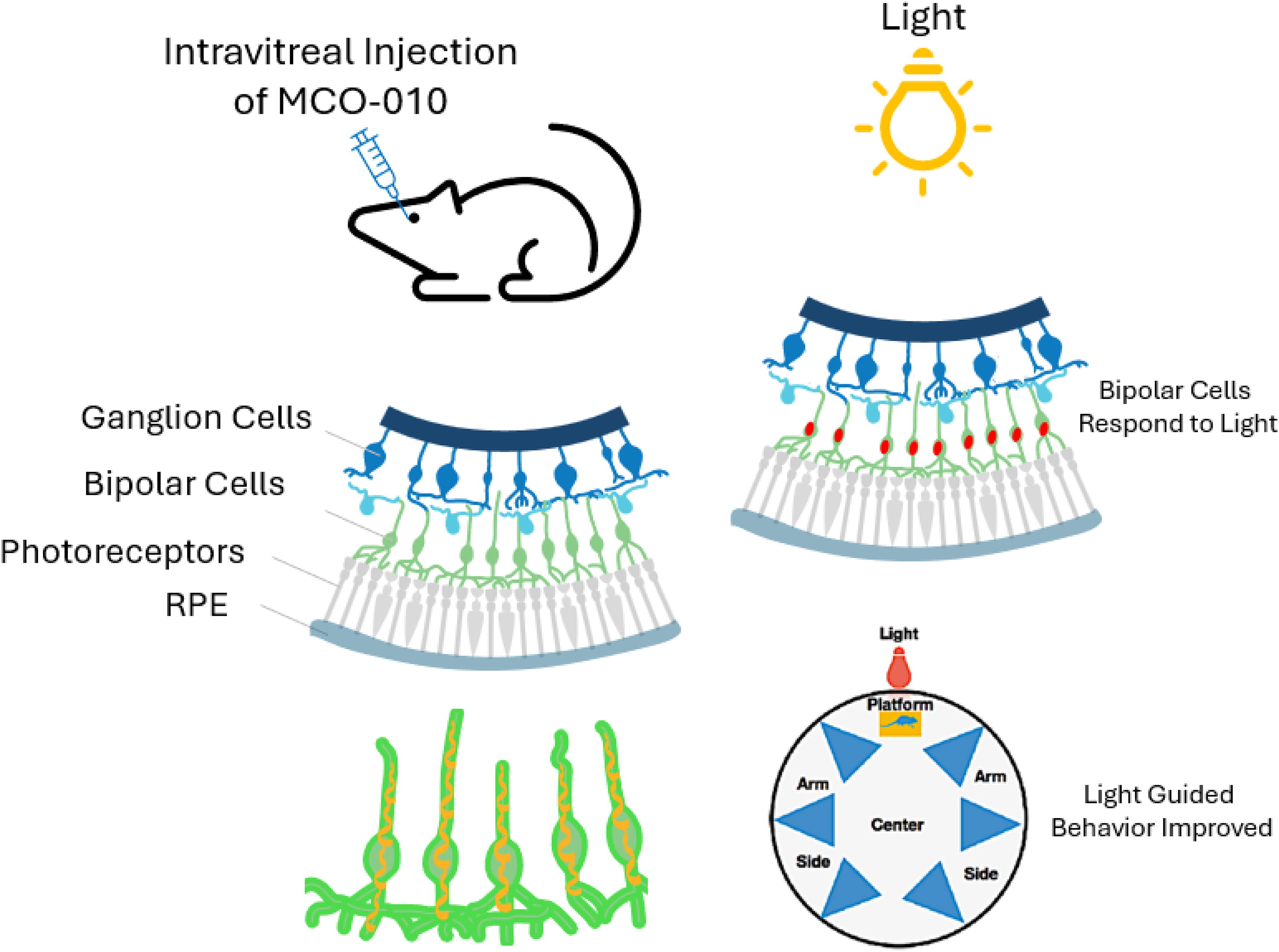
A schematic showing the experimental design of transducing MCO-010 into rd12 mouse bipolar cells to render them light-sensitive (red soma of cells in top right Fig.) in the presence/absence of dysfunctional/degenerated photoreceptors. The transduced bipolar cells (bottom left) are now able to communicate with the RGCs to transmit visual signals down the optic nerve to the visual centers of the brain. The bottom right figure illustrates the water-maze used to assess the visually guided navigational ability of the mice to see the dimly lit platform, and to swim towards it and board it. The faster they find and board the platform the better their vision.
Randomization and masking
Mice were selected randomly, and group number/cage and number/mice were assigned prior to injection based on restricted randomization. The cages were housed in a random order on the shelves and the injections were performed in a random order. To avoid bias, the imaging experiments and analysis were performed by individuals masked to the treatments. After the randomized allocation of animals to the treatments, animals, samples, and treatments remained coded until data analysis.
Optical coherence tomographic imaging
OCT imaging is a standard ophthalmical assessment tool that provides quantitative measurements of the anterior segment and retina instead of subjective evaluation. Optical sectioning/imaging using spectral domain-OCT (SD-OCT) was carried out to monitor any changes in ocular structures due to intravitreal injection of MCO-010 or vehicle AAV2-control. Animals were anesthetized using a mixture of ketamine/xylazine/acepromazine or by connecting to the isoflurane unit. For dilating the pupil, a drop of tropicamide was topically applied onto the ocular surface of the eye. SD-OCT images of the retina after intravitreal MCO-010 injection in the mice were compared with the images before injection. ImageJ software was used to analyze the SD-OCT images as previously described.18,19
Hematoxylin and eosin staining od retinal sections
Standard hematoxylin and eosin (H&E) staining was used to identify different types of cells and tissues of the back of the eye to assess the integrity of the retinal structure in control (AAV2-injected) and MCO-010-injected mice during the studies.
Water maze visually guided behavioral assessment
rd12 mice (4–5 months old; up to 5/group) were tested for their ability to swim, locate, and board a distant ambient white light dimly lit platform once released from the side arms of a water maze (Fig. 1). Those mice that needed > 40 s to reach and board the platform were selected. Each time the mouse arrived at the lit platform; it was left on the platform to recover from swimming for ∼30 s as its reward. Video recording was stopped once the mice found the platform or 60 s after dropping the mice in the water to prevent the mice from getting tired of swimming. Two groups of mice were randomly selected. One group received the intravitreal AAV2-control vector injection, while the second group received the MCO-010 injection. The time taken by the control and MCO-010-injected mice to board the platform was video recorded at 4, 8-, 12-, 16-, and 20-weeks postinjection of AAV2 control vector or MCO-010. Each mouse was tested at least 3 times per time point with the mean value being recorded.18,19
Assessment of ERG response
After overnight dark adaptation of the mice, the mice were anesthetized, the pupil dilated using tropicamide, and measurements were made 10 min after application of tropicamide. Following Diagnosys-Espion 10 Scotopic electroretinographic (ERG) protocol and Colorburst stimulation, ERG responses from mice at baseline and at 4-, 8-, 12-, 16-, and 20-weeks after MCO-010 injection were recorded using the corneal silver electrode. Light flashes were elicited by a white LED stimulator providing stimulus intensity fixed at 7 × 10¹⁴ photons/cm2/s with a stimulus rate of 1 Hz and stimulus duration of 1 ms. Signals were amplified through a Built-In Bias Drive Amplifier, Analog-to-digital converters (ADCs) with a built-in programmable gain amplifier. A high-pass filter at 0.1 Hz and a low-pass filter at 250 Hz with a 60 Hz notch filter. Signal acquisitions were performed at 1 kHz sampling rate. ERG from a sequence of 15 strobe light flashes was averaged to obtain the final waveform. 31 After completion of the experiment, the electrodes from the animal were removed. Then, each mouse was intraperitoneally injected with sterilized saline and placed in a heated pad until it fully recovered. Mouse eyes were kept moist with a hydrating eye ointment.
Data handling and statistics
GraphPad Prism was used to analyze the data. The data were plotted as mean ± S.D. Statistically significant difference analyses were carried out using two-tailed t-tests. P < 0.05 was considered statistically significant.
Results
Safety
None of the mice displayed any untoward behavioral responses whether they received the control AAV2-vector or MCO-010 intravitreal injections, thereby indicating the overall safety of the treatments. Furthermore, the retinal structure and ERGs of the mice were not adversely affected by the treatments as recorded at baseline and at 8 and 16 weeks post intravitreal injections (see below).
Structural changes in retina
Assessment of the structure of the retina by SD-OCT (Fig. 2A and 2B) showed that in the AAV2-injected control mice, the retinal thickness started to decline from 12 to 24 weeks postinjection (Fig. 2C). In contrast, there was no apparent decrease in retinal thickness in mice that received the MCO-010 over the same time period. These results were supported further by the H & E staining of retinas obtained from AAV2-injected and MCO-010-injected rd12 mice (Fig. 2D). These collective data indicated the overall safety of the viral transduction process and that of the MCO-010 transgene product in the recipient retinal cells of these animals.
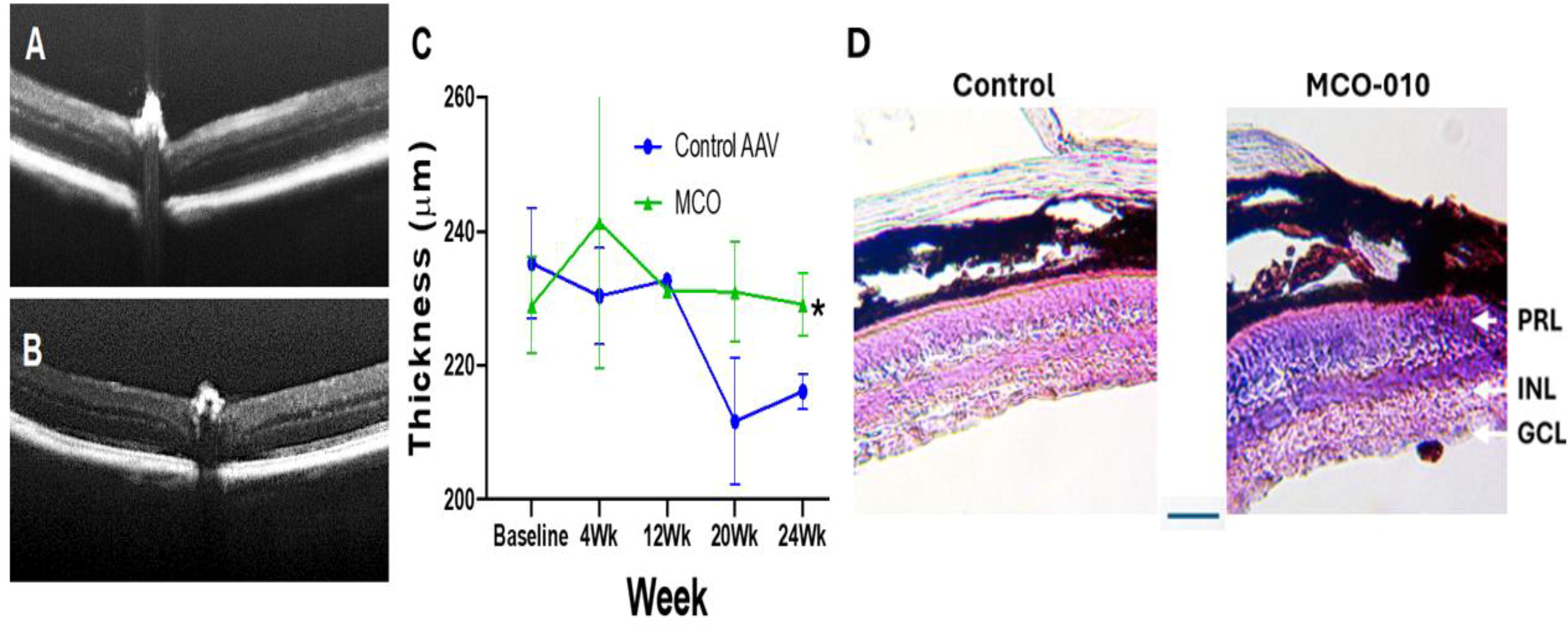
Longitudinal monitoring of retinas of RPE65 rd12 mice with SD-OCT shows stabilization of retinal structure in the MCO-010 injected group. OCT B-scan image
Retinal function assessed by ERG
ERG responses following light stimulation in control AAV2-injected mice did not change from baseline and when assessed at 8-, and 16-weeks postinjection. However, in rd12 mice that received MCO-010, the b-wave amplitude did not change from baseline at 8 weeks but appeared to be slightly elevated at 16-weeks after the MCO-010 injection, but this was not statistically significant compared to controls (Fig. 3).
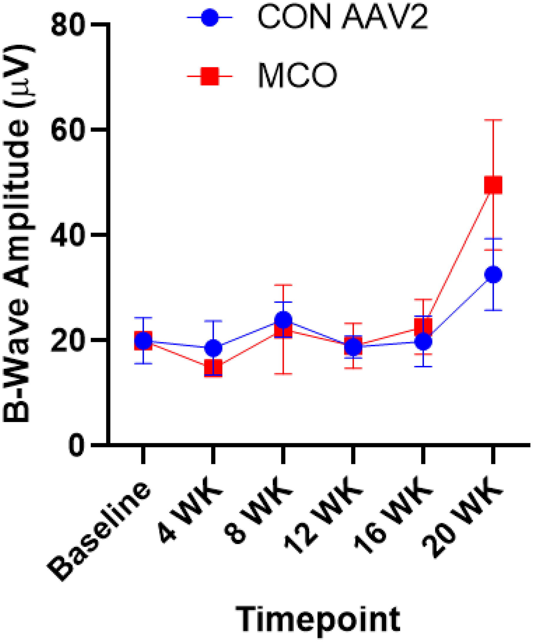
Safety of control AAV2 (CON AAV2) and MCO-010 (MCO) intravitreal injections in retinal function measurements. Essentially, both the control AAV2 (blue line) and MCO-010 (red line) injections helped maintain stable b-wave amplitudes in the rd12 mice postinjection (n = 4/group up to week-16 and n = 3/group after 16 weeks), indicating absence of adverse effects on retinal function. These data were supported by the H & E staining of retinal sections which showed overall similarly maintained retinal structure integrity in both sets of mice, although the AAV2-injected mouse retina was thinner and displayed a somewhat disorganized pattern as compared with the MCO-010-injected retina of another rd12 mouse (Fig. 2D).
Visually guided behavioral assessment
AAV2-control injected rd12 mice required > 30–60 s to reach the dimly lit platform after being released from the side-arm of the water maze (Fig. 4A) from baseline to 20 weeks following the intravitreal injection (Fig. 4B). Although over time, the control mice learned to navigate to the platform faster than at the baseline time-point (red line in Fig. 4B), they were significantly slower than the MCO-010-treated mice at all time periods after 4-weeks up to 16-weeks postinjection (P < 0.01) (Figs. 4B-4D). Specifically, Figures 4C and 4D show the times the two groups of mice needed to find and board the platform 4-weeks after receiving the control AAV2 vector or MCO-010 (**P < 0.01; n = 4/group). Additionally, the MCO-010-treated rd12 mice also exhibited improved vision since they also quickly found and boarded the platform much faster than their AAV2-injected counterparts (P < 0.0024, n = 4/group), with some mice only requiring 4 s to board the platform. Videos demonstrating such improved visually guided behavior of the MCO-010-treated mice, and the AAV-injected mice are presented in the Supplementary Data S1 of this paper and are available to reviewers and readers.
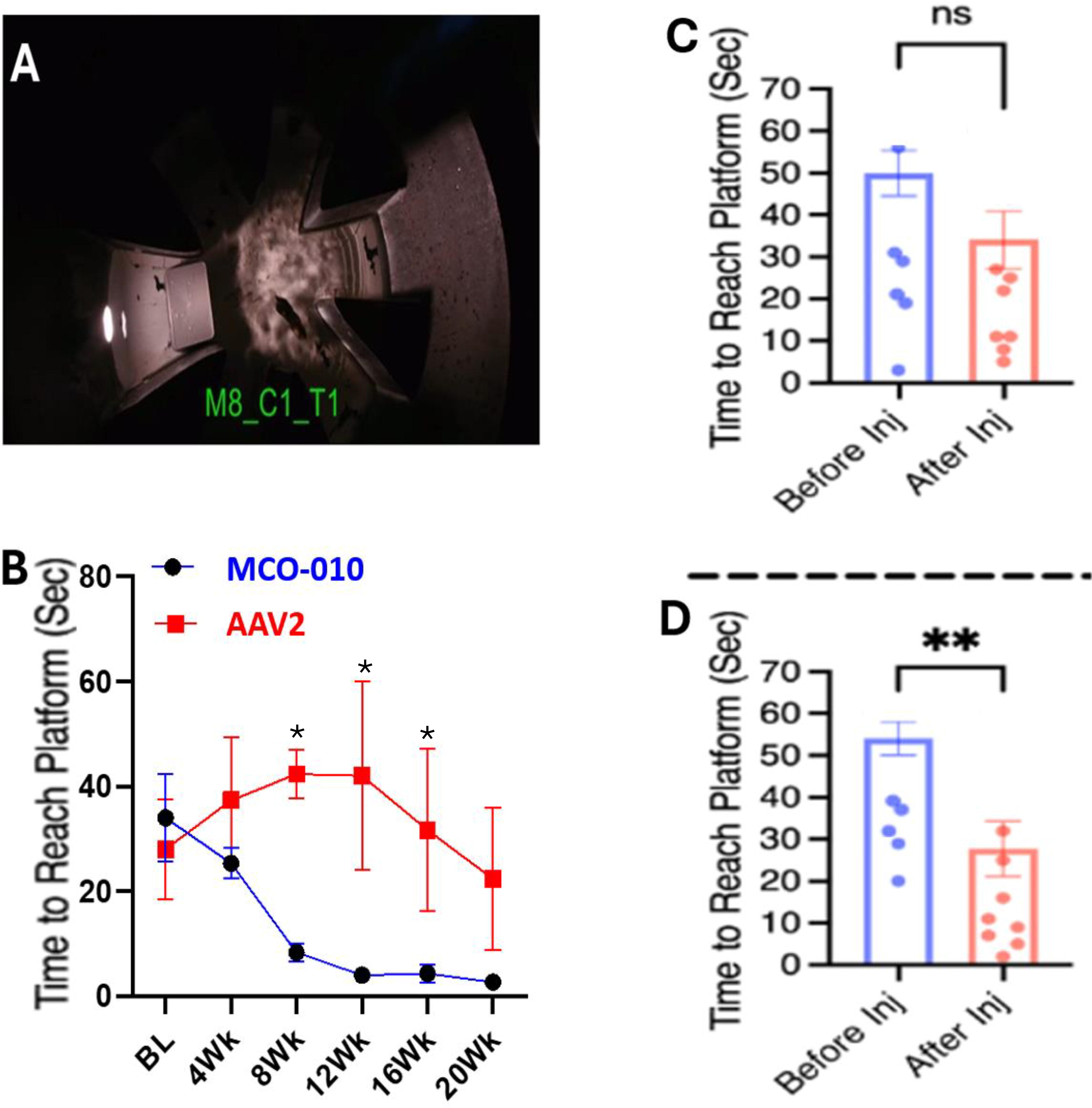
Intravitreal injection of MCO-010 in rd12 mice led to significant improvement in ambient light-guided navigational behavior. Mouse swimming in a water-maze after being released from the side-arm is shown in A. In
Conclusions
As in patients suffering from LCA,4,5 the rd12 mouse exhibits an absence of the critical retinoid isomerase resulting in elevated levels of retinyl esters in RPE cells which begin to die.14,30 Consequential depletion of 11-cis-retinaldehyde and rhodopsin in the photoreceptors causes their degeneration.14,30 During development of the latter pathologies, the inner retinal cells such as bipolar, amacrine, horizontal, and RGCs remain mostly unperturbed and remain functionally viable as shown in mutant mice,9,10,15,34 dystrophic dogs, 35 and in postmortem eyes of RP patients.8,11,24 Whilst gene therapy in general and optogenetic-based treatment specifically offer the potential for vision restoration, realizing this in a clinical setting still represents a major challenge. Therefore, animal models of IRDs10,15,19,20,30 representative of the human conditions serve as important steppingstones towards such clinical translation.5,6,13,23 Accordingly, we currently utilized rd12 mice,14,30 which display retinal pathological features of LCA,4,5,23 to study the safety and efficacy of the optogenetic therapeutic agent MCO-010.
The current studies demonstrated the overall safety and in vivo efficacy of MCO-010 in rd12 mice based on retinal structure, electrophysiological, and visually guided behavioral parameters. As recently reported in mouse models of RP,18,19 it appears that incorporation of the optogenetic triad of light-sensitive opsins via MCO-010 into bipolar cells stabilized the retinal architecture and attenuated the retinal thinning that occurred in mice that only received the AAV2-vector. 36 Whilst the mechanism of action of MCO-010 in slowing down the loss of retinal cells in the LCA-bearing mice remains to be determined, the stabilization or increased light-activated ERG responses in the MCO-010-treated mice, being activity-dependent, may be partially responsible for these observations. This phenomenon appears to be related to various chemical/electrical activity-dependent cellular and molecular mechanisms and associated neuronal plasticity, 37 partially attributed to the release of endogenous growth factors such as brain-derived neurotrophic factor.38–40 However, further studies are needed to confirm whether this happens in the current mouse model of LCA.
The key observations from the current studies pertain to the restoration of functional vision in the rd12 mice following the optogenetic gene therapy treatment using MCO-010. Thus, the statistically significant improvement in the visually guided navigational capabilities of the treated rd12 mice began 4 weeks after bilateral MCO-010 intravitreal injection and continued out to 20 weeks. Interestingly, similar results have been previously reported for vision restoration in mice suffering from other IRDs such as rd1 and rd10 mice expressing RP-like pathologies.18,19,36 Moreover, intravitreally delivered MCO-010 has demonstrated vision improvements in human subjects afflicted with RP. 23 Taken together, the current in vivo efficacy of MCO-010 in stabilizing retinal structure and preventing further decline in retinal thinning underscores its therapeutic potential. Additionally, the continued retinal function (ERG responses) and vision improvement observed in the rd12 mice highlight the ability of MCO-010-transduced bipolar cells to rescue and restore vision in mice with the LCA-like disease. Likewise, whereas other optogenetic gene therapies have employed a single opsin (with fixed sensitivity to a single wavelength of light) to transduce mostly RGCs,4,13,16,21 the current MCO-010 transduces mainly bipolar cells which are far numerous than RGCs, and it utilizes the signal amplification properties of three different opsins to capture all wavelengths of white light.18–20,22 Optogenetic gene therapy, especially using MCO-010, therefore has the potential to help ameliorate and treat numerous IRDs,1,4,33,41,42 including LCA,4,6,23 in a mutation-agnostic manner.
Footnotes
Author Disclosure Statement
All authors are either employees of Nanoscope Therapeutics Inc (NAS, SM) or employees of Nanoscope Technologies LLC (SB, AD, MC, SK, SM). All authors contributed towards the experimental design, execution of the experiments, data gathering and analyses, and drafting and editing of the article.
Funding Information
The reported studies were funded via internal financing.

References
Supplementary Material
Please find the following supplemental material available below.
For Open Access articles published under a Creative Commons License, all supplemental material carries the same license as the article it is associated with.
For non-Open Access articles published, all supplemental material carries a non-exclusive license, and permission requests for re-use of supplemental material or any part of supplemental material shall be sent directly to the copyright owner as specified in the copyright notice associated with the article.
