




Abstract
Purpose:
A dexamethasone canalicular insert, Dextenza®, has been used to treat anterior inflammation of the eye, including keratoconjunctivitis sicca. This study aimed to investigate the tear pharmacokinetics and systemic exposure of dexamethasone in New Zealand White rabbits after inserting the preparation into the punctum and to correlate the data with the in vitro dissolution test.
Methods:
Dextenza was inserted into the punctum of rabbits. A paper strip serially collected tears, and dexamethasone concentrations in tears and plasma were measured using HPLC-MS/MS. The time courses of tear and plasma dexamethasone concentrations were characterized.
Results:
The release of dexamethasone from the insert to tears in rabbits was completed in ∼ 5 days, much faster than in humans and dogs (30 days). The time course of plasma dexamethasone concentration was fully characterized, in contrast to the fact that systemic exposure was merely observed in the other species. The present results might be attributed to the anatomical structure of the lacrimal sac beneath rabbits’ canaliculi. The in vitro dissolution pattern represented an excellent correlation with the in vivo release in tears.
Conclusions:
We first examined the pharmacokinetic study of the canalicular insert in rabbits, which could be applied to the other insert studies in the species.

Introduction
The decrease in the secretion of tears progresses with age and is also common in postmenopausal women. 1 When dry eyes progress, the secretion of proinflammatory cytokines such as interleukin-1, interleukin-6, and tumor necrosis factor increases in epithelial cells of the cornea and conjunctiva, appearing in high concentrations in tears. 2 When those cytokines stimulate the production of matrix metalloproteinase-9, it is known to result in the pathological consequences of keratoconjunctivitis sicca by dissolution of tight junction protein. 3
Although corticosteroid drugs can increase intraocular pressure and augment the incidence of cataracts when used for a long time, they are widely used in the treatment of diabetic retinopathy, macular edema, and non-infective uveitis, including dry eyes and allergic conjunctivitis. The drugs contract peripheral blood vessels, inhibit cellular infiltration and fibroblast proliferation through anti-inflammatory action, and stabilize cellular membranes. 4 Among steroid drugs used for anterior eye disease, dexamethasone is widely used as eye drops. However, it is known that only about 5% of the amount administered reaches the eye tissue, 5 which is attributed to inaccurate administration, loss due to blinking, nasolacrimal drainage, and low coronal permissibility. 6 Therefore, sustained drug administration can be carried out through intracanalicular insertion using punctal plug preparations and safe, biodegradable polymers. A dexamethasone insert using polyethylene glycol (PEG) hydrogel is used in the clinic. The preparation swells when it comes into contact with tears, and dexamethasone is gradually released and continuously exposed to the anterior segment of the eyes. Meanwhile, PEG hydrogel is removed through the nasolacrimal duct system.7–9
The pharmacokinetic and toxicological studies of the plugs using animals can produce important information when developing new drugs and formulations and comparing them with the previous preparations. The research for dexamethasone inserts has mainly used beagle dogs,10–11 but rabbits were used in this study. Rabbits are herbivores, and their gastrointestinal tract differs from rodents, dogs, and primates, which is not well used in general pharmacokinetic studies. However, they have many beneficial consequences for eye studies. As the thickness of the lens of the rabbit is doubled compared with that of the human, the volume of the vitreous humor is only about a third. 12 Nevertheless, the rabbit eyes are big enough to study various implants applied to humans. 13 The amount of tears secreted, the residual amount of tears, and the rates of aqueous humor generation and rotation are similar to those of humans. In addition, the advantage of rabbits is that they occupy less space compared with beagle dogs, the cost of testing is quite low, and they are extremely quiet.
In this study, the pharmacokinetics of dexamethasone were examined in tear and plasma after rabbits were administered a dexamethasone canalicular insert, Dextenza®. The findings revealed that dexamethasone disappeared faster from tears in rabbits than in dogs and humans, a significant discovery. While systemic exposure in the 2 species was insignificant, rabbits showed sufficient exposure to characterize the plasma concentrations over time. In addition, in vitro dissolution represented a good correlation with the in vivo release in tears estimated from the time course of tear dexamethasone concentration.
Methods
Materials
Dexamethasone, HPLC-grade methanol, and acetonitrile were purchased from Sigma-Aldrich Korea (Seoul, Korea). Dextenza and Schirmer tear test strips were obtained from Sangmyung Innovation Co., Ltd. (Seoul, Korea) and BK Pharm Co., Ltd. (Gyeonggi-do, Korea), respectively. Alcon Korea (Seoul, Korea) provided a balanced salt solution.
Animal study
Four 3-month-old young adult New Zealand White rabbits were used. The animal room’s temperature and humidity were maintained at 23 ± 3°C and 50 ± 10%, and the light, with an illumination level of 300 Lux, was turned off for 12 h. After intravenous anesthesia with alfaxalone (3 mg/kg) and xylazine (4 mg/kg), Dextenza was inserted into the canaliculi of both eyes using sterilized forceps. Approximately 200 μL of blood was taken from the ear veins before and after administration at 5, 10, 24, 48, 72, 96, and 120 h, placed in a heparinized tube to prevent coagulation, and then centrifuged to separate the plasma. At the same time, tears were collected using a Schirmer test strip. We ensured that the strip’s scale became wet by >1 cm, placed them in a separate tube, and stored them at −70°C until analysis along with the plasma samples, maintaining the integrity of the samples for accurate results.
The study protocol was approved by the Institutional Animal Care and Use Committees at Chung-Ang University (Approved No. 202401030131), and their recommendations for animal care and handling were followed and adhered to the Association for Research in Vision and Ophthalmology statement.
Pharmacokinetic parameters were obtained directly from time courses of plasma and tear dexamethasone concentrations by a model-independent analysis and were represented as mean and relative standard deviation (RSD, %).
Quantification of dexamethasone in plasma and tear strips
Plasma concentration was measured using previously published literature following a slight modification. Briefly, 90 μL of acetonitrile, including an internal standard (IS, diclofenac 100 ng/mL), was added to 30 μL of plasma and mixed vigorously for 10 s. After centrifugation at 13,000 g for 10 min, 5 μL of the supernatant was injected into an HPLC-MS/MS system (API 5000 LC-MS/MS system, AB Sciex, Toronto, Canada). 13
The method proposed by Glogowski et al. for determining the analyte in tears was modified. 14 After cutting 1 cm of the tear-soaked area of a Schirmer tear strip with scissors, 200 μL acetonitrile with the IS was added. After 10 min sonication, the solution was centrifuged, and 5 μL of the supernatant was used for analysis. The concentrations of dexamethasone in actual plasma and tear samples were determined by the calibration curves prepared in advance using the same pretreatment procedures.
Calibration standards for quantifying the dexamethasone concentrations in plasma and tear were prepared as follows. Dexamethasone was dissolved in methanol at 1 mg/mL and serially diluted to prepare a standard solution of 0.01–10 μg/mL. Ten microliters of each solution were added to 90 μL of BSS solution to obtain 1–1,000 ng/mL of calibration standard solutions. After wetting a Schirmer strip to a length of >1 cm, 1 cm was cut and placed in a new tube. Acetonitrile (200 μL), including the IS, was added. Thereafter, the samples were treated using the same procedure described above. For calibration standards for plasma, 5 μL of the standard solution was spiked to 45 μL of drug-free plasma, and a 3-fold volume of acetonitrile (150 μL) was added for clean-up. Calibration curves were obtained using the peak area ratios of dexamethasone and the IS versus corresponding concentrations.
Quality controls at 1, 3, 200, and 700 ng/mL were prepared to validate the analytical methods in plasma and tear, and the accuracy and precision were measured at 3 replicates. Recovery was calculated by comparing the peak areas of quality controls spiked before the clean-up process to those spiked after pretreatment.
In vitro dissolution test
A dissolution test was carried out based on the review document of FDA approval (NDA 208742 review #3, November 28, 2018). 15 One hundred milliliters of phosphate buffered saline (pH 4.0) were maintained at 37.0 ± 0.3°C in a 125 mL polyethylene bottle. Six inserts of Dextenza were placed into each chamber, and the bottle was gently inverted and swirled to ensure that all inserts were wet. At each sampling point, 1 mL of the supernatant was taken and replaced with the same warmed buffer solution at 37°C. Dexamethasone concentration in each sample was quantified by using the analytical method for plasma described above: 90 μL of the IS solution was added to 30 μL of the in vitro samples.
Results
Determination methods of dexamethasone in rabbit tear and plasma
The calibration curves for quantifying dexamethasone in plasma and tear-soaked Schirmer strips showed good linearity (r2 > 0.998) in the 1–1,000 ng/mL range. The extraction recovery from plasma and the strips was 85% and 93%, respectively, indicating that the extraction method was suitable. The intra- and inter-day precisions of plasma QC samples were <10.5% and 12.4%, and the accuracies were 94.5%–104.5% and 90.7%–108.3%, respectively. The intra- and inter-day precisions of tear QC samples were <12.2% and 13.5%, and the accuracies were 91.8%–94.5% and 89.4%–112.5%, respectively. Therefore, the analytical method used in this study was suitable for quantifying the drug in the biological samples.
Animal study
Figure 1 shows the concentrations measured in tear (left) and plasma (right) over time after 0.4 mg dexamethasone canalicular insert was inserted into both puncta of rabbits. The pharmacokinetic parameters are listed in Table 1.
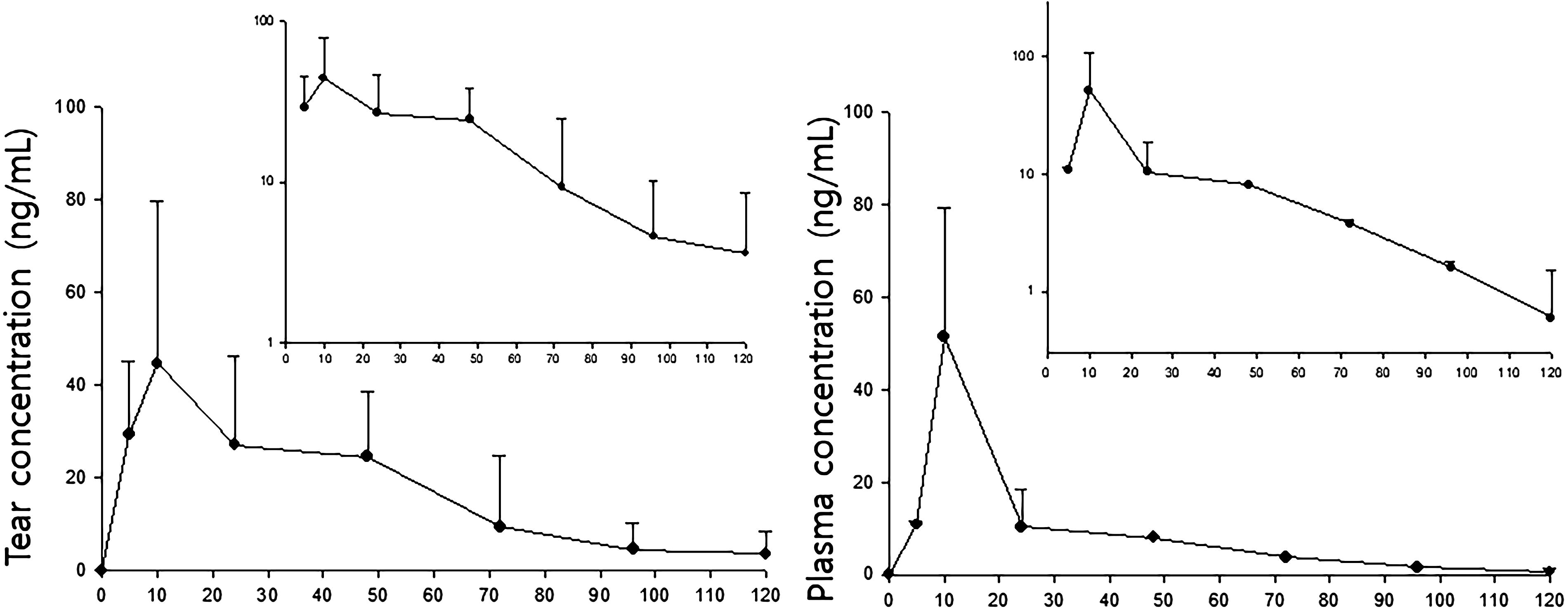
Time courses of tear (left, n = 8) and plasma (right, n = 4) dexamethasone concentrations after an intracanalicular insertion of Dextenza® in New Zealand White rabbits. Each point indicates the mean and standard deviations, and the inserts are semi-log plots.
Pharmacokinetic Parameters of Dexamethasone Intracanalicular Inserts in Rabbit Tear (n = 8) and Plasma (n = 4)
Data are mean (RSD, %).
RSD, relative standard deviation.
In tears, the average peak concentration of 42 ng/mL (RSD, 72%) was reached at 17 h (RSD, 122%) after administration, and it was eliminated with a half-life of 25 h (RSD, 36%). The area under the tear concentration–time curve (AUC) by 120 h was 2,094 ng·h/mL (RSD, 109%), and the total clearance in tears was 0.44 L/h/F (RSD, 94%). On the contrary, in plasma, the maximum concentration was 51 ng/mL (RSD, 55%) at 10 h, and it disappeared with a half-life of 30 h (RSD, 18%). AUC120h was 1,084 ng·h/mL (RSD, 69%), and the total clearance in plasma was 0.48 L/h/F (RSD, 69%).
In vivo and in vitro release test
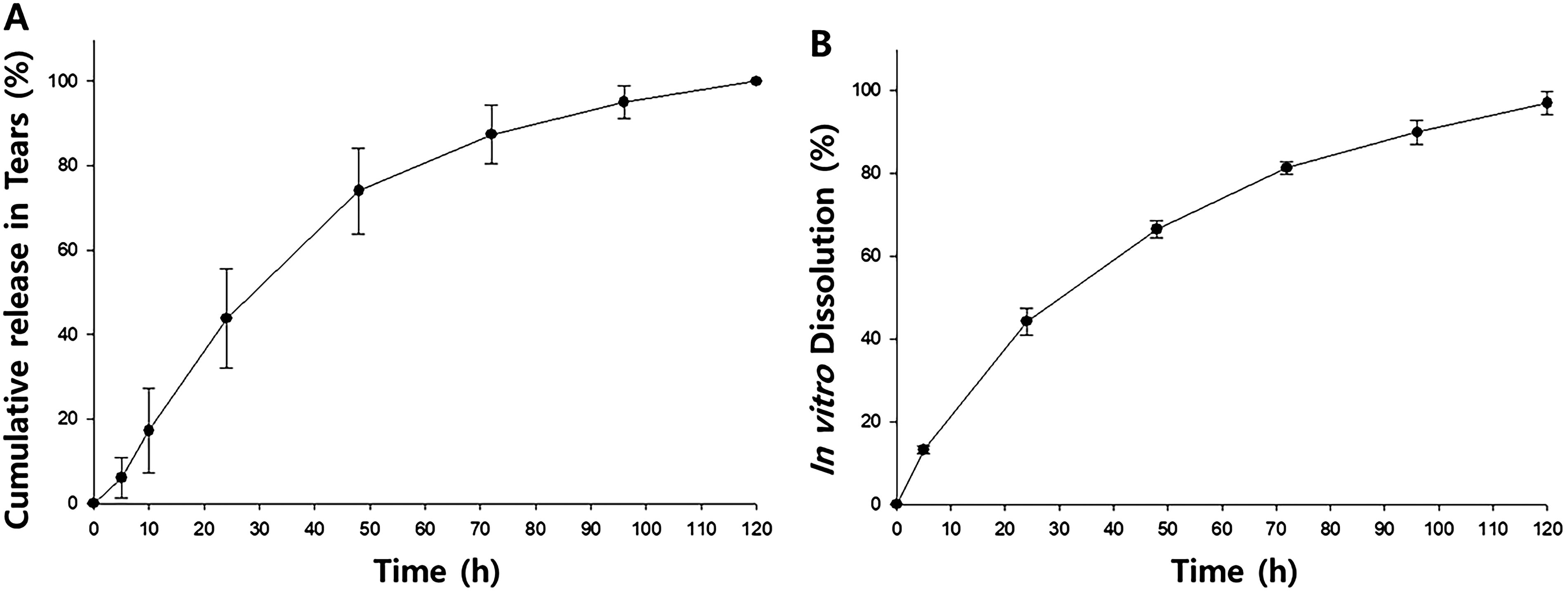
Figure 2A depicts the time course of the cumulative release of dexamethasone in rabbit tears following the insertion of Dextenza in each punctum. The in vivo release was obtained by dividing the AUC at each time by the AUC120h, representing the relative release percentage of dexamethasone from the insert into tears. Figure 2B represents the in vitro dissolution test and the release completed by 120 h.
Discussion
In order to analyze drugs in tears, it is necessary to collect an appropriate amount of tears. A literature directly collected several microliters of tears from the lower lid of anesthetized rabbits. However, considering the sensitivity of the analysis, we modified the method used by Glogowski et al. 14 They collected tears using a Schirmer tear strip, and 1 mL of a 1:1 mixture of acetonitrile and water was added per 10 mg of tears to extract loteprednol. A pretreatment method for better sensitivity was required to determine the concentration of tears over time. Therefore, tears were collected sufficiently to wet >1 cm Schirmer tear strip, which took 2–3 min. The wetting speed was similar to 5 mm/min in the previous literature. 16 The strip was cut to 1 cm, rolled vertically, and placed in a tube; then, 0.2 mL of acetonitrile was added, followed by sonication for 10 min. The amount of acetonitrile was determined to be sufficient to wet the cut strip. Sonication was also considered to help extract dexamethasone in the strip significantly and showed a better recovery rate than plasma samples.
Although sufficient tears must be obtained to validate the analytical method and create a calibration curve, collecting them directly from actual animals is impossible. Therefore, a balanced salt solution was used to replace tears, which is widely used as a tear substitute in the anterior segment, including cataract surgery.
The intraocular pharmacokinetic study using a dexamethasone intracanalicular insert was conducted on beagle dogs. In the literature reported by Blizzard et al., 10 2 doses, 0.4 mg and 0.7 mg, were administered. In the 0.4 mg dose, the first sample was collected 7 days after administration, showed a concentration of 2,805 ng/mL, and tended to decrease in tears with a zero-order disappearance until the 28th day. On the contrary, the 0.7 mg dose showed 4,370 ng/mL at 6 h and about 3,300 ng/mL on the seventh day. It showed a retard decrease compared with the 0.4 mg dose, and the median value on the 35th day was 830 ng/mL.
The results of this study on rabbits are new and have not been reported in the animal previously. Interestingly, when the same dose of 0.4 mg was administered, very low concentrations were observed in rabbit tears compared with dogs and decreased to below the quantitation limit within 1 week. In beagle dogs, the first sampling after administration was carried out at 6 h, the time to peak concentration in tears, so the absorption pattern could not be observed. In contrast, in rabbits, an increasing pattern of dexamethasone in tears was observed for the first 12 h following administration (Fig. 1A). In addition, time courses of plasma and tear concentrations showed similar patterns. It is hard to find literature on a simultaneous observation of dexamethasone in tears and plasma. A direct comparison may provide limited information since the drug distribution depends on its physicochemical properties. However, for example, after oral administration of minocycline to a patient with trachoma, the tear exposure was about 10% of the systemic one, and a very similar pattern was observed in both tears and plasma: following the peak concentration at 3 h, both concentrations disappeared with a half-life of ∼ 18 h. 17 It should be interesting to compare the tear exposure with the plasma concentration after systemic, for example, intragastric or intravenous, administrations of dexamethasone.
When the preparation is inserted into the inferior lacrimal punctum, the upper part of the insert becomes wetted by tears, and the drug is released. 10 The tear production rate of beagle dogs is >15 mm/min, 16 which is about 3 times faster than that of rabbits, so one may guess that the initial concentration in dogs is higher than in rabbits. However, it is difficult to explain that it is continuously high for >4 times the period by the tear production rate alone. 10 On the contrary, the anatomical structure of the rabbit canaliculus duct may provide a clue. Humans and dogs have a single, thin, long, consistent nasolacrimal duct (Fig. 3). In contrast, rabbits have a funnel-shaped lacrimal sac adjacent to the lower portion of the lacrimal punctum, which is connected to the nasolacrimal duct. 18 Rapid release of dexamethasone from the insert immersed in this lacrimal sac is likely induced, resulting in a short release period of less than a week. Despite the rapid release, the low concentration of tears might be due to distribution throughout the body (Fig. 1B). More detailed studies are needed on the contribution of the lacrimal sac and its distribution into the systemic circulation.
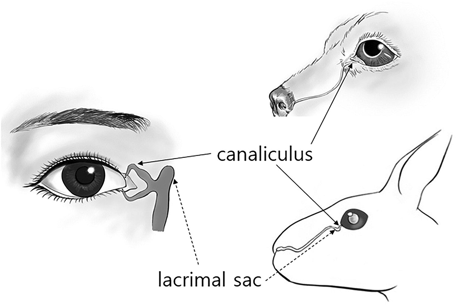
Lacrimal canalicula in humans (left), dogs (top right), and rabbits (bottom right).
No previous literature has been found to measure the tear concentrations after the insertion in humans. Blizzard et al. measured 176 plasma samples from 16 subjects following an intracanalicular administration of the insert. Since only 21 samples were over the lower limit of quantification (50 ng/mL) and ranged from 50 to 810 ng/mL, the individual time course of plasma concentration could not be obtained. 19 Considering that the concentration lasts for about 4 weeks in beagle dog tears and there is no systemic exposure, it should be adequate for treating the anterior segment in beagle dogs because it exists in high concentrations in tears when the insert is used. In addition, the weak distribution in the blood can be an advantage in that there is no need to worry about the steroid’s systemic side effects.
At the very beginning of this work, we expected that an insert in one punctum should not affect the other eye, so the inserts were administered to both puncta in rabbits. However, as mentioned above, probably because of the anatomical characteristics of rabbits, both inserts contributed to the tear concentrations in the opposite eyes each other as well as plasma concentrations. The plasma AUC showed approximately half of the tear AUC in one eye, a significant amount considering the systemic distribution. When one insert is applied to only a punctum in the animal, both tear and plasma concentrations may be lower than the present results. An additional study should be done to clarify.
As shown in Fig. 2A and B, the release pattern of dexamethasone in rabbit tears from the insert is similar to that in the in vitro dissolution chamber. Fifty percent of the release was achieved in about 36 h and the release was completed in 5 days for both in vivo and in vitro. The in vitro dissolution data of the insert shows an excellent correlation (y = 1.03x + 2.55, r2 = 0.9935) with in vivo cumulative release in tears (Fig. 4).
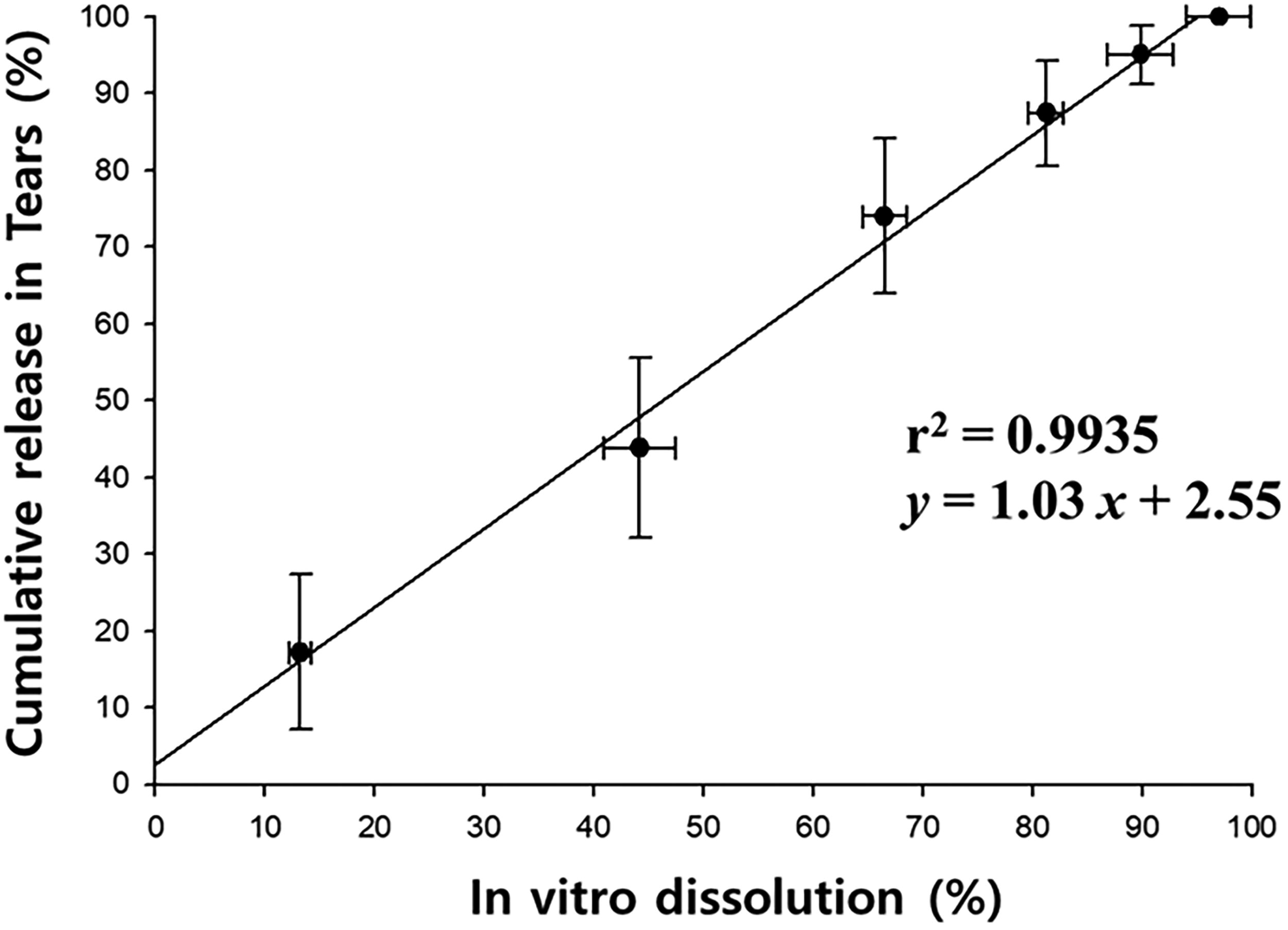
In vitro and in vivo correlation.
Dextenza contains dexamethasone (0.4 mg) in a PEG-based hydrogel conjugated with fluorescein, and its inactive ingredients are 4-arm PEG N-hydroxysuccinimidyl glutarate, trilysine acetate, N-hydroxysuccinimide-fluorescein, sodium phosphate dibasic, and sodium phosphate monobasic. 20 From such a good in vitro and in vivo correlation, rabbits could be a useful animal model to predict the release profiles in tears regarding the dissolution data of dexamethasone insert preparations with various compositions of ingredients.
This study did not measure adverse effects such as increased intraocular pressure due to the steroid insert. However, in a previous study, no significant adverse effects were observed when the same dose was used in beagles. 10 Since the concentration in rabbit tears was even lower at the same dose, local adverse effects could be negligible. However, the adverse effects of repeated use should be considered since systemic exposure was observed in rabbits, albeit at low concentrations.
The present results from 8 eyes in 4 rabbits may not be sufficient to reflect the pharmacokinetics of dexamethasone inserts in the animal. A small number of data could generally lead to large variances, which may be due to inter- and intra-individual differences. A study by Blizzards et al., 10 which used a different number of subjects in the same study design, can provide insight into the variability of the preparation owing to the number of animals. When dexamethasone 0.4 mg and 0.7 mg insert were administered to 6 and 18–33 puncta in beagle dogs, respectively, the average individual variance recalculated by RSD at each observation time was 15% for the formal dose and 10% for the latter. However, the variance decreased with increasing the number of individuals, and no statistical significance was found. Individual variances may be less affected by the sample size for a topical agent such as the insert. A high variance may be attributed to the species specificity of rabbits and the systemic distribution not seen in beagle dogs. Further study using more rabbits should be needed.
Summary
In conclusion, the tear pharmacokinetics and systemic exposure of dexamethasone were fully characterized in rabbits for the first time following a punctum insertion of a dexamethasone insert, Dextenza. The drug’s release profile in tears correlates well with an in vitro dissolution test. In contrast to humans and dogs, dexamethasone was systemically exposed in rabbits. The rabbit could be useful for carrying out a canalicular insert study.
Footnotes
Acknowledgment
The authors thank Sangmyung Innovation Co. Ltd. for their support during the study.
Authors’ Contributions
M.M.: Methodology and data curation. J.W.: Methodology, visualization, and data curation. W.K.: Conceptualization, writing, and review.
Author Disclosure Statement
The authors declare no conflicts of interest.
Funding Information
This work was supported by a grant (22183MFDS366) from the Ministry of Food and Drug Safety of South Korea in 2022–2025 and Chung-Ang University Research grants in 2020.