
Abstract
Abstract
Background:
Biliary atresia (BA) is the progressive inflammatory obstruction and fibro-obliteration of all or part of the extrahepatic biliary tree and the intrahepatic bile ducts and has its onset exclusively within the first several months of life. This study was undertaken to present the value of diagnostic laparoscopy in infants with prolonged jaundice and technique for laparoscopic cholangiography.
Methods:
A 5-mm umbilical trocar was introduced to create a port for a 30-degree laparoscope. If the gallbladder was of good size, the fundus was exteriorized through the right subcostal trocar site and a catheter was inserted into the gallbladder for cholangiography, following partial dissection from the liver bed, if required. If the gallbladder was atretic, the fundus was not exteriorized and a laparotomy was performed and cholangiography was abandoned, because the lumen of an atretic gallbladder was usually not fully patent.
Results:
At laparoscopy, 12 patients had good-sized gallbladders and minimal-to-mild liver fibrosis. They underwent cholangiography via the exteriorized fundus, and infantile hepatitis syndrome (HIS) or cholestatic syndrome (CS) in 8 cases, BA in 2 cases, and biliary hypoplasia (CBDH) in 2 cases were identified. Five patients' gallbladders dissected from the liver bed underwent cholangiography, and BA in 3 cases and CBDH in 2 cases were identified. The remaining 21 had atretic gallbladders and varying degrees of liver fibrosis, so cholangiography via the exteriorized fundus was abandoned and converted to open Kasai portoenterostomy.
Conclusions:
Laparoscopy-assisted cholangiography is a simple, accurate, and safe method in the diagnosis of prolonged jaundice in infants and allows the anatomic structure of the biliary tree to be obtained accurately with minimal surgical intervention.
Introduction
Materials and Methods
Patients
From October 2003 to February 2008, 38 infants with prolonged jaundice were referred to us from outpatients or pediatricians. Fifteen males and 23 females, with an average age of 74 days (range, 27–140), were all admitted with progressing severe skin icterus, acholic stool, and dark tea-like urine. Physical examination indicated moderate or seriously icteric skin or sclera and hard, irregular liver, which swelled in different degrees. The liver function examination indicated total bilirubin, conjugated bilirubin, and unconjugated bilirubin, glutamic oxaloacetic transaminase and glutamic pyruvic transminase were enhanced distinctly. The enrollment critera in our series included and icterus gradually aggravated 3–4 weeks after birth; consistant acholic stool; traditional diagnositic examination (i.e., physical examination, blood biochemical examination, ultrasonography, hepatobiliary scintigraphy, MRCP, etc.) failed to get a definitive diagnosis; and needed to exclude BA. We had a routine laparoscopic cholangioraphy before laparotomy or Kasai procedure.
Diagnostic technique
Under general anesthesia, an umbilical incision was used for the initial introduction of a 5-mm trocar to create a port for a 30-degree laparoscope, which helped to visualize the situation of the gallbladder and extrahepatic bile ducts. Damaging of umbilical vessels should be avoided at this time. Carbon dioxide (CO2) was insufflated to between 6 and 10 mm Hg. A 5-mm right subcostal incision was made and a 5-mm trocar inserted, if the extrahepatic bile ducts existed and the gallbladder appeared normal. The gallbladder fundus was exteriorized through the incision, with no injuries, with forceps (Figs. 1 and 2), following CO2 eduction and the fixing of an inserted catheter by, double knots. The bile duct was swash by gradually inputting 10 mL of 0.9% saline. We repeated this procedure 2–4 times if it was expedited until no resistance existed. In order to swash the intrahepatic bile ducts, the inferior segment of common bile duct was controlled, with no injuries, with forceps, and the above procedure was repeated with suitable force 1–2 times to rinse the intrahepatic bile ducts. After these procedures, we injected 76% meglumine diatrizoate and took pictures. If the gallbladder was shriveled, a 5- or 3-mm trocar was put 1–2 cm below the xyphoid and the gallbladder was dissected from the liver bed partially, and we retained the integrity of the vascular and gallbladder duct, so that it could be exteriorized from the right-upper quadrant incision. If the gallbladder was atretic or fibrous and the liver appeared fibrotic, BA was more likely to be the cause, and rather than persevere with laparoscopy-assisted cholangiography, as above, we progressed directly to a laparotomy. To the patients diagnosed as HIS or CS, the catheter was indwelling after rinsing the bile duct, so that the bile duct could be rinsed repeatedly after operation. A biopsy of the lower edge of the liver (5 × 5 mm) was taken routinely for histopathologic examination. The liver surface was coagulated and we took out the tissue through the trocar.
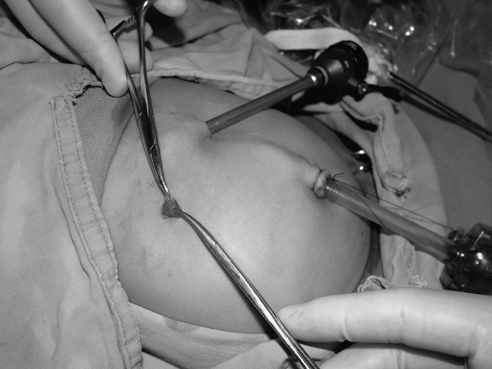
The gallbladder fundus was exteriorized through the right-upper quadrant incision.

The catheter was inserted into the gallbladder for cholangiography.
Results
Thirty-eight patients obtained a clear diagnosis through laparoscopy without any complications and with very little blood lost. Twelve infants were found to have good-sized gallbladders and minimal or mild liver fibrosis. Cholangiography via the exteriorized gallbladder showed a well-developed bile duct in 8 cases, which were diagnosed as HIS or CS (Fig. 3), hypoplastic biliary ducts in 2 cases, and a patent common bile duct with atresia of the common hepatic duct and intrahepatic ducts in 2 cases. The 8 cases diagnosed as HIS or CS underwent rinsing of the bile ducts and tube drainage of the gallbladder. The 2 cases diagnosed with biliary hypoplasia underwent tube drainage of the gallbladder, and the 2 cases diagnosed with BA underwent Kasai portoenterostomy. The shriveled gallbladders in 5 cases needed to be dissociated from the liver mesentery, so that they can be exteriorized for cholangiography. Two cases were diagnosed as biliary hypoplasia (Fig. 4), and the other 3 cases were diagnosed as BA for the extra- and intrahepatic bile ducts were not visualized after standing catheter injecting contrast medium to the gallbladder (Fig. 5). The remaining 21 infants had atretic gallbladders and different degrees of liver fibrosis. Laparotomy was transferred, and we attempted cholangiography intraoperatively. However, we still could not place a fine catheter into the atretic gallbladder, because the gallbladder had no lumen. In these cases, cholangiography was abandoned and Kasai portoenterostomy was performed.
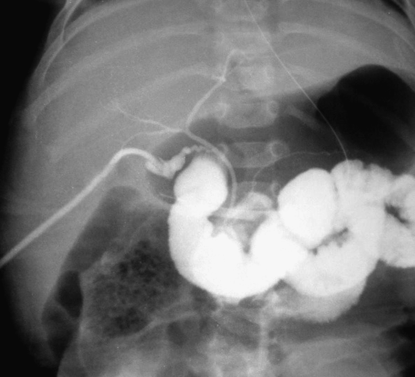
Extra- and intrahepatic bile ducts can be visualized.

Hypoplastic intrahepatic bile ducts can be visualized.
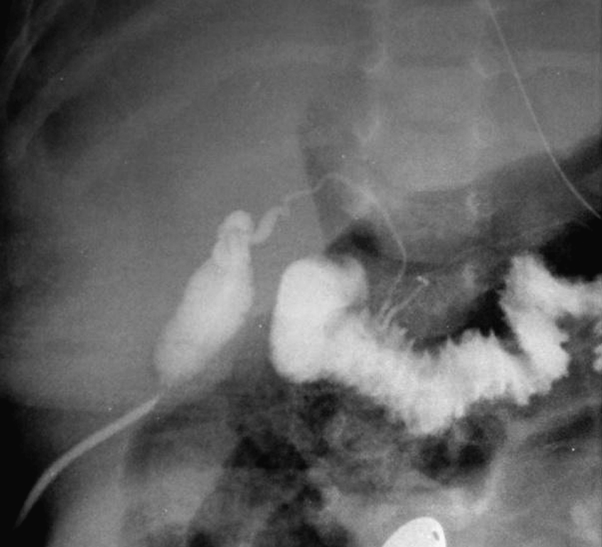
Intrahepatic bile ducts cannot be visualized.
Discussion
The most important differential diagnosis in infants with prolonged jaundice is between HIS (including CS) and BA (including CBDH). At the early stage of infantile hepatitis, about 20% of patients developed entirely cholestatic syndrome. Thus, they appeared with almost the same symptoms, but the treatment and prognosis was definitely different. The prevalence of BA in China is high, and the difficulty of an early diagnosis usually leads to a definitive diagnosis later. Most of the patients performed laparotomy while getting a cirrhotic liver. Some scholars have suggested that all infants with cholestatic jaundice should perform laparotomy with open intraoperative cholangiography, so that the BA patients can have an early surgical treatment, while some others disagreed about this opinion, insisting that laparotomy should be postponed to 4 months later, because it is highly invasive if the cause of the prolonged jaundice is not BA (such as HIS). 5 But, laparoscopy-assisted cholangiography can help to make an early definitive diagnosis and avoid too much invasiveness to infants with HIS.
There are three techniques of laparoscopy-assisted cholangiography that have been reported.6–8 The first method was to insert a catheter into the gallbladder directly under laparoscopy to perform cholangiography. The second was to insert a catheter into the gallbladder via a transhepatic route guided by laparoscopy. The third was to insert a catheter into the exteriorized gallbladder via an abdominal incision route guided by laparoscopy. However, based on our experience, it is usually very difficult to insert a catheter into an atretic gallbladder, even in the case of a laparotomy for open intraoperative cholangiography. Twenty-one cases in our study appeared with a fibrotic atretic gallbladder under laparoscopic vision. Some gallbladders had no lumen or only a latent lumen, while some others contained little white or yellow serous fluid. When gallbladders needed to be cut open, we still could not place a fine catheter into the atretic gallbladders. Therefore, the transhepatic route appears to be very difficult, with an increased risk for bleeding and bile leakage into the peritoneal cavity. Also, laparoscopy-assisted percutaneous needle cholangiography is not technically feasible and is bound to be time-consuming.
Adult laparoscopic cholangiography is a mature endoscopic technique and is usually performed after cholosectomy, while to infants, it is much more difficult to put the catheter into the slim duct of the gallbladder within the abdomen when performing laparoscopic cholangiography. Meanwhile, infants have a thin abdominal wall and it is easy to exteriorize the gallbladder fundus with the guide of laparoscopy, which simplified the procedure and usually led to success. In our technique, we use laparoscopy to check the size of the gallbladder, hepatic color, and the degree of liver fibrosis carefully and exteriorize the fundus of the gallbladder through the 5-mm right subcostal incision, following a partial dissection from the liver bed, if required. The catheter is inserted into the gallbladder outside the abdomen, allowing the fundus to be manipulated directly, which greatly simplifies the whole procedure. Moreover, it can minimize the risk for bile leakage and bleeding. Twelve infants in our series had a normal-sized gallbladder visualized laparoscopically and exteriorized with successful cholangiography. The hypoplastic gallbladder, in some infants, cannot be easily exteriorized out of the abdomen and requires dissection from the gallbladder mesentery without damaging the vessels, and then, the surgeon must retrieve the fundus of the gallbladder out of the abdomen for cholangiography. Meanwhile, attention should be paid to diminishing the aeroperitoneum and suction of ascites during the procedure of the exteriorizing process. This procedure was applied to 5 cases in our series. The indications of this method include prolonged jaundice in neonates or infants, and the contraindications include heart and liver function insufficiency and advanced stage of liver cirrosis with coagulation dysfunction.
As for the patients diagnosed as HIS or CS after cholangiography, we put tube drainage through the right subcostal incision after rinsing the bile ducts, so that the bile ducts could be rinsed repeatedly after the operation. Cholestatic syndrome comes from a virus infection or metabolic abnormality and can cause obstructive jaundice in infants. Laparoscopy has shown great superiority for those patients with CS, because simple bile duct rinsing can cure most of the patients.9–11 Eight CS patients in our series succeeded avoiding the invasiveness of laparotomy, and their jaundice disappeared after half a year of follow-up. Four cases were diagnosed as hypoplastic bile ducts, and cholangiography showed patent intrahepatic bile ducts. In 2 cases, the inner diameter of the common bile ducts were 2.5 and 2.8 mm separately; after bile-duct rinsing, the jaundice disappeared after 6 and 8 months of follow-up. Twenty-six cases were diagnosed as BA, 2 of them had a well-developed gallbladder, and the contrast medium could reach to duodenum smoothly but could not visualize intrahepatic bile ducts and were thus diagnosed as intrahepatic BA; 3 of them had a hypoplastic gallbladder, which can be placed a catheter, but the extrahepatic and intrahepatic bile ducts could not be visualized. The other 21 cases had an atretic gallbladder, which failed to hold the catheter and were diagnosed as extrahepatic or intrahepatic BA; for these, a laparotomy was performed directly with Kasai portoenterostomy.
Many methods are available for the differential diagnosis of prolonged jaundice in neonates and infants, with a 40∼87% definitive diagnostic rate,1–4,12 and chlangiography was regarded as the most ideal procedure for the definitive diagnosis.12,13 Open cholangiography has a tremendous level of invasiveness and may not be suitable for all patients with jaundice. ERCP was restricted in the diagnosis of infants because of the instruments and technology. Therefore, we considered laparoscopic cholangiography as a “gold standard” method for the early diagnosis of neonates and infants who had prolonged jaundice. Technically, our laparoscopy-assisted cholangiography is simple, safe, and minimally invasive. We believe our protocol is also more efficient, practical, and less time-consuming, because at laparoscopy, most infants who do not have BA will have reasonably good-sized gallbladders, while those with atretic gallbladders will almost certainly have BA and can be proceeded to Kasai portoenterostomy.
Conclusions
Based on our experience, laparoscopic cholangioraphy should be undertaken if symptoms do not improve after traditional treatment as neonatal hepatitis for 2 weeks and if jaundice is aggravated. An early laparoscopic examination can rapidly differentiate surgical and nonsurgical cases, and management can be planned accordingly.
Footnotes
Disclosure Statement
No competing financial interests exist.