
Abstract
Abstract
This study aimed to explore and evaluate the feasibility and safety of laparoscopic adnexal surgery using a two-port technique with a multichannel port, using a wound retractor, as previously reported. A series of patients undergoing two-port laparoscopy for a benign pelvic mass were enrolled in this study. To perform two-port laparoscopic surgery, the ancillary 5-mm trocar was inserted at the left iliac fossa under laparoscopic view after umbilical trocar insertion. The inserted umbilical trocar was removed and the skin incision was extended about 1.5 cm with index-finger–passable width. An Alexis® wound retractor XS (Applied Medical, Santa Rancha Margarita, CA) was inserted through the umbilical wound. Two trocars were inserted into two fingers of a no. 6 surgical rubber glove and ligated with rubber bands. The wrist portion of the rubber glove covered the wound retractor, and the edge of the wound retractor was clamped with three Babcock clamps to prevent carbon-dioxide gas leakage. Both a 10-mm laparoscope and atraumatic forceps were inserted through the umbilical multichannel port. Laparoscopic adnexal surgery was performed in the usual manner. A total of 19 patients were enrolled. The operative procedures were adnexectomy (n = 8), myomectomy (n = 1), and ovarian cystectomy and/or salpingectomy (n = 10). There were no operative complications, conversion to laparotomy, or additional trocar insertions. The mean operation time was 81.3 ± 28.7 min. The pathologic diagnosis were mature cystic teratoma (n = 6), benign cyst (n = 4), endometrial cyst (n = 3), serous cystadenoma (n = 3), mucinous cystadenoma (n = 1), leiomyoma (n = 1), and tubo-ovarian abscess (n = 1). The mean postoperative hospital stay was 4.0 ± 1.3 days. This two-port method seems to be safe and needs no additional cost from the use of the conventional laparoscopic instruments. It is also cosmetically effective and highly appreciated by patients, leaving minimal abdominal scarring.
Introduction
Materials and Methods
Patients
Between June 2008 and December 2008, we performed two-port laparoscopic adnexal surgery by using a multichannel port with an Alexis® wound retractor XS (Applied Medical, Rancho Santa Margarita, CA) in 19 consecutive cases (Table 1). The patient mean age and parity were 34.8 ± 9.9 years and 1.2 ± 1.1, respectively. The mean body mass index (BMI) of the patients was 21.1 ± 2.6 kg/m2. The operative indications were pelvic masses. The mean diameter of the masses was 8.3 ± 4.7 cm, measured with preoperative ultrasonography (US) or abdominopelvic computed tomography (CT) scans. The largest mass of these cases was an approximately 25-cm mature cystic teratoma in a 17-year-old girl. Eleven patients had no history of abdominal operation. Eight patients had a history of previous abdominal operations: repeat cesarean section (n = 5), appendectomy (n = 1), oophorectomy (n = 1), or tubal ligation (n = 1).
Preoperative assessments
Patients underwent preoperative ultrasonography and determination of serum CA125 and CA19-9. Most patients underwent preoperative abdominopelvic CT scans to survey other abnormal findings in the pelvic and abdominal cavity.
Surgical techniques
Preoperative preparation
The patients were admitted the day before surgery; operative permission was received from all patients. We explained the possibility of the conversion to laparotomy in cases of severe adhesion, malignancy, and limited operative visual field. A Fleet enema was given at 7
A Foley catheter was inserted into the patients' urethra, and a Kronner Manipujector® uterine manipulator (Cooper Surgical, Trumbull, CT) was inserted vaginally. A uterine manipulator was not used in the adolescent patients and young women with no coital history. The surgical instruments used for the procedure were 10-mm, 0-degree laparoscopes, bipolar forceps, atraumatic forceps, monopolar hook, toothed grasper, monopolar scissor, laparoscopic needle holder, and a suction-irrigation system.
Multichannel port preparation
A small longitudinal skin incision was performed on the umbilicus, and a pneumoperitoneum was obtained with a Veress needle. A 10-mm trocar was inserted into the supraumbilical area and a 10-mm, 0-degree laparoscope was inserted through the trocar. We inspected the pelvic anatomy carefully to decide whether the conversion to laparotomy was required. To perform two-port adnexal surgery, the ancillary 5-mm trocar was inserted at the left iliac fossa under laparoscopic view. The inserted umbilical trocar was removed, and the skin incision was extended by approximately 1.5 cm in width for index-finger passage. The skin incision on the umbilicus was extended toward the upper margin of the umbilicus to decrease abdominal scarring. An Alexis wound retractor XS was inserted through the extended umbilical wound (Fig. 1). Two trocars (two 12-mm trocars or one 5-mm and one 12-mm trocar) were inserted into two fingers of a no. 6 surgical rubber glove and ligated with rubber bands; the other three fingers of the glove were ligated with each other. The wrist portion of the glove covered the wound retractor, and the edge of the wound retractor was clamped with three Babcock clamps for enhancement to prevent carbon-dioxide gas leakage. A 10-mm laparoscope and atraumatic forceps were inserted through the umbilical multichannel port (Fig. 2). The 5-mm trocar at the left iliac fossa was used as the main port for the procedures.
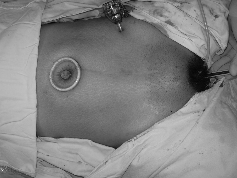
The Alexis® wound retractor XS (Applied Medical, Rancho Santa Margarita, CA) was inserted in the umbilical area with a left ancillary trocar insertion (patient 16).
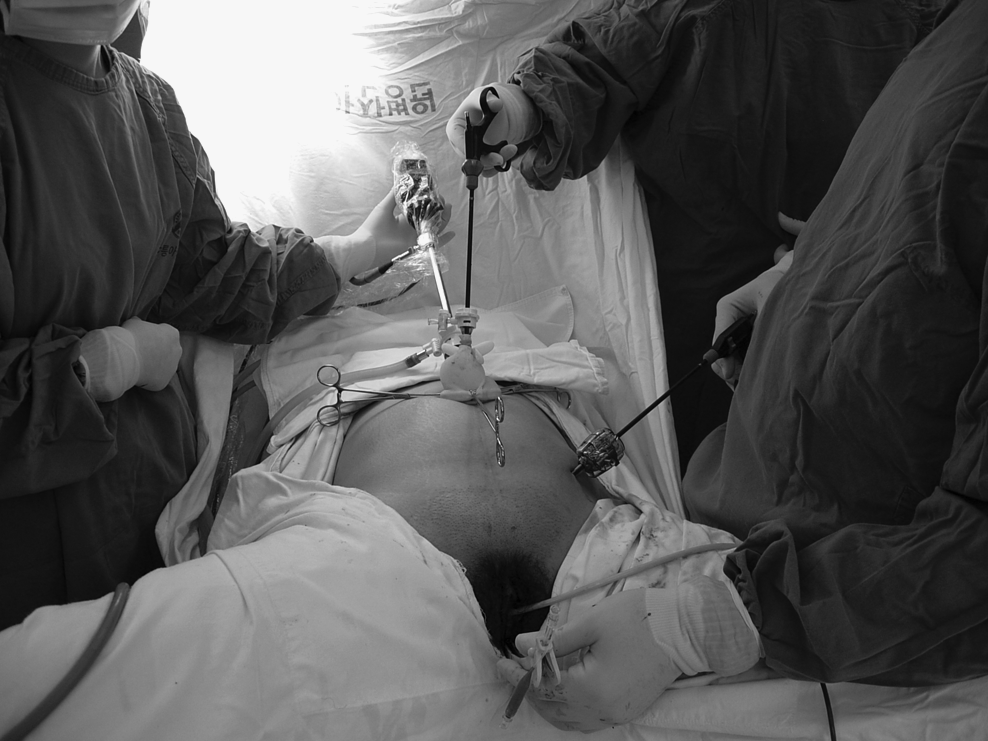
Two trocars were inserted into two fingers of a no. 6 surgical rubber glove and ligated with rubber bands; the other three fingers of the glove were ligated with each other. The wrist portion of the glove covered the wound retractor, and the edge of the wound retractor was clamped with three Babcock clamps for enhancement to prevent carbon-dioxide gas leakage (patient 16).
Adnexal surgery procedure
The pelvic masses were mostly ovarian cysts and tumors. The procedures performed were adnexectomy, cystectomy, or myomectomy in the case of pediculated subserosal myoma confused with an adnexal mass. To perform the adnexectomy, the ipsilateral round ligament was divided with a monopolar hook after coagulation with bipolar forceps. The avascular triangle zone of the ipsilateral broad ligament, made by the round ligament, infundibulopelvic ligament, and external iliac vessels, was desiccated with electrosurgical devices and penetrated to ligate the infundibulopelvic ligament. The infundibulopelvic ligament was ligated with an extracorporeal, Vicryl-modified Roeder knot used with an Endoknot® (Ethicon, Somerville, NJ) and religated with an Endoloop® (Ethicon, Somerville, NJ). To perform a cystectomy, the cystic surface was excised with electrosurgical devices; a cystic wall dissection was performed with a laparoscopic dissector, and bleeding control was performed.
The specimen was removed through an Alexis wound retractor XS of the umbilical port (Fig. 3). Large cystic masses were aspirated with a suction-irrigation system after puncture on the cystic surface exposed through the wound retractor and punctured site was sutured and ligated to minimalize the cystic fluid leakage. The collapsed masses could be easily removed through the wound retractor. The laparoscopic endopouch bag was used for the removal of specimen, if needed. After specimen removal, saline irrigation was performed. The drainage bag was connected though the left 5-mm port site, and the wounds were sutured.

The excised specimen was removed through the Alexis® wound retractor XS (Applied Medical, Rancho Santa Margarita, CA) in the umbilical port (patient 16).
Results
Nineteen patients were enrolled in the study (Table 1). The operative indications were pelvic masses. The operative procedures were adnexectomy (n = 8), myomectomy (n = 1), ovarian cystectomy, and/or salpingectomy (n = 10). There were no operative complications, conversion of the operation to laparotomy, or additional trocar insertion. All patients exhibited uneventful recovery periods and were discharged within 4 days postoperatively. The mean operation time was 81.3 ± 28.7 minutes. The longest operation time was 135 minutes, but the operation time tended to shorten as more cases were done. The pathologic diagnoses were mature cystic teratoma (n = 6), benign cyst (n = 4), endometrial cyst (n = 3), serous cystadenoma (n = 3), mucinous cystadenoma (n = 1), leiomyoma (n = 1), and tubo-ovarian abscess (n = 1). Eight patients had an ovarian cystic mass associated with adnexal torsion. The mean postoperative hospital stay was 4.0 ± 1.3 days. Most patients were not discharged if they did not want to be. The longest postoperative hospital stay was 8 days; in this case, the patient was treated with antibiotics for pelvic inflammatory disease associated with a tubo-ovarian abscess. The mean hemoglobin change between the preoperative hemoglobin and the postoperative 2-day hemoglobin was 2.1 ± 0.9 g/dL. The mean time of first passage of gas was 28.3 ± 7.7 hour. The postoperative wound pain was evaluated with a 100-mm visual analog scale at postoperative days 1 and 2. The mean of pain scales at postoperative days 1 and 2 were 38.6 ± 21.5 and 20.6 ± 19.4, respectively (Table 2). 2 The clinical follow-ups were performed on 2, 6, and 10 weeks after surgery. In all cases, no wound problem was observed (Fig. 4).
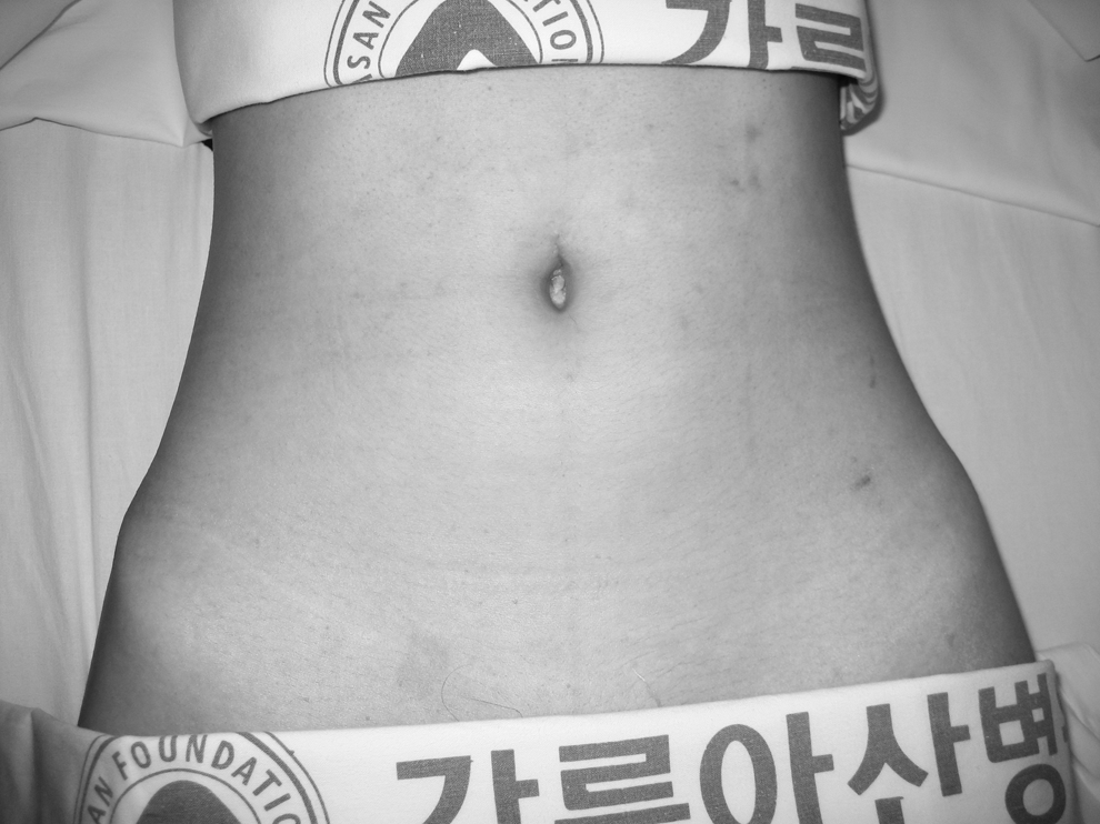
Abdominal wound scar after 5 weeks postoperatively (patient 2).
BOC, bilateral ovarian cystectomy; LOC, left ovarian cystectomy; BTL, bilateral tubal ligation; LS, left salpingectomy; RSO, right salpingo-oophorectomy; LSO, left salpingo-oophorectomy.
Hemoglobin change was the difference between the preoperative hemoglobin and the postoperative 2-day hemoglobin.
Postoperative pain was evaluated with a 100-mm visual analog scale at 1 and 2 days postoperatively.
Discussion
Many benign ovarian tumors have been removed under laparoscopy. The literature shows that many surgeons perform laparoscopy to remove benign ovarian masses. Some researchers compared laparoscopy with traditional laparotomy for dermoid cysts and suggested that laparoscopic management of dermoid cysts is safe, cost effective, and provides patients with the benefits of a shorter hospital stay and recovery time. 3 But, in large pelvic masses, laparotomy has been preferred to laparoscopy because of the possibility of malignancy and limitation of the surgical field. Because ovarian cysts are common in adolescence and during reproductive years, many surgeons have tried to perform ovarian cystectomies to preserve ovarian function. But, this procedure increases the incidence of intraoperative cyst rupture. Due to the risk of aseptic peritonitis associated with the intraoperative rupture of a dermoid cyst, many surgeons are reluctant to remove the lesions laparoscopically. However, some researchers reported laparoscopic excision of dermoid cysts with controlled intraoperative spillage. They suggested that intraoperative spillage of dermoid cysts need be not associated with morbidity as long as vigorous lavage is performed. 4 For this reason, we have tried to perform adnexectomies and cystectomies with a minimal leakage of cystic fluid by using laparoscopic endopouch bags or puncture-site ligations after cyst aspiration. After the removal of the mass, a pelvic-cavity lavage with normal saline was performed.
We reported three-port laparoscopy in 2 cases of large pelvic masses with the extension of the left iliac fossa port. The size of the extension port was about 2.5 cm and the cystic surface was exposed and aspirated. After cyst aspiration, the cyst puncture site was sutured and ligated. A 12-mm trocar was inserted and ligated in the extended port with a full-layer suture using Vicryl® 1.0 (Ethicon, Somerville, NJ). In this technique, conventional laparoscopy can be performed while maintaining a pneumoperitoneum. However, the disadvantage of this technique was that the left iliac fossa wound scar was relatively large and decreased the cosmetic effect.5,6 An extension of the port site was performed to remove large cystic tissues. With a conventional 5- or 10-mm port size, it is a difficult, time-consuming procedure to remove a large amount of cystic tissues. For this reason, the extension of the lower abdominal port site may be needed with a corresponding increase in the postoperative wound scar, especially in cases of huge pelvic masses. However, the extension of the umbilical port site would provide less exposure for the postoperative wound scar, resulting in minimal abdominal wound scarring. In our cases, the mean diameter of the masses was 8.3 ± 4.7 cm, including the largest mass, which was an approximately 25-cm mature cystic teratoma. Therefore, this two-port technique, using a multichannel port, has an advantage in cases of benign large pelvic masses.
Laparoscopy is a minimally invasive operative procedure that is performed worldwide. One advantage is less postoperative pain than from a laparotomy because of the smaller skin incisions. As a result, the hospital stay is shorter and the cosmetic effect is superior. However, multiple puncture wounds performed in conventional laparoscopy may decrease patients' satisfaction with the cosmetic effect. In a prospective, randomized, controlled trial of two- versus four-port laparoscopic cholecystectomy, the researchers recommended a two-port procedure in elective laparoscopic cholecystectomy because it resulted in less port-site pain while retaining a similar clinical outcome. It also resulted in fewer surgical scars than a four-port laparoscopy. 7 Some researchers reported the comparison of two- and three-port laparoscopic appendectomy. 8 They concluded that two-port laparoscopically assisted appendectomy is safe and effective because of less anesthesia time and no Endoloop, Endoclip, or Endo-GIA. In a study on the outcome of a randomized trial comparing laparoscopic and minilaparotomy cholecystectomy, no differences were found between the two in long-term outcomes for either abdominal pain or cosmetic satisfaction. 9 Multipuncture wound scars from laparoscopy and the minimalized postoperative wound scars of minilaparotomy were found to be equivalent in terms of patient satisfaction.
In our series, postoperative outcomes were evaluated, including operation time, hemoglobin change, first passage of gas, pain scales, and postoperative hospital stays (Table 2). Compared with the group of cases (n = 54) who underwent conventional three-port laparoscopy for pelvic masses in our clinic from March 2008 to August 2008, pain scales for postoperative day 1 (POD1) were 38.6 ± 21.6 vs. 40.7 ± 21.3 and for POD 2 were 20.6 ± 19.4 vs. 26.8 ± 18.7. The mean of the pain scales in the two-port group were less than those of the conventional three-port group, but the differences were statistically insignificant, according to a Mann-Whitney test (POD#1, P = 0.690; POD#2, P = 0.095). As this comparison has some limitation in the number of study groups and design, prospective, randomized, case-controlled studies are still needed.
The mean postoperative hospital stay was 3.9 ± 1.3 days in our cases. This result is relatively longer than that of the cases in hospitals in other countries. In Korea, patients tend to want to stay at the hospital for 4 or 5 days after an operation. This situation may be due to the relatively low medical expenses in Korea and patients' anxiety about short hospital stays. With these reasons, most patients were not discharged if they did not want to leave.
Considering the trend of minimally invasive surgery, conventional laparoscopy, using multiple punctures, may invoke an adverse effect in term of cost effectiveness and reduction of postoperative pain. Because multiple punctures induce multiple postoperative wound scars in the patient's abdomen, the patient's cosmetic satisfaction may be much lower than expected. Some researchers reported one-port ovarian cystectomy, one-port salpingectomy, one-port hysterectomy, and supracervical hysterectomy in gynecologic surgery.10–12 The one-port operative procedure was usually performed by using a laparoscope with an operating channel for electrosurgical devices or it used traction sutures. Excised specimens were removed through the extended umbilical port site. Laparoscopic devices through a 5-mm operating channel have a limited range of motion because of coupling with the laparoscope; therefore, it is hard to perform complex procedures with operating channel devices. Rao et al. performed 20 cases of single-port laparoscopic cholecystectomy by using an R-port system and graspers with an angulated shaft. The investigators used the port-closure needle or a 5-mm extra epigastric port to ensure the visual field of operation. 13
Many cases of laparoscopy have been performed with the use of complex procedures, such as sutures and ligations. Moreover, a drainage tube needs to be inserted in the pelvic cavity for postoperative bleeding. For these reasons, two-port laparoscopy is considered a safer operative technique than the one-port procedure with using the present laparoscopic instruments. Some researchers introduced a quasiscarless adnexal operation by using two trocars and suggested that the use of the two-trocar technique is safe and highly appreciated by patients because it does not leave visible abdominal scars. 14
We used an Alexis wound retractor XS for the multichannel port, applying the method used by Ryu et al. 15 The wound retractor was inserted, extending the umbilical wound to about 1.5 cm, and the wrist portion of a no. 6 rubber glove covered the wound retractor with two trocars inserted in the fingers of the glove. The edge of the wound retractor was clamped with three Babcock clamps to prevent carbon-dioxide leakage with the motion of the laparoscope and laparoscopic instruments. With this method, a pnemoperitoneum was maintained during the operation and we could gain a multichannel port. Takeda et al. performed gasless laparoscopic-assisted adnexal surgery with a wound retractor for a large ovarian cyst. In this technique, three or four ports were used with the subcutaneous abdominal wall-lift method. 16 Postoperative abdominal wounds were the same or larger than conventional laparoscopy in this method.
Gynecologic surgery with a wound retractor was reported in the case of a large leiomyoma operation performed with hand-assisted laparoscopy in 1999. In this case, the investigators operated with the surgeon's hand in the air-sealing hand-access system. 17 It permits more rapid removal of the specimen than conventional laparoscopy does, resulting in a reduced operating time. 18 In our experience, a large ovarian mass of a 17-year-old girl was removed easily through the wound retractor port, saving operation time. Therefore, this technique has been shown for potentially wide application to adnexal surgery, including the cases of large ovarian cysts.
Conclusions
The main disadvantages of this two-port technique are that it is a slightly time-consuming procedure, and the laparoscopic instruments and laparoscope in the umbilical port have a limited range of the motion because of coupling. In addition, this technique needs a wound retractor and has limited application in cases of malignancy and extreme obesity. The cosmetic effect is the main advantage of this two-port technique, and no additional cost is required due to the use of three trocars, a wound retractor, and conventional laparoscopic instruments, compared with a conventional four-port laparoscopy. However, in single-port surgery, a flexible scope or other instruments are needed, and this method can be performed in selective cases. Therefore, the two-port method appears safe and cosmetically effective, especially in case of large, benign pelvic masses. This technique also does not incur additional costs, as conventional laparoscopic instruments are used. This technique could be performed safely in most cases in adnexal surgery, including for a large, benign cystic mass, with minimal abdominal wound scarring.
Footnotes
Acknowledgment
This operation technique was presented at the poster session of the 9th Annual Congress of the Asia Pacific Association for Gynecologic Endoscopy and Minimally Invasive Therapy on April 24, 2008, in Daegu, Korea (APAGE 2008 Korea).
Disclosure Statement
No competing financial interests exist.