
Abstract
Abstract
Introduction:
Bile leakage is a common complication of cholecystectomy. The aim of this study was to compare endoscopic sphincterotomy (EST) and biliary stenting (BS) in the treatment of bile leaks after cholecystectomy.
Patients and Methods:
Twenty-seven patients with bile leakage following cholecystectomy underwent endoscopic retrograde cholangiography (ERCP). Patients were randomized into two groups (EST and BS ± EST), according to the initial therapeutic endoscopic intervention. The patients were allocated into subgroups once more, according to diameter of the common bile duct (CBD). Outcomes and efficacy of BS and EST on fistula closure and the time to fistula closure were investigated.
Results:
The median time between cholecystectomy and ERCP in the EST and BS groups was 6.45 ± 3.41 and 4.50 ± 1.99 days, respectively. The mean daily amount of biliary leakage in the EST and BS groups was 376.92 ± 243.77 and 441.07 ± 216.08 cc/day, respectively. The diameter of the distal part of CBD in the EST and BS groups was 9.07 ± 3.84 and 8.28 ± 4.04 mm, respectively. Mean fistula closure was achieved in 6.45 ± 3.41 in 11 of 13 patients in the EST group and 4.50 ± 1.99 days in the BS group in all patients. However, mean time of closure was significantly shorter in the BS 4.71 ± 2.14 group, compared to EST (9.67 ± 2.51), among patients with distal CBD diameter (≤8 mm).
Conclusions:
BS seems to be a more effective method than EST in the management of postcholecystectomy among patients with bile leakage and without CBD dilatation. BS might be the first-line treatment among such patients. However, further prospective, randomized, clinical trials regarding CBD dilatation are warranted.
Introduction
Diagnosis is usually made upon the detection of bile drainage through the drain or the manifestation, on ultrasonography or computerized tomography (CT), of an intra-abdominal collection or a biloma formation. The primary management of the patient with bile leakage consists of the drainage of the collection and treatment of the bile leakage. Bile leaks can be managed either conservatively or through percutaneous drainage, or endoscopically or by surgical intervention. Endoscopic retrograde cholangiopancreatography (ERCP) is an effective, safe method for the diagnosis and management of bile leakages after cholecystectomy. Once the patient is stabilized, ERCP is usually performed to determine the localization and the severity of the injury. Further, ERCP has become an established, effective treatment for bile leakage without need for further surgery.5,9,12 The management of fistulae due to major bile duct injuries is surgery. Morbidity and mortality rates are high for this kind of surgical intervention. The majority of fistulae caused by minor bile duct injuries can be managed by ERCP. Endoscopic treatment modalities include endoscopic sphincterotomy (EST), biliary stenting (BS), and nasobiliary drainage (NBD).3,9,11,13 The aim of these treatment modalities is to facilitate biliary flow in to the duodenum through the elimination of the duodenobiliary pressure gradient.5,9
It has been reported that fistula treatment by endoscopic approaches has 70%–100% success rates.3,5,9,11,13 However, no controlled, comparative, clinical trials between EST, BS, and NBD, to determine the optimal method, have been reported. We have taken into consideration that stasis due to possible ampullary edema secondary to EST increases the risk of post-ERCP pancreatitis, and that stents with wider diameters positively influence the fistula closure. The aim of this study was to compare EST and BS in the treatment of bile leakage following cholecystectomy and influence of the diameter of the distal bile duct on endoscopic recovery of fistula.
Patients and Methods
From January 2000 to December 2003, 27 patients who had undergone ERCP for bile leakage after cholecystectomy and were candidates for endoscopic treatment for their minor injuries were included in our study. The study was conducted after authorization of the local ethics committee, and informed consents of the patients were obtained. Of these patients, 25 were referred to our Endoscopy Department from other university or public training hospitals and 2 were operated at our hospital. Patients were informed about the indications and possible complications during the course of ERCP before the procedure, and informed consents were obtained. None of the patients received nonsteroidal anti-inflammatory drugs or prophylactic antibiotics. After sedation, the duodenum was visualized by a side-viewing duodenoscope (Fujinon ED 201 and 250 × 5T; Wayne, NJ; or Olympus 240; Tokyo, Japan), and hyoscine butylbromide was then administered to decrease peristalsis.
Patients were randomized into two groups (EST and BS ± EST), according to the initial therapeutic endoscopic intervention. The EST group consisted of cholelithiasis patients who underwent EST (EST group, n = 13). The BS group consisted of cholelithiasis patients who underwent EST initially and followed by BS after stone removal, and those without any gallstone who underwent BS (BS group, n = 14). EST and BS patients were randomized into two subgroups, according to the distal diameter of the CBD (≤8 and >8 mm). For the distal diameter of CBD measurement, the two marked opposite points in the lumen wall were measured in millimeters from the computerized digital images transferred by Medgate software (Medgate, Toronto, Ontario, Canada). The success of the procedure was determined according to fistula closure and time to closure, complications, and required surgical intervention.
Selective cannulation of the CBD was attempted with the standard cannula and a guidewire. Great attention was paid to avoid cannulation of the pancreatic duct. If the selective cannulation of the CBD failed, a small, precut sphincterotomy with a needle-knife was performed. After deep cannulation of the CBD, serial cholangiographic examinations were obtained to confirm the location of bile leakage (Fig. 1). A relevant sphincterotomy was performed with the standard sphincterotome, and blended current was used. The CBD was evaluated by a Dormia basket and balloon catheter, and all stones were extracted to the duodenum in the presence of stones in the CBD. In the EST group, patients who did not demonstrate a significant decrease in fistula output 5–7 days after the procedure underwent BS as a second intervention. Patients without choledocholitiasis in group 2 did not undergo EST after the localization of the bile leakage was determined. Those with choledocholitiasis underwent EST and BS after stone extraction. Bile leakages were treated by the insertion of 10-F, 6-cm plastic stents to lower the pressure of the biliary system by bypassing the biliary sphincter. Short stents were used and bridging the bile leakage site was avoided.
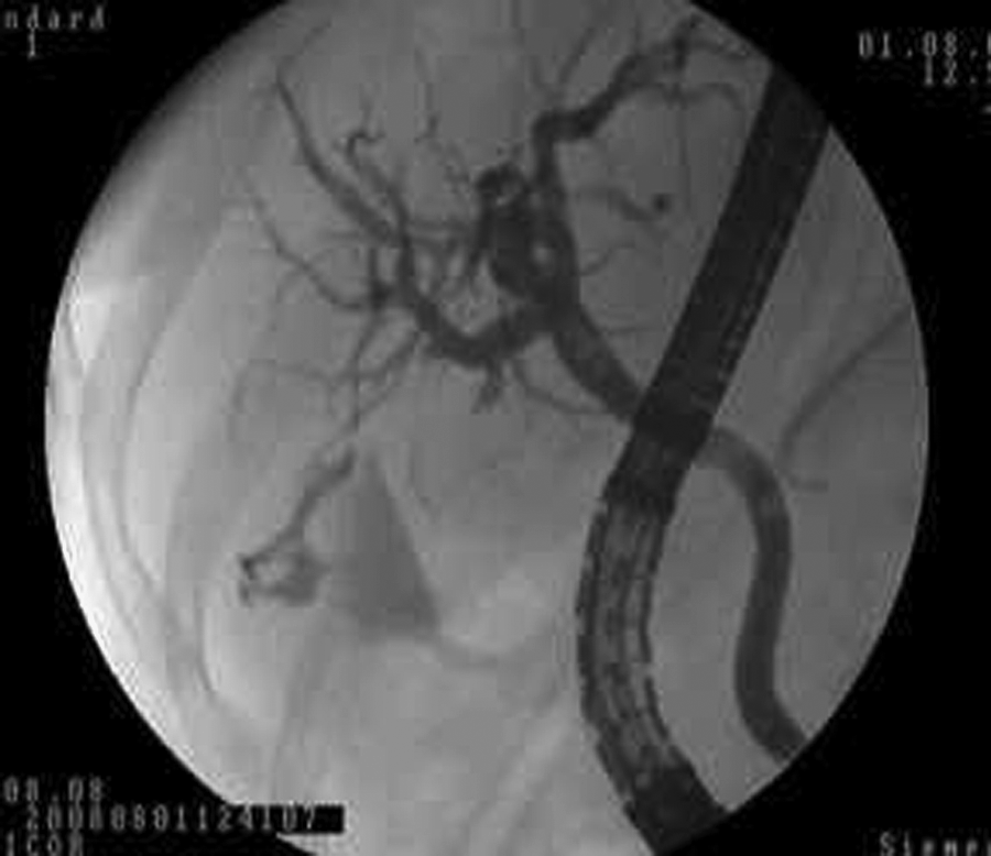
Bile leakage from the duct of Luscka in the patient with postoperative persistent bile fistula.
The stents were removed after 3–6 weeks, when the fistula had totally ameliorated. Patients were evaluated for complications 2 hours, on the first day, and at the end of the first month after the procedure. Complaints, physical examination findings, complete blood counts, and serum amylase values were recorded. Post-ERCP complications were defined according to the consensus criteria reported by Cotton et al. 14 Both groups and their subgroups were compared according to time to postoperative ERCP, bile output from the fistula, distal diameter of CBD, and time to fistula closure.
Statistical analysis
NCSS 2007 and PASS 2008 Statistical Software (Kaysville, UT) was used for statistical calculations. The Student's t- and Mann-Whitney U tests for quantitative calculations, chi-square for qualitative calculations, as well as descriptive analysis, were used. The Pearson correlation test was used to valuate the relationship between the parameters. The results were evaluated with a 95% confidence interval.
Results
The study population consisted of 27 patients (17 women and 10 men), and the mean age of the group was 52.63 ± 15.34 years. Four patients from the EST group and 5 from the BS group had undergone an open cholecystectomy operation. Bile leakage was mostly found at the site of the cystic duct stump. Stones in the CBD were detected in 5 cases in the EST group and 4 cases in the BS group. Stones were extracted into the duodenum in all patients.
Patients' characteristics are demonstrated in Table 1. There were no differences between mean age, sex, distal common bile duct diameter, bile output from the fistula (cc/day), and time to postoperative ERCP (P > 0.05). There was no correlation between fistula bile output and closure time (r = −0.078; P = 0.820) or fistula bile output and diameter of common bile duct (P = 0.366) in the EST group. There was no correlation between fistula bile output and closure time (r = 0.297; P = 0.302) or fistula bile output and diameter of common bile duct (P = 0.926) in the BS group, either. Time to closure (median 10 days) was increased among patients with diameter of the common bile duct of ≤8 mm (average ± SD: 9.67 ± 2.51), compared to patients with a diameter of the common bile duct of >8 mm (average ± SD: 5.25 ± 2.96) (P = 0.049). Time to fistula closure was similar between the groups with respect to diameter of common bile duct in the BS group (P = 0.744) (Table 2).
Student t-test.
Chi-square test.
EST, endoscopic sphincterotomy; BS, biliary stenting; SD, standard deviation; OC, open cholecystectomy; LC, laparoscopic cholecystectomy; CBD, common bile duct.
Mann-Whitney U test.
P < 0.05.
EST, endoscopic sphincterotomy; BS, biliary stenting; SD, standard deviation.
There was no difference between fistula bile output, CBD diameter, closure time, and time to postoperative ERCP among patients with a common bile duct diameter of ≤8 and >8 mm subgroups of the EST and BS groups (P > 0.05). Median fistula closure time among patients with a diameter of the common bile duct of ≤8 mm in the EST group (10 days) was different from those of the BS group (5 days) (P = 0.028). The results of the groups are shown in Table 3, according to distal diameter of the common bile duct. Bile leakage improved in 11patients (84.6%) in the EST group, while all patients (100%) in the BS group were healed; however, this difference was not significant. Two cases in the EST group underwent BS due to failure of fistula closure and completely recovered. BS with ERCP was applied to 2 patients for a second time in the EST group, and the fistula closure was accomplished, but increased the number of the procedures. None of the patients in either group had mortality or morbidity related to ERCP.
Mann-Whitney U test.
P < 0.05.
EST, endoscopic sphincterotomy; BS, biliary stenting; SD, standard deviation.
Discussion
Therapy options for bile leakage following a cholecystectomy include rapid diagnosis and percutaneous drainage and early management with surgical or endoscopic interventions. Estimated morbidity and mortality rates for surgical management of bile leakage following a conventional cholecystectomy are as high as 11 and 37%, respectively.15,16 However, the endoscopic approach to the diagnosis and treatment of bile leakage provides more successful results without significant morbidity and mortality rates.17–20 The aim of endoscopic therapy is to eliminate the pressure gradient between the common bile duct and duodenum by decreasing the pressure of the sphincter of Oddi. Bile drainage into the duodenum, instead of leakage and closure of the leakage, has been obtained by this approach.5,10,12 Endoscopic interventions through biliary sphincterotomy alone, BS with or without a sphincterotomy, and NBD with or without a sphincterotomy, use of self-expanding covered metal stents, and the recent use of biodegradable stents have been very effective in the management of all kinds of biliary leaks. All endoscopic techniques are based on the principle that eliminating the rise in pressure inside the bile duct by promoting decompression in the form of stent placement/sphincterotomy promotes healing of bile leaks.3,5,10,12,14,21–25
NBD is one of the endoscopic treatment modalities for bile leaks. The advantages of NBD are that it can be removed without performing a new endoscopy, and that cholangiography can be performed without a new endoscopic intervention. Disadvantages of NBD include discomfort for the patient, fluid loss, electrolyte imbalance, and, possibly, prolonged hospitalization time.10,25 Experimental studies have shown that BS is superior to EST, in terms of the decline in the duodenobiliary gradient and in obtaining fistula closure in a shorter period of time. However, no prospective, randomized, clinical trials comparing these two methods have been reported.7,10,24 In the experimental canine models, Marks et al. found that CBD pressure was significantly lower in the BS applied group than the sphincterotomy group, and bile leakage closure time was significantly shorter in BS application group.13,24 In a similar study, no difference was observed in efficacy and in time for the treatment of bile leak by sphincterotomy with endoprosthesis or endoprosthesis alone in patients with bile leak after surgery. Thus, the study suggested that sphincterotomy with endoprosthesis or endoprosthesis alone is equally effective in the management of postoperative bile leak. 26 Another study suggested that equalizing biliary and duodenal pressures with a short transpapillary stent is an effective therapy for bile leaks. 27 We have also applied the short stent without bridging.
Bile duct injuries are classified according to the presence of stricture and localization. 28 The localization of leakage is shown in Table 1. It is known that bridged stent application is more successful among patients with strictures. 29 We used short stent in our patients because they were of types A and B. 28
A relationship between bile output of the bile leakage and closure time was demonstrated previously. Among patients with low-grade leakage, EST resulted in 91% of the patients, whereas BS placement among patients with high-grade leakage resulted in closure of bile leaks in all patients. 30 The fistula bile output and time to intervention were similar in the EST and BS groups, so that the subgroups defined with respect to diameter of CBD improved our results. Although a therapeutic-approach decision was made according to the flow rate, the effectiveness of the treatment was evaluated with different parameters.12,26
The most important disadvantage of the BS procedure is the requirement of a second intervention to remove the stent. The removal of the stent generally takes 30 minutes, and the majority of patients have minimal stress, if mild sedation is administered. 23 Complications of stent placement include biliary obstruction, cholangitis, migration of the stent, and pancreatitis. 21 Stricture development is a complication of treatment of biliary leakage and is seen less after BS, compared to NBD. 29
Complications of EST are classified as early- and late-term complications. Early complications of EST include bleeding, duodenal perforation, and pancreatitis. 20 Late complications are still not clear. Sherman et al. reported that the performance of EST on individuals with nondilated bile ducts leads to higher complication rates. 31 Reasons for failure of EST are incomplete cutting of the sphincter muscle or postsphincterotomy edema, spasm, or stenosis.25,31,32 The local inflammatory process following the cutting of the sphincter by electrocoagulation prevents the sudden decrease in biliary pressure. 18
Conclusions
We have performed a subgroup analysis, according to the presence of distal CBD dilatation, in order to see if fistula resolution differs among EST and BS groups. With previous studies based on flow-rate, incision-site support therapy with both EST and BS, the optimal treatment modality is yet to be defined. Our study had a different approach, as it was based on procedure choice, according to the diameter of CBD. In this study, BS was found to be superior to EST, in terms of fistula closure, in patients without dilatation in the distal common bile duct, although our patient population was small. This might be due to marked temporary stasis in the nondilated distal common bile duct as a result of the post-EST inflammatory process. Our study indicates that BS seems to be a more effective method than EST in the management of bile leakage in postcholecystectomy patients without CBD dilatation. BS might be the first-line treatment among these patients. However, further prospective, randomized, clinical trials regarding CBD dilatation are warranted.
Footnotes
Disclosure Statement
No competing financial interests exist.
This study was presented as an oral presentation at the 12th EAES Congress, June 9–12, 2004, in Barcelona, Spain.