




Abstract
Abstract
Introduction:
Portal vein thrombosis (PVT) could be a life-threatening complication after splenectomy if not diagnosed promptly and treated properly. Risk factors of PVT are not completely clarified. Spleen size and underlying hematologic diseases are main potential risk factors for this complication. Laparoscopic surgery might increase the risk of developing PVT, as it reduces the blood flow in the portal system due to the pneumoperitoneum but, on the other hand, it seems to be associated with less postoperative modifications of coagulation parameters than open surgery, thus preventing PVT itself. The authors reviewed their series on open and laparoscopic splenectomies, pointing out their experience on PVT and discussing their surveillance and prophylaxis programs to prevent this complication.
Materials and Methods:
In this series, the authors report their experience on postsplenectomy PVT in 162 patients who have been splenectomised (102 operated on laparoscopically and 60 by open surgery).
Results:
PVT was clinically observed in 1 case out of 60 open splenectomies and in 3 cases out of 102 laparoscopic procedures. Patients were treated with conservative anticoagulation therapy. In one case, additional ileal resection was needed. Mortality was 0%.
Conclusion:
Low-molecular-weight heparin should be administered to all patients who have been splenectomised, especially if they are at high risk of PVT. If symptoms appear, patients need to be treated with high-dose heparin followed, after at least 3 weeks, by oral anticoagulant therapy.
Introduction
The actual incidence of postsplenectomy PVT is not clearly determined, ranging between 0.7% and 80%.2,7 Retrospective studies report an incidence of <2% but are not reliable, because most cases are not identified unless they become clinically evident. 1
Thrombosis can occur even as late as 2–4 months after splenectomy5,6 and, secondary to various degrees of portal vein system involvement, signs of this complication may vary from a subclinical radiologically detected lesion to a lethal event. 8 Recently, with the advent and the spread of laparoscopic procedures for the removal of the spleen, the interest in PVT has increased, as it has been suspected that a high incidence of this complication can occur in the laparoscopic setting. 4 In this article, the authors analyze their experience with postsplenectomy PVT in patients operated on with open or laparoscopic removal of the spleen. Incidence after open and laparoscopic procedures have been compared, and additional risk factors related to laparoscopic technique are discussed. According to the present study, recommendation of surveillance and treatment strategies are finally reported.
Materials and Methods
The charts of 162 patients operated on with splenectomies between January 1998 and September 2009 in the Department of General Surgery, Laparoscopic Surgery Unit of the University of Catania, were retrospectively reviewed. Ninety (55.5%) were women and 72 (44.5%) were men. Patients' age ranged from 8 to 83 years.
Sixty patients underwent open splenectomy, whereas the remaining 102 cases were splenectomised with laparoscopic technique.
Starting from May 2001, laparoscopic procedure for removal of the spleen was the treatment of choice; and open splenectomy was performed only when laparoscopic surgery was excluded, because of a massive splenomegaly preoperatively assessed by ultrasound or computed tomography (CT) scan.
Before the operation, all patients were submitted to hematologic routine test, electrocardiography, chest X-ray, ultrasound examination, and/or abdominal CT scan. Spleen size assessed by radiologists ranged from 10 to 14 cm in benign splenic diseases and from 18 to 24 cm in cases of malignant disease. Search for accessory spleen was also accomplished with CT scan.
Hemophylus influentiae, meningococcal and pneumococcal vaccinations, if not previously received, were administered at least 2 weeks before surgery.
Laparoscopic splenectomies were performed with patients lying in the right lateral position with the left flank elevated about 30° above the operating table. The trocars are positioned in the mid-clavicular line 10 mm above the transverse umbilical line (Hasson trocar), in the subcostal area laterally to the mid-clavicular line (5 mm trocar), in the left flank the level of the transverse umbilical line (12 mm trocar), and subcostal area in the anterior axillary line (5 mm trocar). After introduction of the trocars, diagnostic laparoscopy and detection of accessory spleens were accomplished. The procedure started with division of the gastrosplenic ligament with the short gastric vessels by means of ultrasonic dissector (Ultracision Harmonic Scalpel; Ethicon Endosurgery, Inc., Cincinnati, OH). At that moment, splenic artery was recognized, mobilized, and double clipped, to avoid bleeding during the following steps of the procedure. The convex surface of the spleen was then accurately dissected from the lateral abdominal wall and from the diaphragm. At that point, hanging the spleen, the splenic hilum was approached from the anterior aspect. Splenic artery and vein were clipped and divided, taking care not to damage pancreatic tail, using a stapler device (Endo GIA Vascular Stapler; Ethicon Endosurgery, Inc.). The spleen was then inserted in a plastic bag (endobag), which was pulled through the abdominal umbilical access, and was crushed and removed piece by piece. In malignant cases, the spleen was pulled out of the abdominal cavity through an accessory abdominal incision.
Patients postoperatively received antibiotic prophylaxis for 3 weeks.
In the last 20 cases, all the patients who were splenectomised were subjected to prophylaxis with a daily single dose of low-weight-molecular heparin.
Results
Indications for splenectomy are indicated in Table 1. In Table 2, associated surgical procedures performed in addition to splenectomy are reported. Accessory spleens detected preoperatively by CT scan in nine patients were removed during the surgical procedure. Mean operative time was 65 minutes in both laparoscopic and open procedures, ranging from 40 minutes to 1 hour and a half. No mortality was reported. In 12 patients (11.8%), laparoscopic splenectomy was converted to open technique (conversion rate: 4.3%).
Department of Surgery, Laparoscopic Surgery Unit, University of Catania, Policlinico V.Emanuele Hospital, Catania, Italy.
Department of Surgery, Laparoscopic Surgery Unit, University of Catania, Policlinico V.Emanuele Hospital, Catania, Italy.
PVT was diagnosed in 4 patients (2.5% out of all splenectomies), of whom 1 (0.7%) had been operated with the traditional open procedure and 3 (1.8%) with laparoscopic procedures. PVT occurred in one patient affected by idiopathic thrombocytopenia, in two cases of splenic lymphoma with marked splenomegaly, and in one patient with myelofibrosis and splenomegaly. Spleen weight was respectively 0.6, 2.5, 3, and 4 kg. Patients complained of symptoms consisting mainly of abdominal pain refractory to medical therapy in postoperative days 10–20. Diagnosis was confirmed by CT scan (Fig. 1). Treatment consisted of high dose of heparin infusion followed by anticoagulation therapy (warfarin for 6 months). In one case, laparotomy was performed; and resection of ischemic ileal segment was accomplished, followed by heparin treatment.
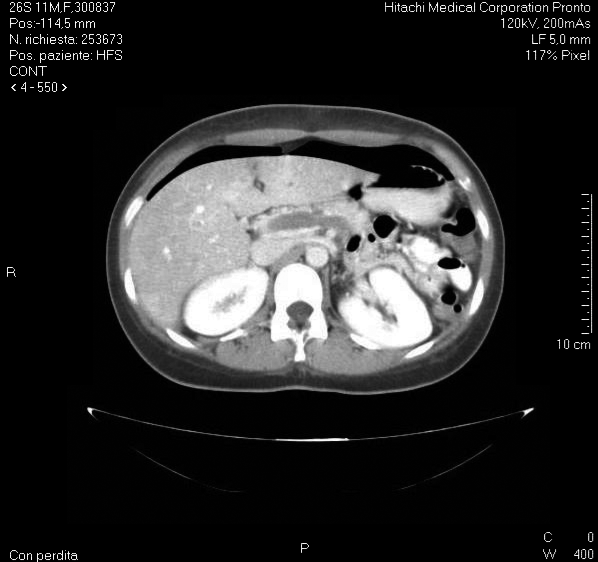
Computed tomography scan in a patient with postsplenectomy portal vein thrombosis.
At a follow-up of 1 year, CT scan showed resolution of PVT and no portal hypertension signs were observed.
Discussion
The incidence of postsplenectomy PVT is not clearly determined. In the literature, it varies from 2% to 10%; and mortality rate could be as high as 4.8%. 8 The low reported incidence may be underestimated due to difficulty in making the diagnosis. 2 Although early reports suggested a low incidence of this complication, modern radiographic imaging shows that PVT after splenectomy is not uncommon. 6
Risk factors in the development of PVT are related to any factor influencing the Virchow triad, 8 consisting of portal vein stasis, hypercoagulability state, and endothelium damage. Hematologic diseases requiring removal of the spleen may increase the risk for PVT. Myeloproliferative disorders, lymphoproliferative diseases, and splenomegaly have showed a high incidence of PVT, which can be, in these conditions, as high as 50% of all operated cases. 7
Laparoscopy has been evaluated as a risk factor for postoperative PVT in patients whose spleen is removed by this mini-invasive approach. Laparoscopic splenectomy, as a matter of fact, might increase the risk of developing PVT, as it reduces blood flow in the portal system veins during the performance of the procedure, due to the pneumoperitoneum. On the other hand, however, laparoscopy seems to be associated with less intraoperative and postoperative modifications of coagulation and fibrinolytic parameters than open surgery, conditions that might prevent vein thrombosis itself. 9
In the series reported in the literature, however, the extent to which laparoscopy is responsible for PVT remains unclear. Only few reports have been published on this issue. In the laparoscopic splenectomy series reported by Romano et al. 5 , in 7 cases out of 38 patients, PVT was demonstrated in a protocol of imaging surveillance using a Doppler ultrasound method. The complication was symptomatic in 4 cases (10.8%), an incidence that seems to be higher when compared with other studies in which splenectomy has been performed with an open approach.
On the contrary, in the 12 laparoscopic splenectomies reported by Valeri, 10 only 1 patient (0.08%) with idiopathic thrombocytopenia developed PVT. Our data have shown an incidence of 2.4% of PVT when either laparoscopic or open splenectomies are considered together. The incidence was slightly higher after laparoscopic removal of the spleen, as, in our series, in only one case after open surgery compared with three cases after laparoscopic procedures this complication occurred.
Clinical symptoms may be insidious, and progression can lead to intestinal infarction and portal hypertension. Clinical pictures may vary greatly but usually the major complaint is abdominal pain requiring re-hospitalization of the patients without clear subjective or objective abdominal signs, sometimes associated with fever. Decreased appetite, vague abdominal pain, nausea, and malaise may be initial symptoms that can be associated with abnormal lab results such as leukocytosis and elevated platelet counts. 11 Low fever is a constant finding not necessarily accompanied by leukocytosis. It is not connected to other septic events and its nature is unclear. Ascites is predominately present when the entire portal vein and its major branches are clotted and the collateral network is immature. Therefore, PVT should be suspected in patients with fever or abdominal pain after splenectomy or leukocytosis. 2 Other patients reported atipical symptoms such as ileus, fever, or diarrhea. 3
The above-mentioned symptoms may develop on day 10 and 15 after surgery (mean interval of 11.8 ± 4.6 days postoperatively 11 ), respectively. The short hospital stay after laparoscopic approach could even contribute to the difficulty of early detection of this condition and it could delay the early diagnosis of this condition unless routine imaging controls are planned after discharge. 6 In our experience, PVT occurred after the patients were discharged from the hospital, requiring their re-admission. Abdominal pain, resistant to any pain medication, was the predominant symptom. In one case, it was associated with signs of ileum and for this reason the patient was readmitted at night time and re-operated with resection of a segment of ileum that was the interest of intestinal infarction. We think that abdominal pain resistant to pain medication, not explained by any other clinical reason, in patients who were operated on with splenectomy could induce the surgeon to readmit the patients and to suspect a PVT, starting immediately an appropriate anticoagulant therapy.
For the diagnosis, abdominal ultrasonography and color Doppler flow imaging appear to have a reliable sensitivity and specificity rate; they are easy to perform, inexpensive, and repeatable.8,12 Nevertheless, their exact sensitivity and specificity rates have not been reported. 8 Abdominal CT scans with portography and magnetic resonance portography have been proved to be of higher accuracy than abdominal ultrasonography and color Doppler flow imaging, although they are more expensive and cannot provide objective hemodynamic data regarding flow direction and flow pattern. The contrast-enhanced CT scan showed in our cases the best accuracy for the diagnosis of PVT. It must be performed, in our opinion, and according to several authors, when any clinical abdominal pain appears after splenectomy. 13
Some authors suggest, in addition to routine ultrasound examination and CT scan, evalutation of D-dimer serologic levels in cases of splenectomy. 8 D-dimer test is considered of adequate sensitivity and of high negative predictive value in diagnosing thrombotic events,8,14 but the reported low specificity rate is expected because D-dimer levels are elevated in such different conditions as disseminated intravascular coagulation and sepsis.8,15,16 However, the negative predictive value of the test is significant. Negative test results, therefore, will strengthen the diagnostic value of a negative radiologic studies, and the need for more specific studies will be minimal. 8 No treatment strategy for PVT has been established. 17 Due to the risk of intestinal infarction, acute symptomatic PVT requires prompt intervention. 18 Treatment of the postsplenectomy PVT is not standardized. Early treatment is successful in all patients, whereas delayed treatment is ineffective. 2 Some authors treat PVT as any thrombotic episode with heparin followed by warfarin for a period of 4 to 6 months.8,19 When severe thrombocytosis occurs, initial treatment with antiplatelet drugs may prove beneficial. It is, therefore, advisable to administer antiplatelet agents (dipyridamole, acetylsalicylic acid) when severe thrombocytosis occurs postoperatively despite the surgeons' reluctance in their early use.8,20 According to some authors, the use of low molecular weight heparin (LMWH) in therapeutic doses is not clearly justified.8,19 Other authors use LMWH in the case of partial PVT, with patent vessel and asymptomatic patients. In their experience, the clot dissolved in <2 months and normal portal flow was restored. Thrombolysis with the use of tissue plasminogen activator or even thrombectomy has been occasionally reported but is not the standard of care.8,18,21 An approach to thrombotic prophylaxis should be considered in high-risk patients with myeloproliferative disorders and massive splenomegaly. Low-molecular-weight heparins have been proved to be of value in certain cases of antithrombotic prophylaxis.8,22,23 Therefore, perioperative therapy with LMWH followed by low-dose warfarin for the first several weeks after splenectomy or routine antiplatelet therapy with acetylsalicylic acid should be considered in selected patients. Some authors, on the contrary, affirm that subcutaneous heparin therapy alone seems to be unable to prevent the development of PVT in high-risk patients. 24 Our strategy in all the patients who have been splenectomised is now to start a prophylaxis with LMWH. If symptoms appear, LMWH is suspended and treatment with heparin at high dose is started and continued for at least 3 weeks. If abdominal symptoms at this time disappear, oral anticoagulant therapy with warfarin is continued for at least 6 months.
Conclusion
In summary, PVT is a relatively frequent complication that requires a high index of suspicion to establish an early diagnosis and appropriate treatment and prophylaxis.
Mortality is nearly 0%, if PVT is diagnosed in time and treated properly.
Diagnosis is sometimes difficult, but in any patient with abdominal pain after splenectomy PVT seems to be the most probable diagnosis and it needs to be ruled out with CT scan.
In the opinion of the authors, LMWH should be administered to all patients who have been splenectomised, especially if they are at high risk of PVT for their pathology or the size of the spleen. If symptoms appear, patients need to be treated with high-dose heparin followed, after at least 3 weeks, by oral anticoagulant therapy.
Footnotes
Disclosure Statement
No competing financial interests exist.