
Abstract
Abstract
Objective:
To report early and late results of laparoscopic-assisted endorectal Soave pull-through (LAEPT) with a short and V-shaped partial resection muscular cuff for Hirschsprung's disease (HD) over a 10-year period.
Methods:
The clinical courses of 218 patients who underwent modified LAEPT for HD were reviewed. LAEPT was described by the Georgeson technique. The main modifications included less dissection of the bottom of the pelvis, rectal mucosa dissection with a long cuff, coloanal anastomosis with a short cuff, and a V-shaped partial resection in the posterior wall of the muscular cuff.
Results:
From September 1999 to July 2009, 218 patients were operated on by the same surgeon. Ages ranged from 15 days to 12 years old. The aganglionic segment was located in the rectum or sigmoid colon in 176 patients, in the left colon in 38 children, and in the transverse colon in 4 patients. The median operating time was 176 minutes. Conversion to open surgery was required in 2 patients. Bleeding of the left iliac vein occurred in 1 patient, and a 180° twist of the neorectum occured in 2 cases. Median first bowel movement time was 23 hours, and median postoperative hospital stay was 10.4 days. Median daily defecation frequency was 4.6 within 2 weeks and 2.3 at 3 months postoperatively. The immediate postoperative complications included intestine herniation from the trocar site in 2 patients, perianal excoriation in 32 patients, and anastomotic leakage in 3 patients. Follow-up ranging from 6 to 120 months was obtained for 182 patients. Late postoperative complications included postoperative adhesive bowel obstruction (1.1%), enterocolitis (7.7%), anasomostic stenosis (2.2%), constipation (1.6%), and soiling (3.6%). Eighty-seven percent had excellent and good bowel function.
Conclusion:
LAEPT with a short muscular cuff with a V-shaped partial resection in the posterior wall is a safe and effective procedure for HD.
Introduction
Patients and Methods
This is a retrospective review of 218 consecutive patients with HD operated on between September 1999 and July 2009 by the same surgeon. Patients with total colonic aganglionosis were excluded. Preoperative diagnosis was made based on clinical presentation, barium enema study, rectoanal inhibitory reflex, and suction rectal biopsy. The diagnosis was further confirmed by intraoperative frozen biopsy and reconfirmed postoperatively by conventional histopathology on the resected specimen. In newborns and infants, patients were started on preoperative oral and intravenous antibiotics 1 or 2 days before the operation, and one or two episodes of colon irrigation were performed. In older children, classical preparation of the colon consisting of rectal irrigations and intravenous and nonabsorbable enteral antibiotics was initiated 3 days before the operation.
Modification of operative procedure
A three- to four-trocar technique was used. The first 5-mm trocar for the laparoscope was placed through the umbilicus. Seromuscular colonic biopsy specimens were taken to confirm the level of the transitional zone. In patients with classic forms, the peritoneal reflection was incised circumferentially, and mobilization started at the rectosigmoid. This was carried proximally until the colon pedicle was long enough to reach deep into the pelvis without tension. The anterior wall of the rectum was not dissected, and the posterior plane was dissected 2–3 cm distal into the pelvis, hugging the rectal wall. If the transition zone was proximal to the splenic flexure, subtotal colectomy was usually performed, with the preservation of the ileocolic pedicle.
Transanal dissection was started by placing six to eight traction sutures to retract the anus. A circumferential incision was made in the rectal mucosa at 0.5–1 cm proximal to the dentate line using electrocautery. A fine-tipped hemostat was used for the delicate dissection, and electrocautery was only used for coagulation through touching the hemostat (Fig. 1), to prevent injury to the mucosa and internal sphincter. To avoid injury to the ureter, vas deferens, and/or perirectal structures, endorectal dissection was continued upward in a circumferential manner until the level of the laparoscopic dissection. The muscular cuff was inverted and pulled out of the anus. It was then shortened, leaving a muscular cuff of 2–3 cm; with the posterior wall V-shaped partial resection, the pointed end of the V ought to reach the level of anastomosis (Fig. 2). Once the colon was pulled through the anus, the laparoscope was again used to ensure that the bowel was not twisted. The aganglionic and dilated segments were resected. Coloanal anastomosis was performed with interrupted fine absorbable sutures. Digital examination of the anus and anal dilatation were performed 2 weeks postoperatively on all the patients. If there was no anastomotic stenosis, anal dilation was done once or twice a week, lasting for 1–3 months. If there was anastomotic stenosis, anal dilation was done once or twice a day until the stenosis disappeared.
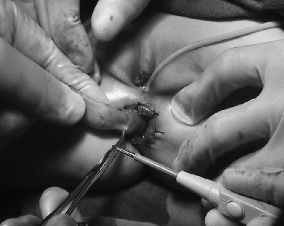
The electrocautery technique. A fine-tipped hemostat was used for the delicate dissection to grip submucosal vessels and tissues, and then electrocautery was only used for coagulation. The cut was through touching the hemostat; there was no direct contact between electrocautery and the rectal mucosa.

The V-shaped partial resection is made in the posterior wall of the rectal seromuscular cuff.
The variables studied included patient's age, surgical procedures, intra- and postoperative complications, and clinical outcomes. Enterocolitis was clinically defined by explosive diarrhea, abdominal distension, and systemic manifestations (i.e., fever) as described in a textbook. 4
Results
In this series, there were 152 boys and 66 girls, ranging in age from 15 days to 12 years (Table 1). The aganglionic segment was located in the rectum or sigmoid colon in 176 patients (80.7%), in the left colon in 38 patients (17.4%), and in the transverse colon in 4 patients (1.8%). Operative time ranged from 80 to 268 minutes (median, 176 minutes). There was no perioperative mortality. Conversion to open surgery was required in two patients because of abdominal distension.
In total there were 182 patients.
Intraoperative complications due to “technical error” occurred in 6 patients (2.8%). These included bleeding of the left iliac vein in 1 patient, difficulty in mobilizing the intraperitoneal colon due to heavy abdominal distension in 2 cases, and a 180° twist of the neorectum in 2 cases.
The time to first bowel movement postoperatively ranged from 8 to 36 hours (median, 23 hours). Hospital stay ranged from 7 to 18 days (median, 10.4 days). During the immediate postoperative period, trocar site hernia occurred in 2 newborns (0.9%). Thirty-two patients (14.7%) had temporary perianal excoriation. Three patients (1.4%) developed anastomotic leakage on postoperative days 5, 5, and 7, respectively, with one patient requiring a colostomy, whereas the other two cases resolved with conservative management. Follow-up was available for 182 patients (83.5%) (range, 6–120 months; median, 68 months). The frequency of daily defecation ranged from 3 to 12 times (median, 4.6 times) within 2 weeks and from 1 to 5 times (median, 2.3 times) at 3 months postoperatively. Two patients who had subtotal colectomy developed postoperative adhesive bowel obstruction, but resolution occurred with conservative treatment. Ten patients were readmitted to the hospital for postoperative enterocolitis, with 1 patient needing diversion for severe enterocolitis; the other 9 resolved on conservative management. Anastomotic stricture was seen in 4 patients, all of whom responded well to anal dilatation. One child who had residual aganglionosis required a second pull-through 13 months after the original operation (Table 2).
In total there were 182 patients.
Postoperative defecation outcomes are presented in Table 3. The 182 patients were divided according to the duration of postoperative follow-up: less than 1 years, less than 3 years, less than 5 years, and over 5 years. Overall, excellent to good continence according to Wingspread score was achieved by 87% (158 of 182) of patients; a fair result occurred in 10% and poor in 3%.
In total there were 182 patients.
Discussion
The laparoscopic approach has several important advantages over the traditional abdominal approach. It provides clear delineation of pelvic structures, faster postoperative recovery, and better cosmetic results.3,5 Laparoscopy can also identify the level of normal innervation, as suggested by the transitional zone. It can avoid twisting and tension of the pull-through segment and overcome the difficulty in dividing mesenteric vessels compared with the pure transanal approach.6,7 During the early learning phase, 2 patients required conversion to the open approach because of difficulty in maintaining an adequate operative space due to a distended proximal colon. Since then, we have routinely put a rectal tube in place to empty gases or liquids under laparoscopic surveillance in patients who had abdominal distension.
One of the most important parts of the LAEPT in transanal mucosectomy requires the exact dissection of the submucosal plane. Some doctors prefer to infiltrate the tissue with saline or diluted epinephrine solutions or to insufflate submucosal pressure-air for easier dissection and hemostatic effect.8–10 As slight bleeding will obscure the operative field, hemostasis needs to be controlled meticulously. Recently, electrocautery combining sharp and/or blunt dissection has been described, but iatrogenic mucosal perforations often occur.3,7 In our experience, rectal mucosa dissection was done by using an electrocautery and a fine-tipped hemostat together. The hemostat grasped the submucosal tissue and tiny vessels; then electrocautery touched the hemostat rather than directly contacted the mucosa to coagulate. This method provided better delineation of the submucosal plane due to the lack of bleeding.
Furthermore, it had the advantage of complete mucosectomy and smooth running and faster dissection without mucosal rupture. Muscular sheath infection is a characteristic complication of the Soave procedure. Damage to the mucosal tube, incomplete mucosal dissection, and infected hematoma after inadequate hemostasis are the main reasons for this complication.11,12
Our results showed that modified LAEPT was associated with a low rate of postoperative complications. Our series showed no muscular sheath infection. Anastomotic leakage was seen only in 3 patients (1.4%) in our series, compared with a rate of 5.6%–11.2% of cases in some series using open surgery13,14 and 1.5%–2.9% of cases using the transanal approach.15,16 Although anastomotic stricture was seen in 4 patients (2.2%), all these patients responded well to anal dilatation. This rate is lower than that reported for open surgery or the transanal approach.11,17 Anastomotic stenosis and leakage are supposed to be important risk factors contributing to enterocolitis. 18 The rate of enterocolitis in our series was 7.7%, compared with a rate of 17.4%–30% of cases using the transanal approach and leaving a long rectal seromuscular sleeve17,19 and a rate of 9.7% of cases using the laparoscopic approach with a short rectal seromuscular sleeve. 20 The relatively low incidence of enterocolitis in the current series may be due to the combined effort of the subjective diagnosis of enterocolitis, the short V-shaped partial resection of the muscular cuff, and anal dilatation as stated above. It is reasonable that other complications such as spasm of the internal sphincters, anastomotic stenosis, and constipation may also be decreased, as shown in Table 2.
We modified on the classical “Georgeson's technique” (the short and V-split cuff). The main modifications included less dissection of the bottom of the pelvis, rectal mucosa dissection with a long cuff, coloanal anastomosis with a short cuff, and a V-shaped partial resection of the posterior wall of the muscular cuff. Li et al. 21 also noticed that the incidence of enterocolitis and rectal stenosis decreased in the short and V-split cuff group in comparison with the long cuff group. Their techniques, different from our modifications, included an oblique mucosectomy and an oblique anastomosis with a short split muscular cuff.
In the laparoscopic colon pull-through, one can remove not only the aganglionic segment and transitional segment, but also the malfunctioned dilated segment. This may further reduce the rate of postoperative enterocolitis, as the bowel segment used in anastomosis has a normal caliber, and the coloanal anastomosis can be easily performed. Furthermore, the V-shaped partially resection of the muscular cuff could increase the space behind the rectum so that the storage function could be retained.
We had always been using suture string to gain exposure to the anus, until 2 years ago when we turned to use Lone Star retractors, which, to our experience, had better anal exposure. Both the methods did little injury to the sphincter and yielded fewer soiling problems. The soiling and increased frequency of bowel movements in several patients may be due to dissection too close to the dentate line or overstretching of the pulled colon,12,22 as in dissection of sigmoid mesocolon by the transanal approach or use of anal retractors. In the laparoscopic procedure, the proximal mesocolonic vessels may easily be divided intraperitoneally; this can avoid the potential impact of stretching of the sphincter for a prolonged time during the anal dissection.
Defecation function was satisfactory in our long-term follow-up. Of the patients, 87% had excellent to good continence according the modified Wingspread scoring system, and there was an improving trend over time. Regarding the urinary system, none of our patients experienced urinary incontinence. This may be mainly attributed to the pelvic dissection closely hugging the rectal wall and rectal mucosa dissection with a long cuff.
In conclusion, LAEPT leaving a short rectal seromuscular cuff and V-shaped partial resection of the posterior wall had satisfactory early and late results in our institution.
Footnotes
Acknowledgments
This research was supported by a grant (2006BAI05A06) from the National 11th Five-Year Plan (China) to support science and technology issues. We thank Dr. Kenneth Wong (Department of Surgery, The University of Hong Kong) for his efforts in editing this manuscript.
Disclosure Statement
No competing financial interests exist.