
Abstract
Abstract
Background and Purpose:
Although cystolitholapaxy is possible in children, the instruments are not available everywhere. For tiny impacted urethral stone, an alternate technique is described.
Case Report:
A 3-year-old boy came with retention of urine and dysuria. On examination: bladder was full, suprapubic region was tender, and a stone could be felt at posterior urethra. We planned push back and suprapubic cystolithotomy. After general anesthesia, the stone was pushed back by instillation of lignocaine jelly into the urethra. Then, a cystoscope was introduced, and a stone was found inside the urinary bladder. Under cystoscopic view, a 5 mm laparoscopic port was inserted into the bladder just above the pubic symphysis in the midline. A 5 mm tissue holding forceps was introduced through this port, and the tiny stone was grasped and brought out along with the port. The port site was closed by a single stitch. A penile catheter was kept for 5 days.
Results:
The boy did well in the postoperative period, and voiding was normal after removal of the catheter.
Conclusions:
Impacted posterior urethral stone can be retrieved by simple percutaneous technique.
Introduction
Case Report
A 3-year-old boy came with dysuria, hematuria, and retention of urine. On examination, the bladder was palpable, and the suprapubic region was tender. A stone could be palpated at the posterior urethra. We planned to push back the stone into the bladder and then to perform suprapubic cystolithotomy. In the operating room, the boy was given general anesthesia. Then, he was positioned supine on the table with both lower limbs suspended down from the foot end of the table. A 10 cc syringe was filled with lignocaine jelly, and the content was pushed through the urethra with little force. The stone in the posterior urethra traveled back into the bladder. An 8.5F 8° pediatric cystoscope was introduced into the bladder, and the stone was visualized (Fig. 1). The bladder cavity was filled with normal saline instilled through the inbuilt channel in the cystoscope. Then, a transverse stab wound was made just above the pubic symphysis, and a 5 mm laparoscopy trocar was introduced into the bladder under cystoscopic view. A toothed 5 mm forceps was introduced through this port (Fig. 2), and the tiny stone was grasped and brought out along with the port (Figs. 3 and 4). The port site was closed with a single stitch, and the bladder was left without sutures. A urethral catheter was kept for 5 days.
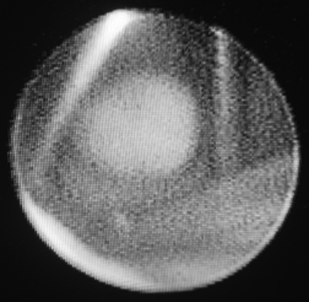
Cystoscopic view of grasping the stone.
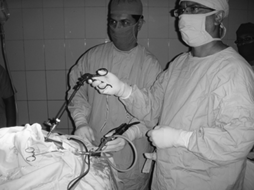
Grasper introduced under cystoscopic guidance.
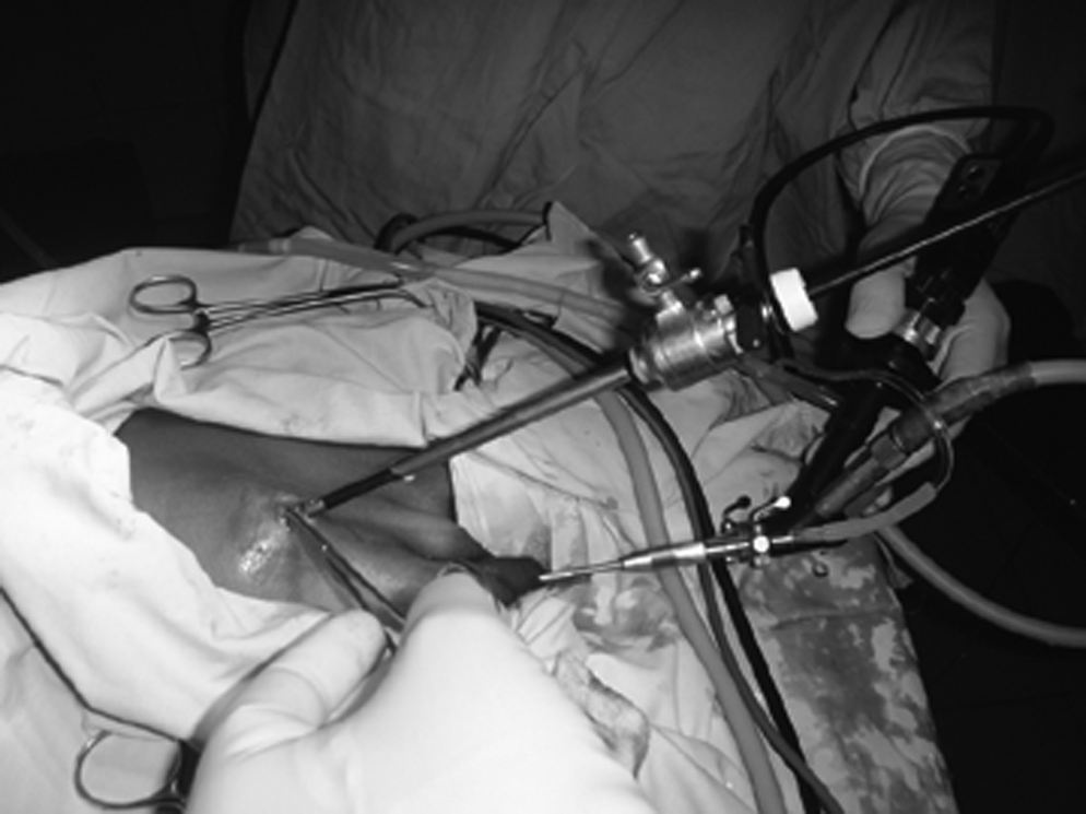
Stone retrieval in progress.

Stone coming out along with grasper.
Results
The total procedure took 18 minutes. The patient was discharged the next day with the catheter in situ, which was removed after 5 days without any problem.
Discussion
Over the years, the management of bladder stones has evolved into minimal invasive approaches from open surgery.8,9 Percutaneous cystolithotomy, extracorporeal shock wave lithotripsy, and transurethral cystolithotomy require special instruments and may prove unsuitable for small caliber urethra in tiny children.2,4,9 The stones that are impacted in the urethra mostly originate in the upper tract, 10 and some originate in the bladder. Some tiny stones traverse all the way to the navicular fossa and need simple meatotomy to be removed. The stones that get stuck in the posterior urethra are ideally treated by push back and cystolithotomy. In the older children, perurethral endoscopic litholapaxy is feasible. However, in small boys, a percutaneous suprapubic approach to the bladder is more suitable, 5 particularly when cystoscopy acts as an adjunct. 11
There are two types of bladder calculi. Primary bladder stones are also called endemic stones and are likely related to nutritional deficiencies. Secondary stones develop from encrustation of foreign bodies or as a result of chronically obstructed and infected urines.12,13 In our case, the urine culture revealed infection with Escherichia coli. Serum calcium level was within normal levels. Preoperative radiography and ultrasonogram revealed a normal urinary tract with no stone elsewhere. We presume this to be an endemic stone and it is unlikely to recur. 13
Conclusions
Cystoscopy-assisted percutaneous removal of impacted posterior urethral stone is a simple and safe technique.
Footnotes
Disclosure Statement
No competing financial interests exist.