
Abstract
Abstract
Background:
The persistent patency of the urachus after birth is a rare anomaly, especially because most of the cases are asymptomatic. The guiding symptom for urachal-umbilical sinus and urachal cyst is the presence of umbilical discharge. Even if today we tend to laparoscopic treatment, in scientific literature there is still no evidence, because the reports are rare and often they are clinical cases.
Methods:
Thirteen patients with a symptomatic urachal pathology were evaluated; 12 of these were affected by umbilical discharge and 1 by periumbilical discomfort. Five of 13 were prospectively treated by laparoscopy and the remaining 8 patients, who had been previously treated with conventional surgery, formed the control group. The authors report the laparoscopic technique used, which allowed the complete excision of the urachus.
Results:
The operation time was lower for the patients treated by conventional surgery (71.9 minutes versus 101 minutes; P=.002), whereas the control pain (P=.05) and, above all, the excised urachus length (11.6 versus 8.7 cm; P=.03) were in favor of the patients treated by laparoscopic surgery. We registered only one recurrence in a patient treated by conventional surgery.
Conclusion:
In the rare international scientific literature, only one study report comparative data, as our study. The results that we obtained seem to be in favor of the laparoscopic procedure, although prospective, randomized trials are needed to get stronger evidence.
Introduction
The first reported case of urachal remnant was in an 18-year-old woman who was identified by Cabrolius in 1550.
1
Since then, the urachal pathology was better described. There are four different conditions:
1. Urachal fistula: the urachus remains patent in its entirety from the bladder to the umbilicus; 2. Urachal cyst: there is an isolated section of the patent urachus, at any height, that is in communication with neither the bladder nor the umbilicus. This is the most common type of urachal remnant; 3. Urachal-umbilical sinus: the patent portion of urachus, often cystic, is in communication with the umbilicus but not the bladder; 4. Vesico-urachal diverticulum: there is a communication between the urachal remnant and the apex of the bladder, but no communication with the umbilicus.
The actual incidence of this condition is difficult to evaluate, because the failure of the urachus to obliterate is not necessarily associated with symptoms, thus often being underestimated. Although a patent urachus is observed in 2% of adult autopsy specimens,1,4 these anomalies account for only 3 in 200,000 general hospital admissions, 4 primarily in patients between 16 and 35 years of age. 5 A urachal cyst, the most common anomaly, occurs in ∼1 in 5000 births.1,2,6,7
We reported our experience with the urachal pathology with umbilical manifestation and focused mainly on the laparoscopic treatment.
Materials and Methods
Between January 2002 and September 2009, we evaluated 13 adult patients (8 females and 5 men) consecutively referred to our department with a clinical diagnosis of urachal pathology with umbilical manifestation. The clinicopathological features of these patients are shown in Table 1. Median age was 22 years (range, 18–51). No patient had urinary discharge from the umbilicus or urinary tract infections.
US, ultrasonography; CT, computed tomography; MR, magnetic resonance; VLS, videolaparoscopy.
All the 13 patients have been surgically treated by excision of an urachal remnant by the same team; no patient underwent additional surgical procedure.
The first 8 patients, which constitute the historical control group, were treated by traditional surgery; the latter 5 patients, constituting the study group, were prospectively treated by laparoscopic technique.
Surgical technique
Open excision
In 7 of 8 patients of the control group, an umbilical-suprapubic incision was performed; 4 of these underwent the excision of a canalicular structure from the umbilicus until the proximity of the bladder, whereas in the remaining 3 patients the excision of the periumbilical urachal cyst and a small cord-like remnant was performed. In only 1 patient, who did not show any umbilical discharge, the excision was limited to the near-umbilicus portion of the urachus where the cyst was located, because no canalicular structures were found in the under umbilicus tract. In all patients, the umbilicus was separated from the underlying aponeurosis and then reconstructed.
Laparoscopic excision
Under general anesthesia, the patient was placed in the Trendelemburg position. A midline longitudinal incision of ∼15 mm was made 5 cm above the umbilicus. A 10-mm trocar was placed using the Hasson open technique, as we usually do. The pneumoperitoneum was performed and a 30° laparoscope was inserted. Two additional trocars (10 and 5 mm) under direct vision were then inserted at the right and left mid clavicular line 2 cm above the transverse umbilical line. In four laparoscopic cases, the urachus was visualized and isolated with the Ultracision® scalpel from the umbilicus down to the bladder, which had been filled with water for better identification. At the level of the bladder wall, the urachus was divided between clips. Afterward through a small incision, a Klemmer forceps was inserted in the abdominal cavity at 1–2 cm under umbilicus where the urachus crosses the aponeurosis (Figs. 1A, 2, and 3). The Klemmer, under laparoscopic vision, with no loss of pneumoperitoneum, allowed to grasp the urachus at a distal point (Fig. 1B) and evert it out of the abdomen (Figs. 1C and 4). Then the urachus was prepared for 1–2 cm in its tract, which runs above the aponeurosis until umbilicus; the last one was entirely detached from its fascial attachment so that total removal of the urachus was performed. In 1 of 4 cases, the careful preparation of urachus until its beginning allowed us to identify further cystic residues near the umbilicus, which were removed together with urachus (Fig. 5).


After laparoscopic preparation of the urachus, the Klemmer forceps is inserted into the abdomen just below the umbilicus.
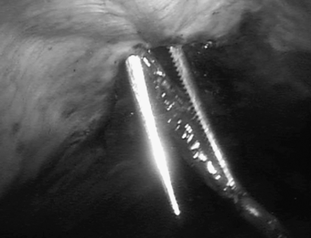
The Klemmer forceps, with no loss of pneumoperitoneum, grasps the distal portion of the urachus within the abdominal cavity.
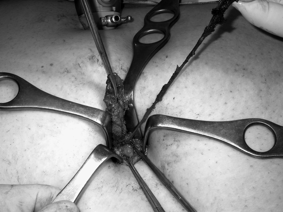
The everted urachus through the periumbilical incision.
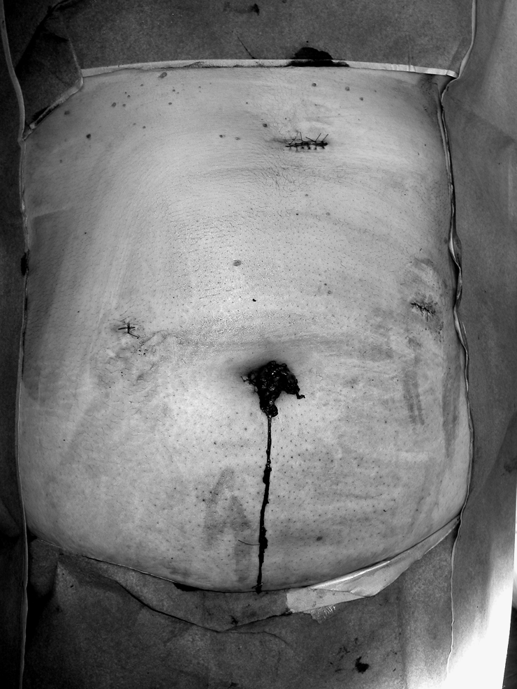
Urachal excision by videolaparoscopy.
In the fifth case, the laparoscopic technique did not allow us to identify any intra-abdominal canalicular structure; however, through an umbilical incision, a cystic periumbilical residue was removed.
The procedure ended with suture of the three port sites and umbilical reconstruction.
For each of the 13 patients, we recorded the operation time, the hospital stay, the intensity of postoperative pain, and the time required for return to full activity. Moreover, for patients in whom the urachal sinus was excised (four treated by traditional technique and four by laparoscopic technique), we also recorded the length of the excised urachus. Pain intensity was recorded by the Numerical Pain Scale, which provides a scale from “0” to “10,” where we considered “0” as no pain and “10” as the worst possible pain; the determination of the pain was carried out in the preoperative period and in the morning following surgery after the first mobilization of the patient.
Comparison between the two groups for operation time, duration of hospital stay, pain intensity, time for return to full activity, and urachal length were carried out through the exact Wilcoxon two-sample test. All statistical tests were two-sided and P<.05 was considered significant.
Results
Ultrasound was the only paraclinical examination performed on all patients. In 12 patients, ultrasound examination showed hypoechogenicity in the periumbilical region, which was suggestive of an urachal pathology, and in only 1 patient with abdominal discomfort, ultrasound allowed the diagnosis of urachal cyst. An abdominal computed tomography (CT) scan was performed on 3 patients with other comorbidities, and in only 2 cases the urachal pathology was confirmed. A patient underwent magnetic resonance (MR), which correctly showed the presence of a periumbilical flogistic process without an urachal-umbilical sinus, which, instead, was found during surgical operation and removed. Fistolography was not performed on the patients.
In 8 patients treated by open technique, the data about operation time, hospital stay, postoperative pain, and return to full activity are reported in Table 2. There was no perioperative mortality. Surgical wound suppuration occurred in 1 patient (Table 1). In the first 4 patients, histological evaluation showed a canalicular urachal remnant, which mainly consisted of muscle and mucosal tissue. The median length of the excised urachus was 8.5 cm (range: 7.8–9 cm). In the other 4 patients, the diagnosis was urachal cyst, supported by histological evaluation, which revealed a cystic wall that presented in 3 cases an epithelial layer and in 1 case mainly granulation tissue secondary to a chronic inflammatory process. The latter case showed also a cord-like urachal remnant with fibrous muscular tissue. Hence, based on the histological findings, the established diagnosis was urachal-umbilical sinus in 4 patients and urachal cyst in the other 4 patients. All the patients underwent follow-up (mean: 48 months; range: 28–60 months). In only 1 of 8 patients we registered a recurrence of the urachal disease at 6 months after surgery.
P<.05 was considered significant.
P=.03 versus conventional excision.
P=.002 versus conventional excision.
P=.53 versus conventional excision.
P=.05 versus conventional excision.
P=.23 versus conventional excision.
NRS, Numerical Pain Scale; m, median.
For 5 patients treated by laparoscopic technique, the data about operation time, hospital stay, postoperative pain, and return to full activity are similarly reported in Table 2. There was no perioperative mortality or morbidity (Table 1).
The histological evaluation revealed in 3 cases a urachal-umbilical sinus; in 1 case the urachal-umbilical sinus was proximally associated with a urachal cyst whose fibrous wall presented an epithelial layer. The median length of the four excised urachus was 11.5 cm (range: 10.8–12.5 cm). In the last case, the diagnosis was urachal cyst only.
All the patients underwent follow-up (mean: 18 months; range: 30–12 months); no patient registered a recurrence of the urachal disease.
The comparison between the two groups for operation time, hospital stay, postoperative pain, return to full activity, and length of urachus is reported in Table 2. Operation time, control pain, and urachus length were statistically different between the two groups.
Discussion
A patent urachus has an outermost muscular layer, a connective tissue layer, and an innermost epithelial layer with some potential secretory function.1,3 It is often silent with no symptoms and did not require any treatment,1,2,4 but in some other cases it produces clinical symptoms. It is easier to diagnose an urachal fistula when it develops in the neonatal period as it is responsible for a leakage of urine through the umbilicus or an vesico-urachal diverticulum, which is responsible for recurrent urinary infections, but it is undoubtedly more difficult to differentiate between urachal-umbilical sinus and urachal cyst based only on the first and chief symptom of seropurulent umbilical discharge produced by a patent portion of the urachus or by the opening in the umbilical region of an infected urachal cyst, which before any fistulization causes unspecific abdominal discomfort and swelling in the abdominal midline.1–3,7 For this reason it is preferred to talk, in generic terms, of “urachal remnants” 1 and make a differentiation between the two conditions when the use of paraclinical diagnostics, but especially surgical treatment and histological examination, 1 as in our experience, can lead to a more precise evaluation.
Diagnostic imaging techniques have not proved so supportive; in fact there is not an “evidence-based” algorithm 5 ; there are different opinions on the diagnostic advantages of ultrasound,4,7 and the role of CT scan, MR, and fistolography is uncertain. For this reason, the actual opinion of many authors, shared by us, is that in case of draining umbilicus, these techniques can be replaced by clinical examination only and by laparoscopic exploration.3,6,7 In our experience, the abdominal ultrasonography, which has been performed in all our patients, resulted in a definitive diagnosis only in the patient with the nonfistulized urachal cyst, whereas in all other cases it only provided a diagnostic orientation, probably because of concomitant phlogosis.
With regard to surgical treatment, the goal is to completely excise all patent urachal remnants, from the umbilicus to the bladder, to avoid recurrence and possible, even if rare (0.01%), 8 malignant transformation later in life. In open surgery, though it is usually dissected as far distally as possible, 5 in the majority of cases, excision is incomplete, 6 also because we think it is often not simple to perform it, especially when the urachus has reduced to an unsteady cord-like fibrous structure. When, on the other hand, there is a noncommunicating urachal condition, as in our experience, only the cyst 4 is often excised.
The first laparoscopic excision of an urachal remnant is not recent and dates back to 1992 9 ; however, because of the rarity of this pathology in young adult, reports on the laparoscopic treatment even today are sporadic in the scientific literature. In 1999, a review 10 reports only 10 cases treated laparoscopically but, to our knowledge, these reports in adult in the last 10 years of literature are not frequent. At present (PubMed search), some authors report single adult cases11–16 and other small series: from the 2 cases of Stroup and Thoman 5 and Castillo et al., 17 to 3 of Permpongkosol et al., 18 4 of Cadeddu et al., 2 5 of Cutting et al., 1 6 of Navarrete et al., 7 and 9 of Nakagawa 19 until the Sanchez' largest series 8 of 14 cases. However, all these authors refer only about patients treated by laparoscopic technique and, after having focused on surgical technique, report on how it is feasible, safe, and effective, particularly with regard for hospital stay, morbidity, convalescence, cosmetics, and not least, the almost complete absence of relapse. None of these studies is comparative with the open technique, and thus, results in terms of effectiveness are at least questionable. In literature prior to ours, to our knowledge, there is only one paper 6 that compares, as we did, traditional and laparoscopic treatments. In fact, Okegawa 6 refers in a series slightly smaller than ours, a statistical significance in favor of the laparoscopic technique for “start to eat” and hospital stay.
We do not want, in our series (Table 2), to discuss again about the well-established benefits of laparoscopy in terms of morbidity, hospital stay, control pain, and return to full activity; in our opinion, instead, the main benefit of laparoscopy is that it provides a better endoabdominal view of the urachus from the umbilicus to the bladder, allowing a complete dissection especially using a “30° laparoscope,” which allows a perfect view of the anterior abdominal wall. Then even if, in our series, the operative time is greater in the laparoscopically treated patients, it seems relevant to underline that the median length of the excised urachus is statistically superior to the traditionally treated patients (Table 2); this datum is not reported in any other series. Further, we stress that our technique allows to completely exteriorize the laparoscopically prepared urachus out of the abdominal cavity up to the umbilicus, allowing the excision of the supra-aponeurotic portion too, so well described by Sanchez. 8 So the not rare recurrences (30% of the cases1,2,8) can be avoided by performing a radical treatment as in our patient affected both by an urachal-umbilical sinus and an urachal cyst. It should also be stressed that with laparoscopy we interrupted the excision of the urachus distally at the bladder dome without opening it, because we believe, as other authors,1,2,7,8 it is not necessary especially if communication is absent.
In our cases, the position of trocars is similar to the position used for the treatment of recurrent inguinal hernia, which in our opinion provides a complete visualization of the whole urachal tract. This technique is similar to that reported by Cadeddu, 2 who, however, uses four trocars, but it differs from the technique reported by other authors1,6,7 who prefer a lateral view.
However, to date, there is still no evidence of the benefits of laparoscopic treatment. For this purpose, prospective, randomized trials may help to clarify issues such as whether to excise the urachus in the event of accidental discovery2,20 or not. Unfortunately, all authors agree that this prospect may be difficult to achieve because of the rarity of this condition.
Footnotes
Acknowledgment
The authors thank Miss. Grazia Strazzeri, who provided illustrations for this work.
Disclosure Statement
No competing financial interests exist.