
Abstract
Abstract
Introduction:
Laparoscopy has become the gold standard in an increasing number of procedures. We analyze the incidence of trocar site hernias (TSH) and determine whether closure of the external fascia prevents onset of TSH and possible complications.
Methods:
We performed a simple-blind randomized trial with two groups, one in which all the orifices were closed by suturing the external fascia of the abdominal wall (group A), and another in which the orifices were left open, closing only the skin (group B). Monitoring for TSH lasted 2 years from the intervention. The trial has been registered at www.clinicaltrials.gov with the clinicaltrials.gov identifier number: NCT01240434.
Results:
A total of 195 patients were randomized. Thirty-three were removed from the study after conversion to open surgery, early open reoperation, or loss to follow-up. The remaining 162 patients comprised the study population, 80 in group A and 82 in group B. We found no differences between the groups regarding basic demographic data, kind of surgery, or topographic distribution of the trocars. Five TSH were diagnosed—four in group A and one in group B (P=.176)—and there was no relation between TSH and trocar size (11 or 12 mm) or location. We found 10 wound infections, 7 in group A and 3 in group B (P=.154).
Conclusion:
Our study suggests that the onset of TSH does not depend on trocar size or location. There is no evidence that suture of the fascial defect prevents the onset of TSH. In addition, we found a trend toward a higher incidence of wound infection among patients in whom the fascia had been sutured.
Introduction
The two main advantages of laparoscopy over conventional surgery are its reduced organ manipulation and minimal abdominal wall insult, both of which are beneficial in terms of postoperative pain. However, disadvantages include a steep learning curve, increased operative time, unnoticed iatrogenic injuries, greater difficulty in controlling complex intraoperative situations, adverse effects in the pneumoperitoneum, and complications arising from the use of trocars.
The main complications associated with the use of trocars are injury to solid or hollow viscera during insertion and trocar site hernias (TSH). Hematomas and wound infections at the insertion site are less serious but more frequent. There has even been a report of necrotizing fasciitis at a trocar insertion site. 1
More than 100 different trocar models are available. 2 One of the most common is the Hasson trocar, which is inserted through an opening in the abdominal wall (air leaks are prevented using an inflated ballon). Other types are directly inserted into the abdomen (cutting trocars) or by separating muscles and aponeurotic planes (blunt trocars). Cutting trocars create a slightly bigger defect than blunt trocars, although they require less insertion strength, 3 thus minimizing the risk of organ injury. 2 Moreover, with cutting trocars, the risk of bleeding and the abdominal wall defect vary with the size and shape of the blades, that is, both are bigger with pyramidal blades (triple) and smaller with flat blades (single).
Incisional hernia is observed in 15% of laparotomies performed in open abdominal surgery; TSH is observed in 0.02%–3.6% of procedures.4–9 In open surgery, 70% of cases are diagnosed during the first year after surgery, although similar data have not been confirmed in TSH. Data on TSH are contradictory: some authors associate this complication with the location of the insertion site,7,10 whereas others associate it with the size of the trocar. 11
Several approaches have been proposed to avoid TSH. These include “Z” trocar insertion or avoiding the linea alba, although the most common has always been suture of the fascia, either under direct vision or by using different techniques4–5,10,12–18 or devices, including placement of preventive mesh in selected cases.19,20 Despite consensus on the safety of not closing the insertion sites of 5-mm trocars, controversy over closing those of trocars measuring 10 mm or larger has persisted. The first reports of TSH once laparoscopy had become generalized21,22 recommended that the fascia be closed in a separate procedure. However, with the appearance of blunt-tip trocars, some authors have started to recommend not suturing the fascial defect.23–25 Despite systematic use of blunt-tip trocars, subsequent articles on TSH26,27 have warned against the risk of compromising the advantages of laparoscopy and again recommended closure of orifices in the fascia.9,11,22,27–32
Depending on whether the fascia needs to be closed, there are three main techniques for suturing the orifices created by trocars: closing only the skin, closing the external layer of the fascia followed by skin closure, and closing all the layers of the abdominal wall, including the peritoneum. A new classification of these techniques has recently been published. 33
Nevertheless, fascial closure is not free from risks, such as inclusion of the suture in a bowel segment, and it is not technically simple, 15 especially in obese patients with a skin wound smaller than 1.5 cm.
The contradictory opinions just set out lead us to believe that the closure of laparoscopy orifices has not been proved to prevent TSH. Therefore, we designed a prospective, random, and single-blind study with two arms and the following working hypothesis: systematic closure of the external fascial layer in laparoscopic trocar orifices does not decrease the risk of TSH. Further, since this approach involves more extensive wound manipulation, it can lead to additional local complications such as increased incidence of surgical wound infection.
Aim
To analyze the incidence of TSH in orifices created by trocars measuring ≥10 mm in diameter, and to determine whether closure of the external fascial layer prevents TSH and potential related complications.
Patients and Methods
This was a single-blind trial including all patients undergoing laparoscopic surgery performed by the same surgical team (5 surgeons, from S-1 to S-5) and for whom at least one “study trocar” (ST) was used. The STs were defined as blunt-tip trocars that were inserted blind, had a diameter of 11 mm or 12 mm, and were not enlarged during surgery. We did not study the Hasson trocar, as the defect it creates is surgeon dependent and not uniform.
We excluded patients presenting factors that affect the scarring process, such as malnutrition (serum proteins <5 g/dL), advanced cancer, perioperative hemodynamic instability, or prolonged treatment with corticosteroids. Orifices that were enlarged for specimen extraction were also excluded, although other orifices in the same patient were included.
We excluded the following patients: those who had converted to open surgery, those who had undergone a second intervention with an open technique during the early postoperative period, or those who had been lost to follow-up. Patients who did not have an ST after surgery (mostly patients with only one ST that had to be enlarged for specimen extraction) were excluded from the trial.
We randomized patients by using a random table from Excel® for Windows XP®.
The trocars used were Endopath Xcel (Ethicon Endo-Surgery), which were 11 and 12 mm in diameter.
The study had two arms: one in which all the ST orifices were closed by suturing the external fascia of the abdominal wall with a number 1 monofilament absorbable suture (Polydioxanone) (group A), and another in which all the orifices of the ST were left open, closing only the skin (group B).
This trial was approved by the corresponding ethics committee. All the patients gave their informed consent to participate in the trial before randomization.
Follow-up involved two postoperative contacts for each patient, the first during the first year after the operation, and the second 2 years after the intervention. These contacts were by phone, and the patient was asked about any lumps or discomfort in the scar area. A positive answer was followed by an appointment for a detailed examination and, if necessary an abdominal ultrasound scan.
Demographics, diagnosis, and surgical technique were analyzed for each patient, as were hospital stay, morbidity, and the number of trocars and STs used with their size and location.
For the topographic analysis, we divided the abdomen into seven areas according to the different fascial layers of the abdominal wall (Fig. 1).
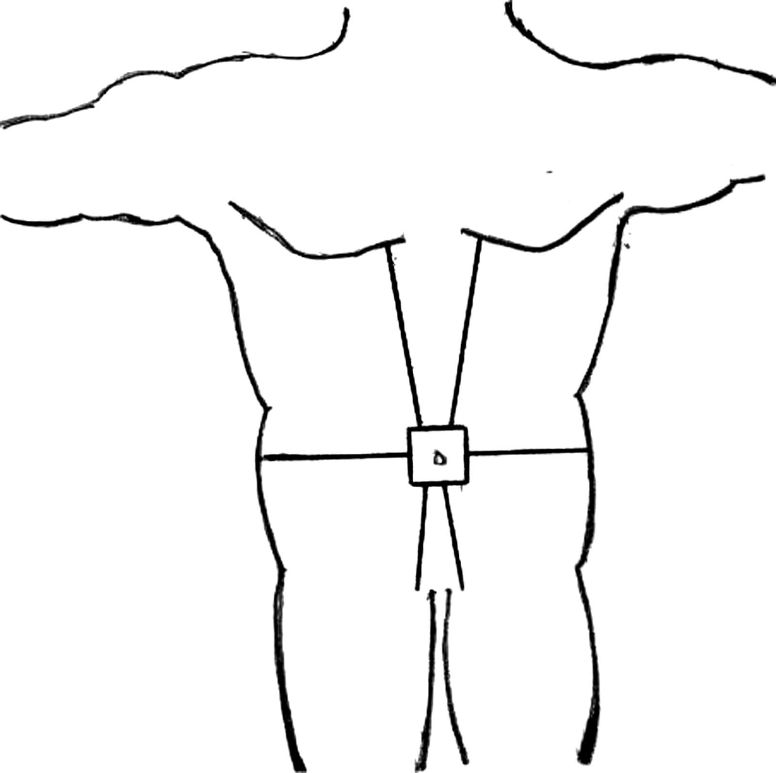
Topographic areas.
The statistical analysis was performed by using SPSS 11.5 for Windows.
The trial has been registered at www.clinicaltrials.gov with the clinicaltrials.gov identifier number: NCT01240434.
Results
A total of 195 patients were randomized. Thirty-three were excluded (9 were converted to open surgery, 2 underwent an early open reoperation, and 22 were lost to follow-up). The final number of patients studied was 162: 80 in group A (closure) and 82 in group B (nonclosure).
The number of patients operated by each surgeon was as follows: S-1 38 patients, S-2 30 patients, S-3 29 patients, S-4 31 patients, and S-5 34 patients.
Demographic data for both groups are presented in Table 1. Appendectomies and cholecystectomies accounted for 79.6% of all procedures, with no differences between the groups (80% vs. 79.3%). There were no differences regarding the kind of surgery (clean, clean-contaminated, contaminated, and dirty) between the groups (P=.44).
With regard to the topographic distribution of trocars (Table 2), as expected, most were inserted in the peri-umbilical area, followed by the upper midline. In both cases, most trocars were 11 mm in diameter. The 12-mm trocars were used mainly in the lateral quadrants. This distribution pattern was similar in both groups.
We observed 10 wound infections, 7 in group A and 3 in group B, although this difference was not statistically significant (P=.154). Regarding the main variable, we found 5 cases of TSH: 4 in group A and 1 in group B (P=.176).
Given the low incidence of TSH, we were unable to compare patients presenting this complication with those who did not, although we do provide a descriptive analysis of the results. Four TSH appeared below 11-mm trocar orifices and 1 below a 12-mm trocar orifice. All the cases presented after clean-contaminated surgery (3 after cholecystectomy, 1 after right hemicolectomy, and 1 after sigmoidectomy). All the cases were diagnosed during the first year after surgery. Three patients underwent uneventful elective surgery, 1 rejected the intervention in spite of our advice, and 1 had the hernia repaired during surgery for liver metastasis. The distribution of hernias according to the surgeon was as follows: S-1 2 cases (6.25%), S-2 1 case (33.3%), S-3 1 case (3.45%), S-4 no cases (0%) and S-5 1 case (2.94%), not showing statistical significance.
The mean age of patients presenting TSH was 70 years (63–77), and this was considerably higher than in patients without hernia (47 years [16–99]). TSH occurred in 3 women and 2 men. The total number of trocars and STs was similar in patients with TSH and in patients without. Only one of the cases of TSH was associated with wound infection. Regarding the location of the TSH, 4 were peri-umbilical, and only 1 was lateral (upper left quadrant). We want to remark that no one hernia was found in any of the “non-study port” sites.
Discussion
The need to suture the fascial defect remaining after insertion of laparoscopic trocars is controversial. To date, several case reports and some articles have analyzed prevention of TSH (using a specific kind of trocar or a special approach to introduce or close the orifices); however, recommendations are based on low-level evidence (expert opinion or case series).4,5,10,12–20
The study of TSH is difficult for several reasons. First, the low incidence of this condition means that any approach requires a large study sample (calculated population of over 2000 patients in this case). This is not possible without a multicenter study, which introduces bias such as the use of different trocar models, suture materials, and the involvement of a larger number of surgeons. Further, most TSH are small, have little aesthetic impact, and are not always symptomatic; therefore, we suspect that diagnosis depends on a meticulous interview and examination by the surgeon. At least 2 of the TSHs were diagnosed during the telephone interview, and the patients subsequently stated that they were not planning any medical consultation for this reason. Although there are no reports of TSH appearing years after the laparoscopy, this fascial defect is expected to behave similar to others, namely, it grows in size over time, with the result that the indication for surgical repair is clear. Our conclusions should be confirmed in further studies with a similar design; however, we believe that our results add to existing knowledge on this condition.
We realize that using a phone interview is an important weakness of the study; however, we believe that this “easy” commitment for the patient increased the adherence to a long term follow-up in patients undergoing simple procedures such as appendectomy or cholecystectomy in the frame of a benign condition.
Other limitation of the study concerns the lack of information given about the body mass index of the patients. These data could not be retrospectively collected, and we believe that they should be taken into account in the design of further studies.
Our data suggest that the suture of the fascial defect created by the introduction of laparoscopic trocars does not prevent onset of TSH, for different reasons. First, this suture is technically difficult, especially in patients with a thick subcutaneous layer, which makes it difficult to visualize the anatomical layers. Second, the orifices left by these trocars are small, not much larger than those left by a flat drain and equivalent to the distance between stitches during closure of a laparotomy. Third, manipulation of the wound, together with the presence of a foreign body, can increase the risk of developing wound infection, which is a risk factor for incisional hernia.
Most cases appeared in the peri-umbilical region, that is, the area where most trocars were inserted. Consequently, it is impossible to provide evidence that the insertion site affects the likelihood of a TSH appearing. The same could be said for trocar size, as the most commonly used type during the study was the 11-mm trocar. The exceptions were those trocars excluded from the study (Hasson trocars) and those enlarged for specimen extraction (the defect is surgeon-dependant and not stable).
Finally, all the TSHs were diagnosed during the first year after surgery, although the average follow up was 2 years and 3 months. Future series could consider a follow-up of 1 year as sufficient, thus considerably shortening the study. Long-term outcome (5 or 10 years) has yet to be studied.
Conclusions
Our data do not give a clear answer to the question as to whether or not it is necessary to suture port sites, but they suggest that TSH does not depend on trocar size (11 or 12 mm), insertion site, or kind of surgery, and that the suture of the fascial defect created by the introduction of laparoscopic trocars does not prevent the onset of TSH. In addition, we found a trend toward a higher incidence of wound infection among patients in whom the fascia was sutured. However, larger studies are needed to obtain statistical significance.
Footnotes
Disclosure Statement
No competing financial interests exist.