
Abstract
Abstract
Background:
The most important criterion in the management of endoscopic retrograde cholangiopancreatography (ERCP)-related perforations is the delineation of the injury pattern. The aim of the present study was to evaluate in a retrospective manner the patients who undergo surgery due to ERCP-related perforations.
Patients and Methods:
Between January 2006 and December 2010, a total of 9209 ERCPs were performed at Turkiye Yuksek Ihtisas Teaching and Research Hospital, Ankara, Turkey. From these, perforation was diagnosed in 52 patients (0.56%). Twenty-four patients (46.2%) underwent surgery. Patients were evaluated according to age, gender, ERCP indication, comorbid disease, the time between diagnosis and perforation, the time between ERCP and surgical intervention, radiological and clinical signs, localization of the perforation, surgical procedure, hospitalization period, and postoperative outcome.
Results:
Twenty-four patients underwent surgery. Thirteen patients (54.1%) had lateral duodenal wall perforation, 4 patients (16.7%) had perforation in the afferent loop (these patients had Billroth-II gastroenterostomy at ERCP admission), 2 patients (8.3%) had bile duct perforation, and 1 patient (4.1%) had esophageal perforation. In 4 patients (16.7%), the localization of the perforation could not be found. Nine patients (37.5%) died in the postoperative period. Six patients had lateral duodenal wall perforation, 2 patients had afferent loop perforation, and one patient had esophagus perforation. Three patients died of nonsurgical reasons (myocardial infarction, serebrovascular occlusion, and cardiac dysrhythmia).
Conclusions:
Duodenal wall perforations have a serious fatal outcome even if early surgical intervention is performed. In contrast to duodenal wall injuries, perivaterian and choledochal injuries have a better outcome.
Introduction
The most important criterion in the management of ERCP-related perforations is the delineation of the injury pattern. In general, it is accepted that lateral or medial duodenal wall perforations should undergo immediate operative intervention.8,9 Late diagnosed postsphincterotomy perforations—particularly duodenal—require surgical drainage, which carries a higher morbidity and mortality rate. 7 However, nonsurgical management may be acceptable for injuries at the sphincter of Oddi and bile duct injuries if an early contrast study demonstrates minimal extravasation or a sealed perforation without associated fluid collection. 9
The aim of the present study was to evaluate in a retrospective manner patients who underwent surgery due to ERCP-related perforations. Patient demographics, indications for ERCP, clinical and radiological signs, localization of the perforations, surgical procedures, and clinical outcomes were analyzed.
Patients and Methods
Between January 2006 and December 2010, a total of 9209 ERCPs were performed at Turkiye Yuksek Ihtisas Teaching and Research Hospital, Ankara, Turkey. Of these, perforation was diagnosed in 52 patients (0.56%), and 24 of these patients (46.2%) underwent surgery. The patients were evaluated by means of age, gender, ERCP indication, comorbid disease, the time between diagnosis and perforation, the time between ERCP and surgical intervention, radiological and clinical signs, localization of the perforation, surgical procedure, hospitalization period, and postoperative outcome. Data of the patients were recorded in a prospective manner.
The time between diagnosis and perforation was classified as during or after ERCP. The time between ERCP and surgical intervention was further classified as early period (≤6 hours) or late period (>6 hours). The perforations were classified according to the site of perforations using the classification previously defined by Stapfer et al. 8
The treatment policy of our institution for ERCP perforations is defined as follows: All injuries resulting with perforation of the gastrointestinal tract are candidates for urgent surgical repair. When perforation occurs during papillotomy for cannulation of the biliary tract (sphincterotomy), a nasobiliary drainage catheter is inserted—if possible—to prevent biliary leakage. The patient's oral intake is stopped, and antibiotic therapy is started. In the presence of postprocedural severe abdominal pain, fever, and signs of peritonitis, surgery is planned initially. Patients for whom conservative management was planned are hospitalized and are closely monitored with physical examination, laboratory tests (white blood cell count, C-reactive protein), and radiological imaging (ultrasonography, computerized tomography). If the abdominal symptoms and signs improve, a duodenography with a water-soluble contrast is obtained, and oral intake is started. The patients are evaluated daily, and surgery is performed for those without any clinical and laboratory improvement. However, the patient's status and the underlying disease such as impacted bile duct stone, gallbladder cancer, and pancreas carcinoma are factors that can influence the extension of the surgical intervention.
Results
Of the 24 patients who underwent surgery, 12 (50%) were male, and the remaining 12 (50%) were female. The mean age was 61.6±14.8 years. The indications for ERCP were bile duct stones in 13 patients (54.1%), malignancy-associated jaundice in 7 patients (29.2%), benign biliary stricture in 3 patients (12.5%), and pancreatic duct stone in 1 patient (4.2%) (Table 1).
ERCP, endoscopic retrograde cholangiopancreatograpy.
Of the patients who underwent surgery, sites of the perforations were lateral duodenal wall in 13 patients (54.1%), afferent loop (these patients had Billroth-II gastroenterostomy at ERCP admission) in 4 patients (16.7%), bile duct in 2 patients (8.3%), and esophagus in 1 patient (4.1%). In 4 patients (16.7%), the localization of the perforation could not be found (Table 2).
Perforation was diagnosed during ERCP in 21 patients (87.5%) and in the postprocedural period in 3 patients (Table 3). Of the patients whose perforation was diagnosed during ERCP, surgery was performed in 18 patients (85.7%) in the early period, whereas 3 patients (14.3%) underwent surgery in the late period. Indications for surgery in the early period were visualization of intraperitoneal structures by endoscopy, existence of extraperitoneal or intraperitoneal free gas and extravasations of contrast fluid on fluoroscopic images during ERCP, determination of intraabdominal or retroperitoneal fluid collection by computed tomography, massive subcutaneous emphysema, and suspected perforation with tight stricture or impacted common bile duct stone (Tables 4–6). Indications for surgery in the late period were signs of peritonitis and radiological findings of intraabdominal fluid collections (Tables 4–6).
CBD, common bile duct; CBDE, common bile duct exploration; CC, cholecystectomy; COLD, chronic obstructive lung disease; CRI, chronic renal insufficiency; CT, computed tomography; D2, second part of duodenum; D3, third part of duodenum; DM, diabetes mellitus; HT, hypertension; ICD, ischemic coronary heart disease; GE, gastroenterostomy.
MI, myocardial infarction.
In patients who underwent surgery in the early period, sites of perforations were the lateral duodenal wall in 9 patients, the afferent loop in 4 patients, the esophagus in 1 patient, and the common bile duct in 1 patient. However, perforation could not be found in 3 patients. Of the 3 patients for whom conservative follow-up was conducted despite the diagnosis of perforation during ERCP and who required subsequent surgical intervention, 2 had perforation in the duodenal wall; however, in the remaining patient, perforation could not be found. Of the patients who underwent surgery in the late period, 2 had perforation in the duodenum; the common bile duct perforation was detected in the remaining patient.
Six of the 13 duodenal wall perforations were treated by duodenal wall repair plus cholecystectomy plus choledochal exploration with T-tube drainage. Two patients underwent duodenal wall repair plus cholecystectomy plus choledochal exploration with T-tube drainage plus pyloric exclusion and gastrojejunostomy, 2 patients underwent duodenal wall repair plus pyloric exclusion and gastrojejunostomy, 2 patients underwent duodenal wall repair plus cholecystectomy plus choledochal exploration and stone extraction with T-tube drainage, and one patient underwent duodenal wall repair+choledochal exploration and stone extraction with T-tube drainage (Table 4). Of the 4 patients in whom perforation could not be found, 2 underwent cholecystectomy plus choledochal exploration and stone extraction with T-tube drainage, 1 patient underwent cholecystectomy and choledochal exploration with T-tube drainage, and the remaining patient underwent explorative laparotomy with retroperitoneal fluid drainage. Two patients had common bile duct perforation: One of them underwent cholecystectomy and choledochal exploration with T-tube drainage, whereas the other patient underwent cholecystectomy plus choledocoduodenostomy (Table 5). Four patients had perforation in the afferent loop: 3 of them underwent primary jejunal repair, and the remaining patient underwent primary jejunal repair with bilioenteric anastomosis. One patient with cervical esophageal perforation underwent primary repair (Table 6).
In the postoperative period, enterocutaneous fistula and intraabdominal abscess developed in 8 (33.3%) patients. Five patients (20.8%) required re-operation because of intraabdominal complications. The perforation sites of these 5 patients in their previous operations were the duodenal wall (3 patients), the common bile duct (1 patient), and the afferent loop (1 patient) (Tables 4–6).
The mean hospitalization period was 13.5±10.69 days. Nine patients (37.5%) died in the postoperative period, and their mean age was 70.4±9.1 years (Table 3). Of these patients, perforation was diagnosed during ERCP in 8 patients and after ERCP in 1 patient. Six patients underwent surgery in the early period and 3 in the late period. Six patients had lateral duodenal wall perforation, 2 patients had afferent loop perforation, and 1 patient had esophagus perforation (Tables 3, 4, and 6). Three patients died from nonsurgical reasons (myocardial infarction, cerebrovascular occlusion, and cardiac dysrhythmia). Of the 6 patients who died from sepsis, 4 had undergone re-operation (Tables 4 and 6).
Discussion
Although ERCP-related perforations are rare, the morbidity and mortality rates are high, especially in patients with duodenal wall perforations.7–9 Late recognition of a duodenal perforation and failure of nonsurgical treatment have a high complication rate with a potentially fatal outcome. However, several studies have shown that in the majority of patients, ERCP perforations can be managed with a nonoperative course because perforations occur more commonly during sphincterotomy in the periampullary region,9–11 and these perforations can frequently be managed conservatively.
Many clinical researches have been working to classify the perforations according to localizations and mechanisms to reach a consensus for treatment of these perforations.7,8 Stapfer et al. 8 pointed out that Type I injuries (duodenal wall perforation) required prompt surgical intervention, whereas Type II (perforation at the sphincter of Oddi) and Type III (common bile duct perforation) could be managed conservatively if minimal extravasation or a sealed perforation only occurred. Furthermore, they did not regard Type IV perforations (retroperitoneal free air) as a real perforation and claimed that these group perforations did not require surgery. Howard et al. 7 classified sphincterotomy perforations as guide wire, periampullary, and duodenal perforations. Guide wire perforations are recognized early and resolve with medical treatment. Periampullary perforations diagnosed early respond to aggressive endoscopic drainage and medical treatment. Late diagnosed postsphincterotomy perforations (particularly duodenal) require surgical drainage. The treatment policy in our institution is conservative follow-up for Type II and III perforations unless intraabdominal fluid collection is defined by computed tomography. Type I and Type II–III perforations with significant peritoneal findings are treated by surgery. In the present study, we found bile duct injury only in two cases among the patients who were operated on for ERCP perforation. Although our 4 patients had radiological and clinical signs of perforation, the localization of the injury could not be found during surgery. These perforations were regarded as perivaterian (Type II) perforations. In this case, conservative treatment seems to be more accurate for most of these perforations as noted in the literature. However, these patients underwent surgery not only for injury but also for the underlying disease, which could not be treated by ERCP.
Perforations are mostly diagnosed during ERCP.6,8 In the present study, 87.5% of the perforations were determined during ERCP. Determination of free retroperitoneal air by fluoroscopy during the ERCP is a pathognomonic sign for perforation (Fig. 1). Another typical sign of perforation is the direct visualization of the intraabdominal organs or retroperitoneal fat tissue during endoscopy (Fig. 2). In cases with duodenal wall perforation, the existence of pneumoperitoneum or retroperitoneal air by fluoroscopy, development of subcutaneous emphysema, and direct visualization of the peritoneum by endoscopy are early pathognomonic signs. In the late period, signs of peritoneal irritation become apparent and remarkable intraabdominal/retroperitoneal air or fluid collection may detected by computed tomography.
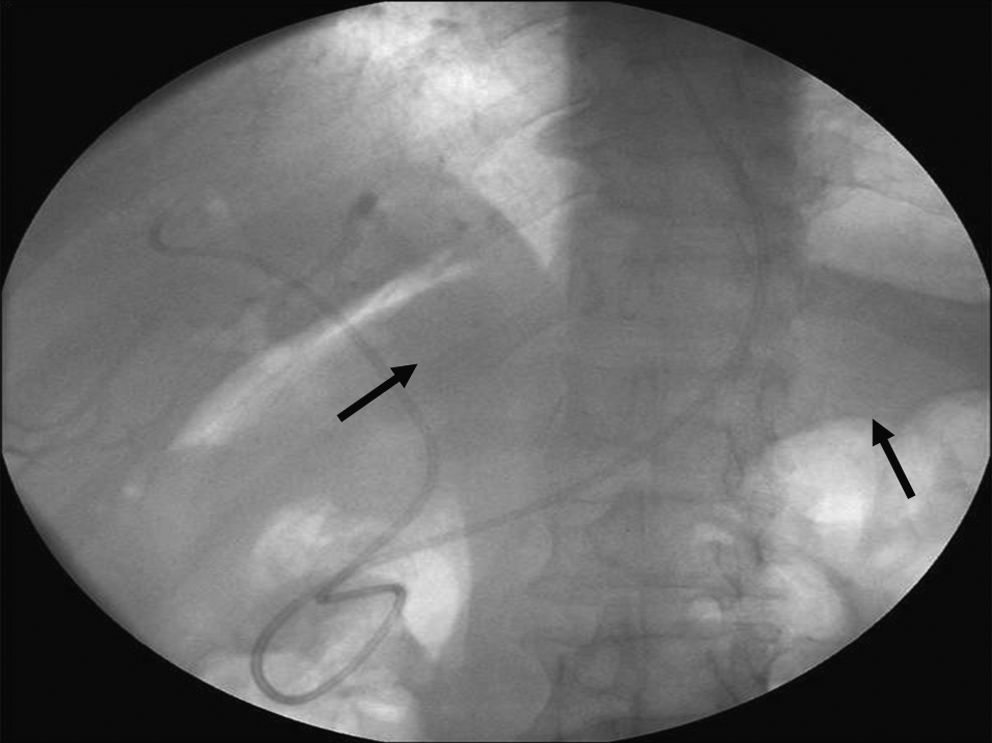
Free retroperitoneal air by fluoroscopy during the ERCP.

The image illustrates direct visualization of the perforation site during endoscopy.
In duodenal wall perforations, primer repair with duodenal diversion by pyloric exclusion with gastrojejunostomy seems to be the optimal procedure.8,12 However, if perforation is diagnosed in the early period without major spillage or contamination of the abdominal cavity with enteric bacteria, 13 only duodenal wall repair may be sufficient. Our treatment policy consists of primary duodenal wall repair with omental patch placement and inserting a T-tube in the common bile duct for biliary drainage if the abdomen is not widely contaminated.
Another ERCP-related perforation is the afferent loop perforation in patients with Billroth-II gastroenterostomy.9,12 ERCP and endoscopic sphincterotomy are technically more difficult in patients with Billroth-II gastrectomy because of the reversed anatomy. 14 In the present study, 4 patients underwent surgery because of afferent loop perforation. Diagnosis of afferent loop perforations is easier because of the direct visualization of the intraabdominal organs by endoscope during ERCP. In addition, 1 patient was operated on for cervical esophageal perforation, which was diagnosed during ERCP by determining subcutaneous emphysema.
Our mortality rate after surgery was 37.5%. This high rate was thought to be related to several factors. A large number of the patients had lateral duodenal wall and afferent loop perforations, and 1 patient died because of esophageal perforation. Most of the patients who died were operated on in the early period. This finding suggests that ERCP perforations directly related to endoscopy have a high mortality risk even if the patients undergo urgent surgery. Second, advanced age, comorbid diseases, and advanced stage tumors may be the other factors that have negative influence on our mortality rate.
In conclusion, perforations directly related to endoscopy have a serious fatal outcome even if early surgical intervention is performed. Perforations in the periampullary region due to sphichterotomy can be managed conservatively and occasionally require surgery. In contrast to duodenal wall injuries, perivaterian and choledochal injuries have a better outcome.
Footnotes
Disclosure Statement
No competing financial interests exist.