
Abstract
Abstract
Background:
Recurrent appendicitis with the appendix tip in the subhepatic area and late presentations of perforated appendicitis sometimes test the skills of the surgeon. Because of dense adhesions and distorted anatomy, trying to do retrograde appendectomy or looking for the appendicular artery may lead to troublesome bleeding and injure the adjacent intestine. Submucosal appendectomy could be an answer in these situations.
Subjects and Methods:
From October 7, 2005 to July 31, 2011, 1589 laparoscopic appendectomies were performed, of which 239 were recurrent or perforated or formed a mass. In 19 of these cases no plane could be established between the appendix and adjacent structures. So an incision was made on the anti-mesenteric wall of the appendix, and the mucosal sleeve was pulled out, leaving the muscular wall. The base was then ligated flush with the cecum and divided distally, leaving the muscular tube. Postoperative management was similar to usual appendectomies.
Results:
Out of 19 cases 13 were male. Ages ranged from 3 to 14 years. Seven cases were perforated, and 12 were recurrent. Submucosal appendectomy was done in all these 19 cases. There was no intraoperative complication. Average operating time was 51 minutes. In 16 cases feeding was tolerated early, and 3 cases suffered from prolonged ileus. Average postoperative hospital stay was 3.47±2.34 days. Follow-up ranged from 3 months to 5 years. Two cases reported occasional abdominal pain, which required re-admission.
Conclusions:
Submucosal appendectomy minimizes complications, obviates the need for conversion, and is a safe option for difficult cases during laparoscopy.
Introduction
Subjects and Methods
From October 7, 2005 to July 31, 2011, laparoscopic appendectomies have been performed in 1589 children, and 239 of them were recurrent or perforated or formed a mass. In 19 of these cases we have used the submucosal dissection technique. Laparoscopy was done with the patient under general endotracheal anesthesia in all these cases. We have used the conventional three-port technique: a supraumbilical port for the camera, one port medial to the left anterior superior iliac spine, and another just above and to the right of the pubic symphysis. For optical viewing, the telescope was 5 mm 30° in children younger than 5 years and 10 mm 30° for those older than 5 years, with an additional 5-mm telescope used during retrieval of appendix in the latter group. The supraumbilical port was introduced by the open technique, and insufflations were done. The CO2 pressure was kept between 10 and 15 mm Hg.
During laparoscopy we have encountered three situations with those 19 cases described in this study. These were as follows: (a) An upturned cecum with the appendix base in a high up position, the appendix tip close to the liver, and the appendix running parallel to the cecum and ascending colon. There was a dense adhesive fibrous layer covering both the appendix and colon, making them inseparable. (b) An engorged appendix curled upon itself running behind the ileum and tip could not be brought into view. (c) A perforated appendix forming a mass in the pelvic cavity that could not be separated from adjacent intestine without risk of injury (Fig. 1). In all these circumstances the appendix could be identified by its tubular shape and noncompressibility when grasped with a fenestrated grasper.
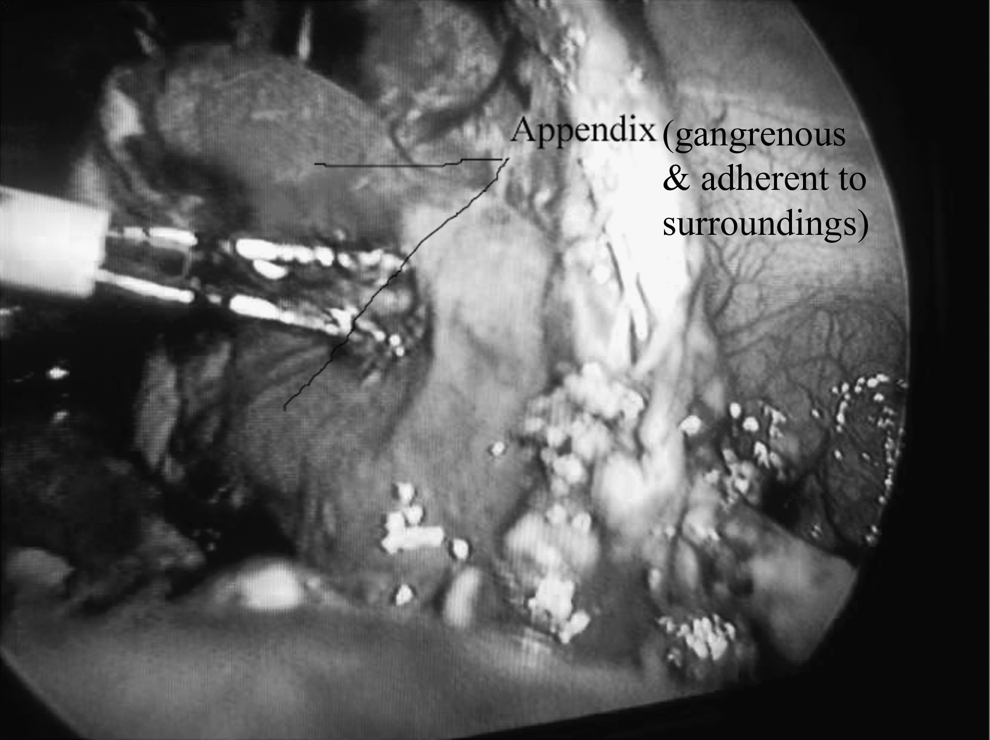
Perforated pelvic appendicitis with the appendix densely adherent to adjacent structures.
An incision was made on the anti-mesenteric wall of the appendix, and the mucosal sleeve was pulled out, leaving the muscular wall. The base of the tube was then ligated flush with the cecum and divided distally. The muscular tube was left alone (Fig. 2). In perforated cases we looked for fecolith. The peritoneal cavity was cleansed with normal saline by irrigation and aspiration. Closed suction drain tubes were kept in the peritoneal cavity in all the cases. Feeding was allowed 6 hours after the operation in the majority of cases and later on in a few cases where there was much handling of the intestines during the operation.

Submucosal dissection in progress with the mucosal tube and muscle cuff clearly definable.
In recurrent appendicitis cases where there was no perforation, our policy was to administer single doses of ceftriaxone and metronidazole at induction and discharge the patient the following morning after removal of the drain. In a few cases with substantial serosanguinous fluid collection in the drainage bag we have waited for 2 or more days. In perforated cases we have kept the drain tube until the culture and sensitivity reports arrived. In these situations ceftriaxone, metronidazole, and amikacin were given for 3 days unless the sensitivity report suggested otherwise.
When the collection in the drainage bag became negligible, feeding was tolerated, and the patient was afebrile, we could discharge them. Patients were followed up at 2 weeks and 6 weeks after discharge, every 3 months thereafter for 1 year, and then yearly. An ultrasonogram was done in 2 cases; those were because of abdominal pain between 3 and 6 months postoperatively and revealed no abnormality.
This retrospective study describes the age, sex, operative technique, postoperative stay, complications, and outcomes. The Ethical Review Committee for Thesis and Research of Chattagram Maa-O-Shishu Hospital Medical College gave permission to conduct this study.
Results
Out of the total of 19 cases 13 were male. Ages ranged from 3 years to 14 years (mean, 9.26±2.98 years). Seven cases were perforated, and 12 were recurrent appendicitis. According to position the cases were retro-ileal (n=2), subcecal (n=2), pelvic (n=3), and retrocecal (n=12) appendices. Submucosal appendectomy was done in all these 19 cases. There was no intraoperative complication (e.g., uncontrolled bleeding or intestinal injury). In 1 case the cecal base was exposed during dissection, and the muscle cuff around it was repaired. Fecolith was found and retrieved in 2 cases. Mean operating time was 51±10.30 minutes (range, 34–71 minutes). In 16 cases feeding was tolerated early, and 3 cases suffered from prolonged ileus. Postoperative hospital stay was 1–8 days (mean, 3.47±2.34 days). The follow-up period ranged from 3 months to 5 years. Although these cases had some abdominal discomfort postoperatively, only 2 cases reported abdominal pain, which required re-admission.
Discussion
Laparoscopy offers a safe and effective alternative to the open technique for complicated appendicitis in terms of postoperative outcomes (e.g., faster recovery, less pain, and fewer complications like wound infections, intra-abdominal abscesses, and reoperations).8,9,11,14,18–20 Our experience is that most of the complicated appendicitis cases can be operated on laparoscopically with reasonable ease and outcomes comparable to those of other published series. The technique we described was an incidental occurrence initially while we were trying to create a plane between the appendix and the colon in a difficult case. Thereafter in the subsequent cases we did it purposefully when the situation demanded.
In 13 of the cases the base of the appendix could be identified first, and the incision was made near the base. In the remaining 6 cases the tip came into view first, and the procedure was started from the apical side. The incision was made using hook cautery, taking care not to perforate the mucosa unless it was perforated already. In perforated cases the task was a bit easier to begin at the site of the perforation. Once the muscle layers were separated, the mucosal tube came into view. Then with the mucosal tube grasped with Babcock forceps by the left hand, the remainder of the dissection of the muscular tube was done using Maryland forceps in the right hand. Care should be taken while dissecting toward the base so as not to expose the cecum, looking meticulously and continuously to the change in caliber of the mucosal tube and releasing the tension intermittently. Minor hemorrhage can occur during the peeling off of the mucosal tube from the small vessels running from the muscle coat to the mucosa. These are easily amenable to bipolar cautery, which we have used in our cases. In 1 of our cases we actually exposed part of the cecal mucosa, which fortunately was detected before the appendix base was tied. So, we tied the appendix at its base and repaired the muscular cuff around the cecal base. The outcome in this case was uneventful. In the remaining 18 cases the muscular cuffs were left as leaving them causes no untoward effect. Those cases that we found in the subhepatic location seemed to be due to recurrent episodes of appendicitis with adhesions causing the cecum to be pulled up along with the appendix. Laparoscopy is a feasible option for appendices in unusual locations, including subhepatic ones also.21,22
There is a tendency toward conversion to the open technique when difficulty is encountered during laparoscopic appendectomy.4,7,16,23 Retrograde appendectomy has been practiced in both open and laparoscopic methods to overcome the problem. 17 “Subserosal appendicular stripping” has been described in recurrent appendicitis with extensive serosal adhesions. 24 Our technique is different in that the entire muscle coat is left behind, and the maneuver is very easy to perform once the mucosal tube can be identified. The appendicular artery and adjacent intestine remain separated and protected from the dissection field by the thick muscle cuff; this technique thereby obviates the need for conversion. “Stump appendicitis” is another problem that can arise after laparoscopy in complicated and recurrent cases, particularly where the basal parts of the appendix remain hidden by dense adhesions.25,26 Submucosal appendectomy also eliminates the risk of stump appendicitis, enabling the dissection as far as the cecal–appendiceal junction without jeopardy.
Postoperative wound infections and intra-abdominal abscesses are less common after laparoscopy.9,12,14 Laparoscopy provides direct visualization during peritoneal toileting and prevents wound contamination, particularly if an endobag or a substitute is used during appendix retrieval.11,12 In the technique that we described, the diameter of the mucosal tube was easily amenable to retrieval through the port without use of bags and without port-site contamination. However, fecolith in 2 perforated cases needed to be retrieved in the finger of a glove used as a bag. For perforated cases we took pus for culture and sensitivity testing, and a thorough peritoneal wash was performed with normal saline before closure, keeping a drain. In other cases there was little hemorrhage from the raw inner surface of the muscular cuff, which was washed with saline, and again a drain was kept for 24 hours. None of our study cases had postoperative wound infection or intra-abdominal abscesses, although we encountered these complications in a few of our other complicated cases.
Laparoscopy has been related to longer operative times for complicated appendicitis in most studies.5,14,21 Also, we did not do any submucosal appendectomy by the open method, and the average operative time for our cases was comparable to that with the open technique for complicated appendicitis. It is our practice to start feeding 6 hours after laparoscopy in the majority of appendicitis surgeries, including perforated ones that did not pose much difficulty during the operation or where much handling of the intestine was not necessary. However, in perforated cases that needed thorough peritoneal washing, we have waited overnight. Three of those patients developed ileus 23 and needed longer time to tolerate feeds.
Postoperative hospital stay for laparoscopy in complicated cases is shorter than with the open method.14,18,19 Some patients in our series needed to stay longer because feeding was tolerated late, and in some cases the sensitivity report suggested a different drug. As our cases needed more tissue handling than with simple appendectomies, probably that was the reason for some discomfort on the right side of the abdomen for a few days postoperatively. However, 2 cases required re-admission during the follow-up period for abdominal pain and improved on conservative treatment. In these cases we did not find anything significant clinically or radiologically.
The limitation of our study is that it is only a descriptive one without comparing the outcomes with those of a control group operated on by the open technique or without submucosal dissection. However, we wanted to highlight our technique of submucosal dissection for a particular group of appendicitis patients.
Conclusions
Submucosal appendectomy is an innovative technique that minimizes intraoperative complications, obviates the need for conversion, eliminates the risk of stump appendicitis, and is a safe option for difficult cases during laparoscopy.
Footnotes
Disclosure Statement
No competing financial interests exist.