
Abstract
Abstract
Introduction:
Thanks to the technical progress in instrumentation, laparoscopic surgery has made considerable advances over the last decade. Various robotic systems have been introduced to assist laparoscopic procedures. A new prototype of miniaturized laparoscope-holder (called the Light Endoscope Robot [LER]) has been developed by the TIMC-IMAG-CNRS Laboratory in Grenoble, France and is now currently marketed by the French company Endocontrol™ (La Tronche, Grenoble). The aim of this pilot study was to assess the LER in clinical practice.
Subjects and Methods:
This was a prospective, single-center study. The LER had already been successfully validated on preclinical laboratory and cadaveric trials. The study was conducted at the Grenoble University Hospital during standardized laparoscopic rectopexies on adult patients. Demographic and operative data and qualitative results were collected prospectively and analyzed retrospectively. All patients provided written informed consent, and the study was approved by the Regional Committee for Medical and Health Research Ethics.
Results:
Between March 2008 and September 2010, 16 adult patients underwent laparoscopic rectopexy assisted by the LER. All the patients were women with an average age of 63.6 years and an average body mass index of 24 kg/m2. The procedure was completed in 15 patients. No conversion to open surgery was required. The postoperative mortality rate was 0%, and a complication occurred in 1 patient. The surgeon graded ease of use as 7±2, global comfort as 8±2, and quality of vision as 8±2.
Conclusions:
This pilot study demonstrated the feasibility, safety, and comfort for the surgeon of the laparoscopic rectopexy assisted by the miniaturized light endoscope-holder LER.
Introduction
Subjects and Methods
Study design
We conducted a prospective study in the framework of biomedical research at the University Hospital of Grenoble to assess the feasibility of use, safety, and operator comfort when using the first marketed version of the miniaturized LER during laparoscopic rectopexy procedures. In all cases the indication for rectopexy was full-thickness rectal prolapse. We selected this operation for the pilot study because it is a replicable surgical procedure, unlike other colorectal procedures. Prior to assessment, all patients provided written informed consent, and the study was approved by the Regional Committee for Medical and Health Research Ethics. The trial was registered at www.ClinicalTrials.gov prior to patient inclusion. Exclusion criteria were as follows: age under 18 years, patient unfit for surgery, and patients with a “hostile abdomen” after previous extensive abdominal surgery. All operations were performed using a standard technique by a single surgeon with several years of experience in advanced minimally invasive techniques in colorectal surgery. Prior to the start of this study, the surgeon had performed three rectopexies using the LER, which gave him some experience in manipulating the robot.
Operative procedure
Two people (the surgeon and one assistant) were usually required to perform LER-assisted laparoscopic rectopexy, although in 1 case the surgeon performed the full rectopexy procedure alone. A standard operating procedure was performed using the promontory fixation technique described initially by Loygue et al. 9 General anesthesia was used, and the patients were placed in the Trendelenburg position. The LER was allowed to contact the patient's abdomen and was fixed to the operating table using an articulated arm. The robotically maneuvered laparoscope (0° angled rigid scope) was inserted through the umbilical trocar. Three other trocars were introduced under visual control. During dissection of the rectum, the hypogastric nerve was spared. The promontory was exposed over a 6-cm2 surface to the right of the rectosigmoid junction. Dissection of the pouch of Douglas exposed the anterior aspect of the lower rectum over a distance of about 5 cm, and in the female patients the posterior aspect of the vagina was totally exposed. Two nonresorbable 15-mm-wide bands were fixed to the anterolateral aspect of the lower rectum. These bands were then placed without tension straight on the promontory to which they were firmly sutured after good reduction of the prolapse was confirmed. Local anesthetic was injected into the peritoneal cavity through the trocars, and the wounds were closed using polyglactin 910 (Vicryl® 0; Ethicon, Somerville, NJ).
Robot description 10–13
The LER was invented and developed by the TIMC-IMAG Laboratory and is now marketed by the company Endocontrol™ (La Tronche, Grenoble) under the name ViKY®. This first marketed version, which is CE certified and Food and Drug Administration approved, is approximately 110 mm in diameter and 75 mm high; it weighs 625 g. It is produced in stainless steel and is fully sterilizable. Its compact nature was designed to avoid hampering the surgeon's movements and to enable both rapid set-up and dismantling. It consists of a miniaturized, compact camera-holder robot whose architecture is based on a rotating circle and an electronic box containing the electricity supply and robot control. The robot is connected to the control box by two cables (Fig. 1). The robot holds and moves an optical trocar (tested diameter, 10 mm) to guide and move the endoscope. Three miniaturized motors are integrated within the device. One motor controls the endoscope insertion depth to a maximum distance of 20 cm along one axis. The second motor enables 360° rotation of the laparoscope around the vertical axis. The third enables 80° tilt of the endoscope from the vertical. Gears and spoked wheels are used to control the rotations, and a cable around a pulley device and held by a spring makes it possible to control the endoscope insertion depth. The control box, which contains motor controllers and software, analyzes the surgeon's orders and translates them to commands for the motors. It contains a touch-panel screen for the user interface. The system is controlled by either voice (Bluetooth® [Bluetooth SIG, Kirkland, WA] microphone supervised by a single footswitch for security) or foot (six-function foot switch). The benefit of associating two complementary interfaces is that the risk of technical failure decreases. It is also possible to move the endoscope manually when necessary. It can be allowed to rest on the patient's abdomen (Fig. 2) but needs to be fixed to the operating table using an articulated arm (Fig. 3). Preclinical tests had given hope that it would be possible to lay the robot directly on the abdomen without fixation, but after several hours of surgery the device left marks on the laboratory animals' skin. Clamping the robot to the operating table made it possible to correct the problem while also improving image stability. Although the robot had been created for use in the dorsal supine position, it offers the possibility to extend the potential positions to include the lateral position.
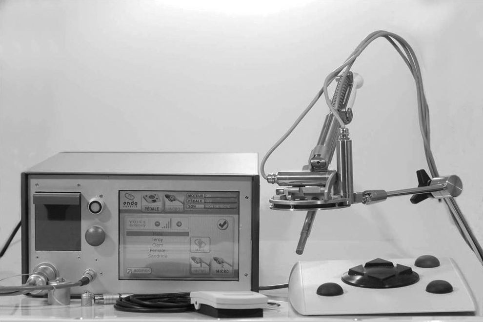
The miniaturized light endoscope-holder ViKY with its control box containing the electricity supply and robot control.
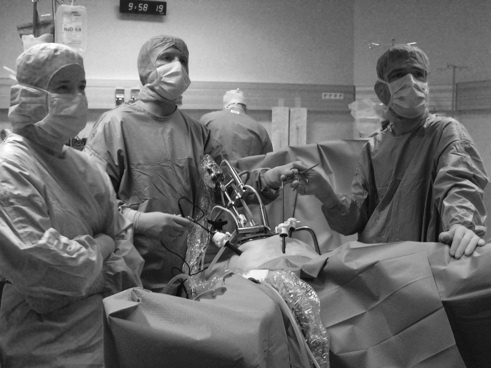
ViKY in use, resting on the patient's abdomen.
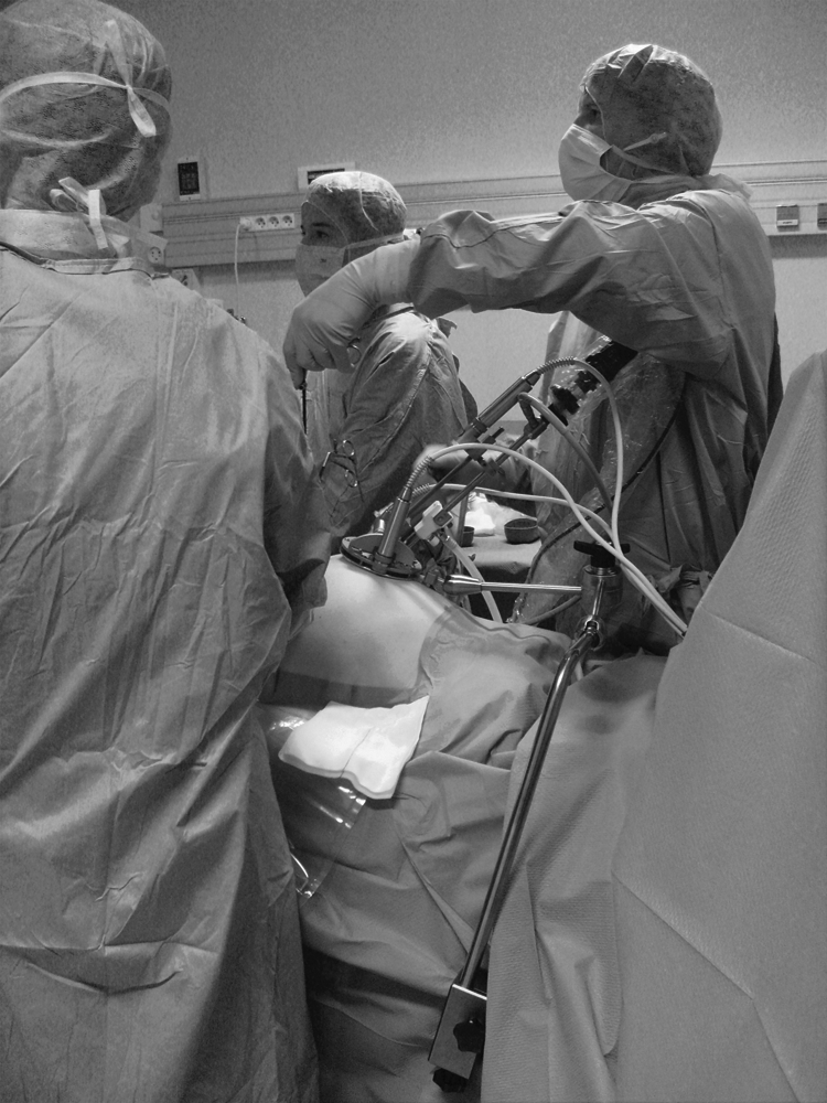
ViKY in use, fixed to the operating table using an articulated arm.
End points
For each procedure the operating time from robot set-up to port removal was recorded together with any robot technical problems or conversion to an open procedure. Median operative time, length of hospitalization, postoperative morbidity and mortality, and functional results were also recorded. At the conclusion of each procedure, three aspects of the surgeon's satisfaction were evaluated by the surgeon himself with a note ranging from 0 (bad) to 10 (excellent). These aspects included easiness of use, overall comfort, and quality of vision. Furthermore, the surgeons had to record any discomfort they experienced with the LER during the rectopexy procedures.
Follow-up
After surgery all patients underwent regular follow-up consultations at 1 month, 6 months, and annually thereafter. The follow-ups included a clinical examination. Median follow-up was 20 months (range, 12–42 months), and no patient was lost to follow-up.
Results
Patient population
Sixteen laparoscopic rectopexy procedures have been performed between March 2008 and September 2010 with the assistance of the LER. The patients were 16 (100%) women with an average age of 63.6±13.8 years. The median preoperative body mass index was 24 kg/m2. Using the American Society of Anesthesiology (ASA) classification systems, 3 patients (18.75%) were classified as “ASA 1,” 9 (56.25%) as “ASA 2,” and 4 (25%) as “ASA 3.”
Operative data
No conversion to open surgery was performed, and the laparoscopic procedure could be completely achieved in 15 patients with the LER. In 1 case, the Bluetooth microphone was damaged after it fell down during the set-up procedure of the robot, and the operation had to be completed manually. In eight procedures (50%) the robot was voice-controlled; it was controlled via a pedal in one procedure and using voice and pedal in seven procedures. The median operative time was 117.5 minutes (range, 93.25–128.8 minutes). The median time to set-up the LER was 15 minutes (range, 10–25 minutes), and these times became progressively shorter as more operations were performed. During the set-up procedures three minor instances of discomfort were recorded by the surgeon: they occurred during the fixation of the passive arm to the table in one instance, the fixation of the robot to the umbilical trocar in another case, and finally during the assemblage to the laparoscope lens in one other instance. The robot never interfered with the positioning of the trocars. Removal of the LER was rapid, as it could be dismantle within a median time of 3 minutes (range, 2–5 minutes). No particular difficulties were recorded by the surgeon during the surgical procedure or during the dismantling of the robot.
There were no intraoperative complications. Among the 15 procedures performed with the assistance of the LER the intraoperative and postoperative mortality rate was 0%. During the postoperative follow-up, one complication (6.25%) was recorded, consisting of a pelvic hematoma (Clavien grade II 14 ) for which the patient was rehospitalized at postoperative Day 7. No surgical treatment was necessary, and the patient could be discharged home after 5 days.
Patients were discharged from the hospital after a median of 3 days (range, 1–4 days). During the follow-up of the patients, no recurrence of the rectal prolapse was diagnosed.
Surgeon's satisfaction
The scores of ease of use, global comfort, and quality of the vision were 7±2, 8±2, and 8±2, respectively, with the LER.
Discussion
Manual manipulation of the laparoscope is a difficulty encountered by surgeons.15–17 Needing both hands free to carry out the operation, the surgeon has to delegate to the assisting surgeon who maneuvers the videocamera and sometimes an additional grasper. Consequently, the surgeon has no direct control over the viewing angle. An assistant without experience or understanding of the surgeon's needs for visualization often provides erroneous views. Holding the camera may also be exhausting in long cases. Because of fatigue and hand tremor the image may become unsteady. These problems tend to diminish the concentration of the surgeon and impede the flow of the operation. 18
Various supporting instruments have been developed to facilitate laparoscopic surgery. Among them, instrument positioners were introduced as an alternative to human camera holders. These instruments assume the task of the surgical assistant and return camera control to the surgeon. They are divided into two main groups: passive positioners (manually positioned by the surgeon) and active positioners (positioned by a robotic device).
The passive positioners such as Endofreeze (Aesculap, Tuttlingen Germany), 19 Endoboy (Geyser-Endobloc, Coudes, France), 20 Martin Arm (Gebrüder Martin GmbH & Co., Tuttlingen), 21 PASSIST (Academic Medical Centre, Amsterdam, The Netherlands), 20 or the Boonpong laparoscopic camera holder 22 are stable, but inherently slower in use compared with robotic systems. 23 The operator must release instruments and interrupt the procedure to make operating field adjustments. This may be particularly difficult at critical points in the operation. 24
Various robotic systems designed to overcome these limitations have been brought to market over recent decades. The first active positioner was the AESOP, followed by the Endoassist, the FIPS endoarm, the LapMan, and the Naviot. The introduction of robotic-assisted endoscopic surgery offers new advantages to the surgeon. Specifically, it allows precise voice-activated or foot control of the robotic camera holder and enables the surgeon to use both hands under normal operating room conditions. The stability of these robotic platforms provides a tremor-free image and better visualization during the entire surgical procedure, compared with the quality of the image obtained with a human camera operator who is more subject to distraction and fatigue, resulting in so-called “tremor view.”
The main drawbacks of these active robotic systems are their high purchase prices and cumbersome sizes, both of which tend to impede their acceptance and widespread use. 25
More recently, the TIMC-IMAG-CNRS Laboratory in France designed a miniaturized light endoscope-holder, called the LER. In comparison with existing robotic camera holders, the compactness of the LER is an advantage—the robot is approximately 110 mm in diameter and 75 mm high and weighs less than 1 kg (625 g). Thus, during the rectopexy procedures, the surgeon never recorded any discomfort due to the size of the LER. The LER never hampered the work space of either the surgeon or his assistants, and it never hindered the surgeon's movements. Furthermore, the 11-cm-diameter base did not require any change in trocar position during the procedures. The only minor discomforts noted by the surgeon were during the set-up procedures. Furthermore, if the operating table is readjusted during surgery, then some robots (e.g., EndoAssist, LapMan) must be realigned, although the LER is fixed to the operating table using an articulated arm.
In cadaveric and animal models the system proved to be efficient, user-friendly, and compatible with existing surgical equipment. 7 However, before widespread use, it was necessary to objectively assess the feasibility, safety, and surgical comfort of the system in various fields of laparoscopic surgery, including urology, gynecology, and digestive surgery. Because the LER had already been evaluated in urologic surgery, 8 our team assessed the LER in rectal surgery during laparoscopic rectopexy procedures. Initial analysis of our results reveals that the use of the LER in rectal surgery is feasible, safe, reliable, and user-friendly in human patients. There was no mortality or intraoperative complications, and postoperative complications were in the normal range (6.25%). Overall, 15 of the 16 robotic procedures could be successfully completed with the assistance of the LER, whereas in 1 patient the preoperative damage to the Bluetooth microphone led to manual completion. No conversion to open surgery was required, and the median operative time was comparable to that for rectopexy procedures without a robot published in the medical literature. 26 Considering the high grades in ease of use, global comfort, and quality of vision, the satisfaction of the surgeon with the LER was very good. Although the lack of a control group makes these results subjective, they are similar to the good scores of operator comfort reported while using the LER in urologic surgery. 8 Furthermore, the LER is easy and quick to set-up and dismantle. The interface used in greater than 50% of the cases was the voice recognition system. It proved reliable in recognizing orders, was unimpeded by the ambient sound environment of the operating room, which was particularly noisy at times, and created none of the problems described with other robots. 27 It should be noted that voice training was required beforehand to ensure effective recognition.
As for robotic endoscope holders in general, the question is whether use of this device is really necessary. Does it really offer the possibility to the surgeon to perform solo surgery without any human assistant? What are the advantages in comparison with conventional procedures? Studies comparing the differences between human and robotic control of the laparoscope have shown the robotic system to be superior.1–3,7 However, very few of these studies were prospective and randomized. Furthermore, a recent Belgian study conducted by researchers in the health technology assessment field underlined the necessity of further comparative, prospective, randomized biomedical research to prove the benefits of robotics in surgery. 28 These results are the first of a more ambitious study whose objective is to randomly compare a human holder and the LER in laparoscopic rectopexy procedures. 29
Footnotes
Acknowledgments
The authors thank Ms. Antonia Giraud for her editorial assistance.
Disclosure Statement
No competing financial interests exist.