
Abstract
Abstract
Although natural orifice specimen extraction is now widely performed, there have been no reports of transoral extraction following laparoscopic gastric resection. This report describes the first transoral specimen extraction in a patient with a gastrointestinal stromal tumor (GIST) of the lesser curvature of the stomach. The clinical data of a patient with a large gastric GIST were reviewed. Totally laparoscopic resection of the gastric lesser curvature was performed using four trocars. The specimen, put in a retrieval bag, was withdrawn via the transgastric and esophageal route. Reconstruction of the stomach was performed using the intracorporeal technique. The procedure was successfully accomplished without intraoperative and postoperative complications. In conclusion, transoral specimen extraction after laparoscopic gastric resection is a safe and feasible operative procedure for selected patients with a large benign gastric tumor.
Introduction
Among the potential natural orifice specimen extraction routes, the transgastric route has never been used for gastric specimen extraction because its usual large volume. To our knowledge this is the first report concerning transoral specimen extraction after a laparoscopic gastric resection for a large gastrointestinal stromal tumor of the stomach.
Case Report
A 68-year old female patient presented with epigastric pain, retrosternal pyrosis, and episodic arrhythmia. An upper gastrointestinal endoscopy showed the presence of a large submucosal mass, infiltrating the lesser gastric curvature (from the cardias to the pylorus) with normal mucosa. A needle biopsy allowed the diagnosis of gastric gastrointestinal stromal tumor (GIST). A computed tomography scan confirmed the presence of a 12-×6-cm mass developing into the gastric wall.
Surgical procedure
The procedure was carried out with the patient under general anesthesia with endotracheal intubation and epidural analgesia. The patient lay on the table in the supine position, with legs apart and 20° head-up tilt. The surgeon operated in the “French” position with the camera assistant on his right. CO2 pneumoperitoneum was induced after umbilical insertion of the first 12-mm trocar by the open technique. Three other ports were then inserted under vision through the abdominal wall: one in the left upper quadrant, one in the right upper quadrant and one in the midline, just below the xiphoid process. 6 The stomach, the liver, and the peritoneal cavity were inspected to rule out adjacent organ invasion, metastasis, and peritoneal seeding. The lesser sac was opened, and the gastrocolic ligament was divided using a 5-mm radiofrequency forceps (LigaSure Advance™; Covidien, Mansfield, MA). A large tumor extended along the lesser curvature, from the cardias to the pylorus (Fig. 1). The left gastric vessels were clipped and divided at their root. The lymph nodes along the celiac axis were dissected. The anterior gastric wall was entered 1 cm away from the tumor, and the mass was resected. The specimen was then put in a bag, introduced through the transabdominal trocar, and morcellated in small fragments. Seven endoloops were then tied along the bag (like a row of sausages) to avoid clustering of the fragments during the specimen removal. The bag was transferred into the open stomach, and a fibrogastroscope was used to retrieve it out through the mouth (Fig. 2). In order to suture the gastric wall, four lifting stitches were tied along the open lesser curvature, and then four endostaples were fired below them (triple-staggered row of a 60-mm Endo GIA Roticulator™; Covidien).

View of the posterior gastric wall with the gastrocolic ligament raised with an instrument. A large lumpy tumor (arrows) extends along the lesser curvature, from the cardias to the pylorus.
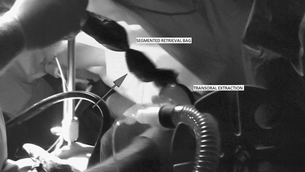
Gastric specimen retrieval through the mouth. After tumor morcellating, the retrieval bag was divided like a row of sausages with several endoloops (arrow).
Postoperative course
Fluid and soft solid diets were introduced, respectively, on postoperative Days 2 and 6. The patient was discharged on postoperative Day 8.
Discussion
The first reported trans-natural orifice specimen extraction was performed in 1993 by Delvaux et al., 7 who described the extraction of the gallbladder and its 4-cm stone via the transvaginal route following laparoscopic cholecystectomy in a woman. In 1996, Kim et al. 8 described transvaginal extraction of the large bowel in 4 patients after a low anterior resection, with excellent results. In 2002, Gill et al. 9 reported vaginal extraction of the intact specimen following laparoscopic radical nephrectomy. Recently, Palanivelu et al. 10 reported transvaginal extraction of the entire large bowel after totally laparoscopic proctocolectomy.
Transvaginal extraction following partial gastrectomy in patients with gastric submucosal tumors was reported in 2009 by Nakajima et al. 11 Because of the usual large volume of gastric specimens, the transgastric route for stomach specimen extraction has never been used.
In our patient we were able to resect the large tumor of the lesser gastric curvature, to withdraw it via the oral route, and to reconstruct the stomach with an intracorporeal suture, with a totally laparoscopic approach.
The main technical difficulties were the large specimen retrieval through the esophagus and the accomplishment of a unique totally intracorporeal anastomosis. The specimen volume reduction was obtained by morcellating the tumor; the retrieval bag was then divided in several not-merging cameras tying seven endoloops.
For the gastrointestinal reconstruction, after a laparoscopic subtotal gastrectomy for cancer, we used to perform an intracorporeal Billroth II retrocolic gastrojejunostomy or a Roux-en-Y reconstruction. 12 In this patient, gastric resection involved a large area of the lesser curvature. We passed four lifting stitches on the lesser curvature, and then we completed the suture by four endostapler applications (60-mm cartridges) below them.
In conclusion, we think that transoral specimen extraction after laparoscopic gastric resection is a safe and feasible operative procedure and can be used also in patients with large benign gastric tumors.
Disclosure Statement
No competing financial interests exist.