
Abstract
Abstract
Background:
Ureteral injury during laparoscopic surgery is rare, but when it occurs, it can be a serious problem. Near-infrared fluorescence (NIRF) with methylene blue (MB) administration is a promising technique for easier and potentially earlier intraoperative visualization of the ureter. Aim of this prospective study was to assess the feasibility of NIRF imaging of the ureter during laparoscopic colorectal surgery, using MB.
Methods:
Patients undergoing laparoscopic colorectal surgery were included and received intravenous injection of MB preoperatively. The ureter was visualized using a laparoscope, which offered both conventional and fluorescence imaging. Intraoperative recognition of the ureter was registered. The precision of ureter distinction with MB imaging was compared to the conventional laparoscopic view.
Results:
Ten patients were included. All procedures were initially performed using a laparoscopic approach. Dose per injection ranged between 0.125 mg/kg and 1.0 mg/kg bodyweight. There were no adverse effects attributable to MB administration. The ureter was successfully detected in five patients, with highest contrast between ureter and surrounding tissue at an administered dose of 0.75–1.0 mg/kg. The fluorescent signal was only picked up after the ureter was already visible in the conventional white light mode.
Conclusion:
Ureteral fluorescence imaging using MB proved to be safe and feasible. However, the present technique does not provide practical advantage over conventional laparoscopic imaging for identification of the ureter during laparoscopic colorectal surgery. Future research is necessary to explore more extensive dose finding, alternative fluorescent dyes, or improvement of the imaging system to make this application clinically beneficial.
Introduction
T
Ureteral injury during surgery is rare, but when it occurs, constitutes a serious problem. The major causes are trauma, iatrogenic injury during open surgery, and injury during catheter or endoscopic intervention. Ureteral injuries are often insidious since they are found only after other, more profound injuries are addressed, which may lead to worse outcomes. Risk factors for iatrogenic injury during colorectal surgery are previous pelvic operations, infection, and inflammatory bowel disease, but most ureteral injuries occur in patients lacking these risk factors. 2 Earlier studies have reported an incidence of 0.1% to 7.6% in colorectal and gynecologic surgery, with more than 80% of cases going unrecognized intraoperatively.3–6 Assimos et al. 6 compared the incidence of iatrogenic ureteral injuries between the prelaparoscopic and laparoscopic era and found that the incidence of iatrogenic ureteral injuries was significantly greater in the latter. Thus, the early detection and prevention of ureteral injury are important. If in any other way than with normal vision and common anatomical knowledge, essential anatomical planes and structures could be identified, this would beneficially influence the safety and efficiency of laparoscopic colorectal surgery.
To help avoid injury, intravenous pyelography, retrograde pyelography, or urologic computed tomography can be performed preoperatively; however, none of these imaging techniques provides real-time guidance during the actual procedure. 7 Another applied method is ureteral stent placement. However, in laparoscopic surgery, the results are not convincing and apart from added operation time, can cause complications itself.2,8,9
Intraoperative fluorescence ureteral identification with preoperative optical dye administration10,11 is a promising new technique for easier and earlier intraoperative visualization of the ureter and could thereby improve the outcome–safety and efficiency–of laparoscopic colorectal surgery.
Matsui et al. 10 showed that the clinically available dye methylene blue (MB) has near-infrared (NIR) fluorescence properties (even in very low doses) that permit real-time intraoperative visualization of the ureters in a pig model in open and laparoscopic procedures. Verbeek et al. 12 were able to visualize the ureters during open human surgery, using MB in combination with a near-infrared fluorescence (NIRF) imaging system.
We hypothesize that the application of the NIRF technique using MB during laparoscopic colorectal surgery can improve the visualization of the ureter, thereby speeding up the time of dissection and identification of the ureter, which increases patient safety by lowering the risk of ureteral injury. Therefore, in this study, we assessed the feasibility of this technique for the detection of ureters in patients undergoing elective laparoscopic colorectal surgery.
Materials and Methods
Patients
This prospective observational study has been approved by the Medical Ethics Committee of the Maastricht University Medical Center and was performed in accordance with the ethical standards of the Helsinki Declaration of 1975. The study was conducted in the Maastricht University Medical Center (MUMC) and registered in the Netherlands National Trial Register: registration number NTR3605 (www.trialregister.nl/trialreg/adminrctview.asp?TC = 3605). Randomly chosen patients meeting the inclusion criteria (Table 1) were enrolled between January 2014 and July 2015. All patients scheduled to undergo laparoscopic colorectal surgery, in which identification of the left ureter was part of the standard procedure, were eligible for participation in the study. All included patients provided written informed consent and the collected data were anonymized.
Intraoperative imaging system
A commercially available laparoscopic fluorescence imaging system (Karl Storz GmbH & CO. KG, Tuttlingen, Germany) was used. This system has been described in more detail previously. 13 For this study, the system had been adapted with a special filter to detect the fluorescent signal of MB, which is at around 670 nm. The equipment included a plasma light guide and a 30-degree 10-mm laparoscope applicable for NIRF imaging. Procedures were digitally recorded.
MB preparation and injection
Methylthionine chloride (Methylene blue [MB]) 10 mg/mL was prepared by the hospital pharmacy of the Catharina Hospital (Eindhoven, the Netherlands).
After intravenous administration, MB is rapidly taken up by the tissues. The majority of the dose is excreted in the urine, usually in the form of leucomethylthioninium chloride. Peak MB absorbance and emission occur at 668 and 688 nm, respectively. 10 An intravenous MB solution for injection was administered preoperatively. Fluorescence imaging was performed at various moments during the phase of surgical dissection in which ureteral identification was relevant.
Surgical technique
The operations were performed by gastrointestinal surgeons with extensive experience in laparoscopic colorectal surgery. MB was administered using a preoperative intravenous injection in a vein of the upper extremity. Doses ranged from 0.25 mg/kg up to 1.0 mg/kg body weight. In the course of the study, these were increased based on previous publications.10,14 The dye administration occurred during the induction of anesthesia and ranged between 40 and 0 minutes before the introduction of the first trocar.
After surgery, the NIRF technique was assessed using an intraoperative registration form and visual recordings. Visual (both conventional and fluorescence) recordings were obtained during the same laparoscopic procedure. Urine from four included patients was collected from the bladder catheter at the end of the surgical procedure for NIRF imaging.
Ex vivo assessment of the NIRF technique
As a quality check of the procedure, in an ex vivo experiment, urine samples of two healthy volunteers were collected and accumulating concentrations of MB diluted in urine were made. Thereafter, fluorescence imaging was performed in a dark room with the camera at a 2 cm proximity of the sample. For each sample, a target to background ratio (TBR) was calculated as explained below. For this calculation, a vial filled with sterile phosphate-buffered saline was used as the background.
Target-to-background ratio
In image-guided surgery research, the target-to-background ratio is commonly used for analyzing recordings. For assessment of the degree of fluorescent illumination, OsiriX 5.0.1 Imaging Software was used. The fluorescent images were analyzed by determining the TBR. The fluorescence intensity of the target was measured as a mean of three point regions of interest in the ureter minus the mean fluorescence intensity of three regions in the immediate surrounding tissue as background, divided by the mean fluorescence intensity of the background. Hence, the following formula was used: TBR = (Fluorescence intensity of Target − Fluorescence intensity of Background)/Fluorescence intensity of Background.
Results
A total of ten patients were included in this study. Patient characteristics and clinical data are summarized in Table 2. Median age was 73.4 years (range 52–87) and median BMI was 27.44 kg/m2 (range 23.24–37.04). All patients were scheduled to undergo elective laparoscopic surgery for colorectal cancer. Administration of the dye ranged from 0 to 40 minutes before the introduction of the first trocar.
NIRF, near-infrared fluorescence.
A dose of 0.125 up to 1.0 mg/kg resulting in a total dose of 8–90 mg per patient was administered preoperatively. The dose was gradually increased during the time span of the study, due to disappointing results of the low doses. In only two of the six patients in which a concentration of 0.125–0.25 mg/kg MB was administered, there was (weak) visualization of the ureter in the fluorescence mode. In the other four, up to 100 minutes after administration, no fluorescent imaging of the ureter could be obtained, even after the detection of the ureter in white light mode. One of these procedures was converted to open procedure (due to intra-abdominal adhesions) before it was possible to analyze the ureter in fluorescence mode.
In one of two patients with a dosing of 0.5 mg/kg, the ureter was detected in fluorescence mode. This occurred with a weak signal and after the ureter had already been identified in white light mode. In the other patient, conversion to an open procedure was performed (because of intra-abdominal adhesions) before actual imaging of the ureter in fluorescence mode could take place.
In the remaining two patients, a concentration of 0.75 and 1 mg/kg showed a clear delineation of the ureter during the pulsatile movement of the urine through the ureter (see example screenshot in Fig. 1). The laparoscope had to be held close (within a few centimeters) to the ureter to obtain this visualization.
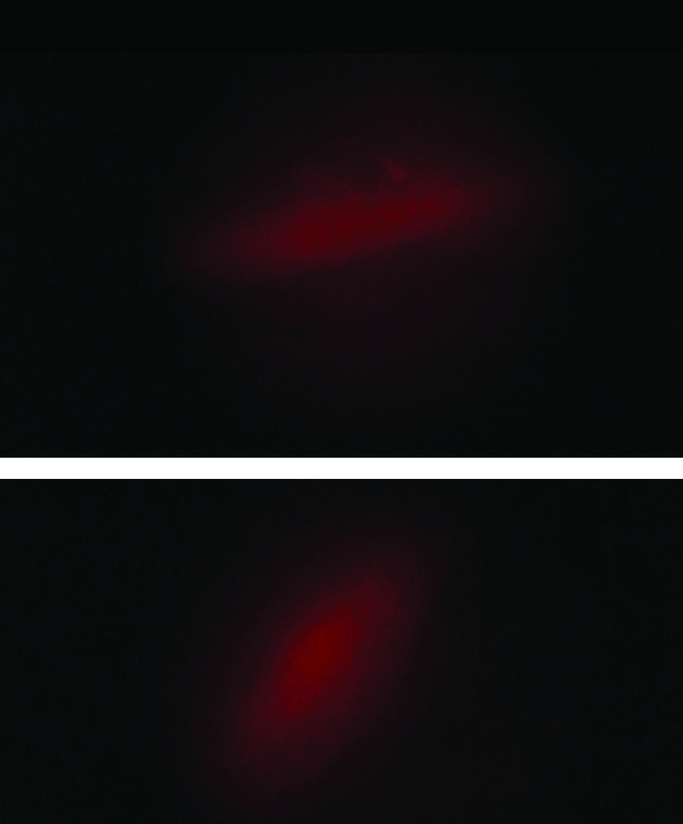
Screenshot of ureter visualization with fluorescence imaging at dosing of Methylene Blue of 1.0 mg/kg (top screenshot) and 0.75 mg/kg (bottom screenshot). In this screenshot, red is the illumination of the ureter and black is the surrounding tissue.
In four consecutive patients (patients number 5–8 in Table 2), urine was collected from the bladder catheter at the end of the procedure. Ex vivo fluorescence imaging with the same camera system as explained in the methods section showed a clear fluorescence signal of all four urine samples with a TBR varying between 6.88 and 9.2 (Table 3).
In the ex vivo experiment, a strong fluorescence signal in a wide range of considerable dilutions was obtained (Table 4). Very high concentrations of MB revealed no signal (TBR <0) and in very high dilutions, only a weak, although still present, signal.
MB, methylene blue; TBR, target-to-background ratio.
Discussion
Various recent studies have paid attention to the use of NIRF imaging in minimally invasive surgery. 15 This report describes a clinical feasibility study investigating the utility of NIRF imaging using MB for ureteral recognition during human laparoscopic colorectal surgery. Previously, promising results were reported with MB in an animal model 10 and in two human studies during open and laparoscopic surgical procedures.12,16 The primary objective of this study was to investigate the feasibility of intraoperative NIRF imaging using MB administration for detection of the left ureter during laparoscopic colorectal surgery.
In this study, it was found that this type of imaging provides additional visual distinction of the left ureter from its surrounding tissues, compared to the conventional laparoscopic view. However, this signal was not strong enough to support the surgeon with enhanced ureteral visualization over the conventional white light mode. When the ureter was identified in fluorescent mode, it could already be seen in white light mode. Another disadvantage is the close proximity (few centimeters) that was required between the endoscope and the ureter to obtain a signal. In the aforementioned previous studies, the authors described a distance of 18 and 30 cm between the camera and the surgical field in open surgery. No distance was given for the laparoscopic experiments. 10
In this study, higher doses of MB resulted in better visualization of the ureter. The dose chosen in the beginning of our study was based on the study of Matsui et al. 10 The better recognition with higher doses is in accordance with the observations of Verbeek et al. 14
The ex vivo experiment with MB dilutions in urine of healthy volunteers showed a strong fluorescence signal in a wide range of dilutions (Table 3), revealing that even very small concentrations of methylene blue in urine can give a clearly visible fluorescence signal. This confirms the possibility to obtain a good signal using the present equipment. Also, in the collected urine from four patients at the end of the surgical procedure, we were able to clearly obtain a fluorescent signal in these urine samples, even though in vivo ureteral detection in these patients was not possible with NIRF imaging. It is hypothesized that the absence of a clear fluorescence signal in the included cases may be influenced by anatomical barriers, such as the ureteral wall thickness and the fatty tissues surrounding the ureter, hindering fluorescence signal detection. Currently, fluorescence imaging at the NIR range of wavelengths has the potential of a better tissue penetration than visible light, up to 10 mm, but the in vivo situation often exceeds this tissue thickness. 17
The results found in this study are not compatible with the results found in the study by Matsui et al. 10 in which the feasibility of methylene blue application in open surgery in 20 pigs was investigated. In all animals, the ureter was identified successfully. A possible explanation for these results is the thinner ureteral wall and subretroperitoneal layer in pigs and less intra-abdominal fat compared to humans. Verbeek et al. found similar results in humans.10,12 However, in their study, they only investigated the feasibility of the use of methylene blue in open surgery. A clear identification of the ureters was observed at doses between 0.25 and 1.0 mg/kg. Perhaps, in the setup of this study, the optimal combination of dye (e.g., dosage and timing of administration) and laparoscopic system has not yet been reached to obtain similar results.
There were no adverse (immediate or delayed) reactions with regard to the clinical outcome following MB administration in this study. However, shortly after the administration of the dye intravenously, a transient decrease of the oxygen saturation was observed, which was measured by a finger pulse oximeter. This phenomenon is known and is caused by the principle of pulse oximetry, which is based on the red and infrared light absorption characteristics of oxygenated and deoxygenated hemoglobin, 18 which is influenced by the transient passage of MB. Preoperatively, the patient should be notified that intravenous MB administration could potentially cause a severe anaphylactic reaction; however, this risk is very low to negligible.
Main limitation of this study is that the study population is too small to draw firm conclusions regarding variables such as optimal timing and dosage of dye administration.
Furthermore, the patients were included over a relatively long time span. This was due to changing of the involved researchers. This is not likely to have had a negative effect on the performance of the study.
In the cases where the ureter was recognized using the fluorescence mode of the laparoscope, recognition was only possible after the ureter had already been identified using the conventional white light mode, suggesting that fluorescence imaging of the ureter using MB does not provide any added clinical value to this procedure. Therefore, this particular imaging modality in this setup is not yet deemed to be suitable in aiding the identification of the ureter during laparoscopic colorectal surgery.
In conclusion, we report the human clinical experience with near-infrared fluorescence imaging and methylene blue during laparoscopic colorectal surgery for intraoperative ureteral detection. Although promising, as indeed visualization of the ureter could be obtained, this imaging modality did not provide added value in the delineation of the ureter because simultaneous with the fluorescent signal, the ureter could be visualized even in conventional white light mode. Future research should focus on exploring alternative fluorescent dyes (for example preclinical dyes 19 ) and optimization of the equipment to make this promising technique clinically relevant.
Footnotes
Disclosure Statement
Dr. M. Al-Taher, MD; Dr. J. van den Bos, MD; Dr. R.M. Schols, MD, PhD; Prof. Dr. N.D. Bouvy, MD, PhD; and Prof. Dr. L.P.S. Stassen, MD, PhD declare that Karl Storz Gmbh has supplied the laparoscopic equipment for this study.