
Abstract
Abstract
Background:
Spigelian hernia represents a rare entity. Traditionally, it was repaired by the open technique. Various laparoscopic techniques have emerged periodically. Most laparoscopic repairs use the technique of placing an intraperitoneal onlay mesh (IPOM). There is currently a tendency to close the hernia defect.
Methods:
A 68-year-old man was admitted to our hospital complaining of vague abdominal pain and discomfort. Physical examination revealed a bulky palpable mass on the left side of the patient. Computed tomography of abdomen revealed a left-sided incarcerated spigelian hernia containing bowel. The aim of this study was to report an educational video providing a successful laparoscopic IPOM-plus technique with muscles approximation of spigelian hernia repair step by step.
Results:
Total operative time was 120 minutes. The postoperative period was uneventful. The patient reported no pain and the cosmetic result was excellent. No bulging or seroma was noted. The patient was discharged on postoperative day 1.
Conclusion:
The IPOM-plus technique may improve the outcome of spigelian hernia repair.
Introduction
S
Traditionally, the repair of spigelian hernia was open anterior herniorrhaphy. The laparoscopic intra-abdominal approach was performed at 1992 by Carter and Mizes. 2 Since then, laparoscopic repair has increased sporadically offering the advantages of minimal access surgery such as reduction of postoperative pain, morbidity, length of hospital stay, and better cosmesis.3–5
Most laparoscopic repairs use the technique of placing an intraperitoneal onlay mesh (IPOM). There is currently a tendency to close the hernia defect. The adoption of this technique seems to improve the outcomes because it results in lower rate of seroma formation and adverse hernia site events. 6
Case Presentation—Surgical Technique
A 68-year-old man with body mass index of 35 kg/m2 and medical history significant for coronary artery disease and diabetes was admitted to our hospital complaining of vague abdominal pain and discomfort. Physical examination revealed a bulky palpable mass of the left side of the patient. Computed tomography of the abdomen revealed a left-sided incarcerated spigelian hernia containing bowel.
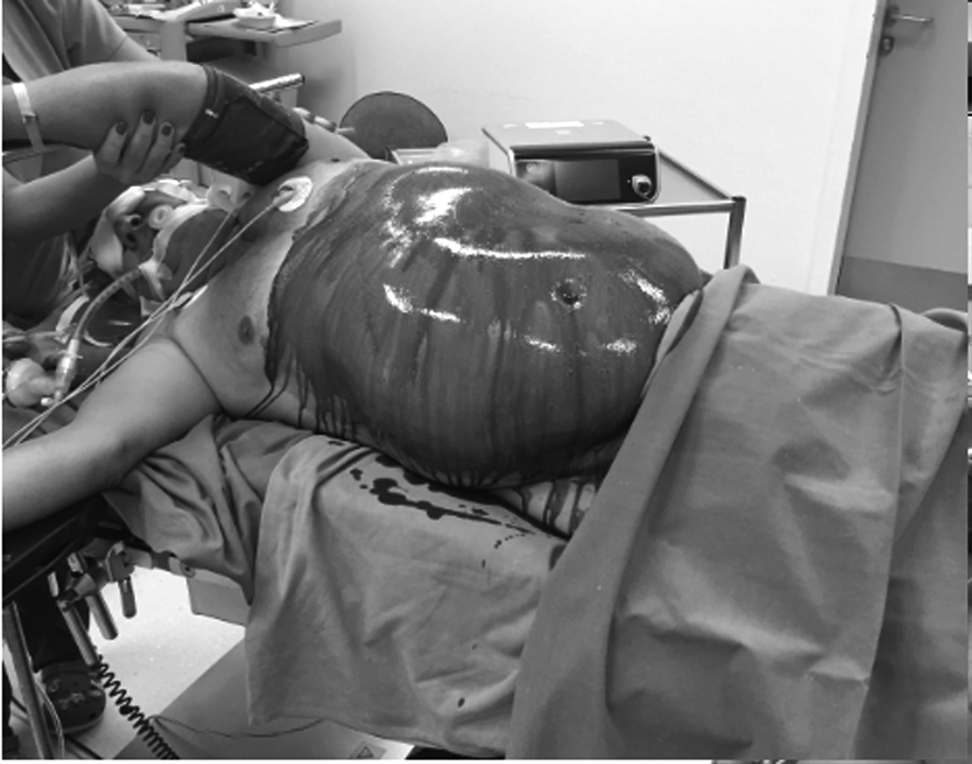
The patient was positioned in the left decubitus position (Fig. 1). A pneumoperitoneum was created using a Veress needle. A 10-mm trocar for the telescope and two additional trocars were inserted (Fig. 2). The spigelian hernia was identified and the sigmoid colon was successfully reduced with gentle traction. The hernia sac was also dissected. The margins of the hernia defect were then determined. Owing to the different muscular layers produced by the spigelian hernia, closure of the defect was decided using intracorporeal interrupted figure of eight sutures with PDS 2-0. Pneumoperitoneum was reduced to 10 mm Hg when sutures were positioned. The polyester composite mesh with an absorbable adhesion prevention membrane (Covamesh™, Biom'up, France) 12 × 12 cm was then inserted through the 10-mm trocar. The size of the mesh was determined by the defect size and a 4 cm overlap in all directions was considered important. The mesh was unfolded, anchored with a stay suture with an endoclose, and secured outside the abdomen with a hemostatic clip. This maneuver allowed the mesh to be positioned flat against the anterior abdominal wall covering completely the hernia defect with appropriate overlap. The mesh was then fixed to the anterior abdominal wall with the double-crown technique of absorbable tacks. The tacks were placed around the entire edge of the mesh providing adequate pressure while securing the spiral tacks (Supplementary Video S1; Supplementary Data are available online at www.liebertpub.com/lap).
Patient positioning.

Trocars position.
The total operative time was 120 minutes. The postoperative period was uneventful. The patient reported no pain and the cosmetic result was excellent (Fig. 3). No bulging or seroma was noted. The patient was discharged on postoperative day 1. At follow-up after 3 months, the patient had good recovery and action and reported no pain.

Final cosmetic result.
Discussion
Spigelian hernias produce two different layers from the aponeuroses of oblique and the transversus muscle. Traditionally, repair consisted of open anterior herniorraphy, using direct muscle approximation, mesh, and prostheses with various personalized adapted techniques. Since the introduction of the intra-abdominal laparoscopic repair in 1992 by Morena-Egea and the extraperitoneal approach in 2002, several sporadic reports have been published. There is paucity of a large series of laparoscopic spigelian hernia repair. Largest data report that only 11.3% of patients had a laparoscopic repair in total, supporting the feasibility and success of the laparoscopic approach. 7
Various laparoscopic techniques have been successfully used for the treatment of spigelian hernias with different advantages. Namely, the IPOM approach was considered the most popular (39.2%), followed by the transabdominal preperitoneal patch (TAPP) approach (26.1%), total extraperitoneal patch (TEP) approach (19%), and laparoscopic suturing techniques (8.3%). 8 The TAPP approach is usually preferred for large, complex, recurrent, and incarcerated hernias, because it allows assessment of the viability of the hernia sac contents. The TEP technique has the advantage of avoiding entry into the peritoneal cavity and thus bowel damage and is recommended particularly on patients who may have intra-abdominal adhesions from previous surgery. 7 Disadvantages include technical expertise, prolongation of operative time, reduced operative field necessitating extensive dissection, missing of small hernia defects, and inability to explore the contents of an incarcerated hernia sac. The IPOM approach represents the most commonly used popular technique in the management of spigelian hernias.
The IPOM is the easiest technique, providing adequate experience in laparoscopic surgery, because it offers the best visibility, requires the least dissection to get a good view of the weak areas, and enables positioning of a large mesh without any problems extending it; however, it has the potential risk of intestinal injuries.7–9 According to the guidelines for laparoscopic ventral hernia repair, the IPOM technique is limited by the fact that it produces a bridged area without musculoaponeurotic coverage, which is functionally adynamic and is responsible for bulging and seroma formation.
Adding sutured repair in the IPOM technique (“augmentation repair” or IPOM-plus technique) may be advantageous because it reduces the hernia size to minimum, eliminates bulging, and decreases the seroma size and incidence, thus lowering potential infection risk. 10 Primary fascial closure in a recent systematic review was associated with lower recurrence rates (0%–5.7% versus 4.8%–16.7%), seroma formation rates (5.6%–11.4% versus 4.3%–27.8%), and bulging than nonclosure. Patients with closure reported satisfaction with the results and better functional status. 11
In our case, the defect size was 5 cm. The hernia content was the sigmoid, but the mobilization was feasible, because few adhesions were found. The closure of the defect was possible without unacceptable tension. The closure of the defect provided a secure reconstruction along with an excellent cosmetic result. No bulging or seroma was noted. The patient reported that he had no pain and was satisfied. Thus, the IPOM-plus technique may improve the outcome of spigelian hernia repair.
Footnotes
Disclosure Statement
No competing financial interests exist.
References
Supplementary Material
Please find the following supplemental material available below.
For Open Access articles published under a Creative Commons License, all supplemental material carries the same license as the article it is associated with.
For non-Open Access articles published, all supplemental material carries a non-exclusive license, and permission requests for re-use of supplemental material or any part of supplemental material shall be sent directly to the copyright owner as specified in the copyright notice associated with the article.