
Abstract
Abstract
Background:
The aim of our study was to report our experience in thoracoscopy in infants and neonates for vascular surgical conditions in neonates and infants and to compare our results to open surgery regarding the short-term outcome.
Patients and Methods:
We retrospectively reviewed all the patients operated in a single institution from 1997 to 2016 for persistent ductus arteriosus (PDA) and vascular ring (VR) anomalies. We compared our thoracoscopic series to a historical control group operated by open surgery. Data collection from charts and office notes included age and weight at surgery, cardiac ultrasound data for PDA, preoperative clinical symptoms for VR, type of surgery, operating time, analgesic treatment requirements, ventilation status during postoperative course, and early complications.
Results:
The thoracoscopic group included 13 PDA (median age and weight at surgery: 34 days and 1800 g) and 11 VR (median age and weight at surgery: 8 months and 7000 g). The thoracoscopic group did not differ in preoperative symptoms and work-up, operating time, ventilation status, length of hospital-stay, and postoperative complications with the group operated on by thoracotomy, for either PDA or VR.
Conclusion:
Our short-term results in thoracoscopic PDA closure and VR anomalies surgery in neonates and infants are comparable to open surgery. Thoracoscopy seems to provide less pain especially for neonates and premature babies and allows to decrease the risk for postoperative chest wall deformities. Long-term outcome is mandatory to confirm these preliminary results.
Introduction
T
The numerous advantages of thoracoscopy tend nowadays to be demonstrated by numerous reports, regarding decreased postoperative pain, shorter hospital stay, decreased incidence of chest wall deformity (including scoliosis and breast deformity), lower incidence of postthoracotomy pain syndrome, and better cosmetic results.1,8,9
However, there is still a need for report series comparing thoracoscopy with the standard thoracotomy regarding effectiveness and morbidity, especially in neonates and premature babies for PDA closure. The aim of our study was to report our thoracoscopic experience of general pediatric surgeons for surgery of such pathology (PDA+VR) and to compare our results to open surgery regarding the short-term outcome.
Patients and Methods
Clinical data
We performed a monocentric retrospective study of all patients operated on in our center from 1997 to 2016 for PDA and VR anomalies. Data collection from charts and office notes included age and weight at surgery, cardiac ultrasound data for PDA, preoperative clinical symptoms for VR, type of surgery (thoracoscopy or thoracotomy), operating time, success rate of the procedure, conversion rate, postoperative analgesic treatment requirements, ventilation status during postoperative course, length of hospital stay, postoperative recurrent nerve palsy, and occurrence of chylothorax. Regarding the thoracoscopic group, PDA surgical closure was indicated for a hemodynamically significant PDA in a neonate and either failure of ibuprofen or if ibuprofen was contraindicated. All VR patients benefited from preoperative laryngotracheal endoscopy and multiple detector computed tomography (MDCT) (See Supplementary Video S1; Supplementary Data are available online at liebertpub.com/lap) scanner with anatomical description of the malformation.
Surgical technique
The thoracoscopic approach for both pathologies used three trocars with optic port placement above the tip of the scapula (Fig. 1): 5 mm/30° trocar for the camera, and 3 or 5 mm for two operator trocars. For PDA surgery, we also used a fourth 3 mm trocar placed in the inferior part of the thorax to push away down the lung. Insufflation pressure was around 5 mmHg, without single lung ventilation. PDA closure was realized with a titan clip, and the VR was divided after intracorporeal knot with nonabsorbable suture. We did not leave any thoracic drain for PDA patients.
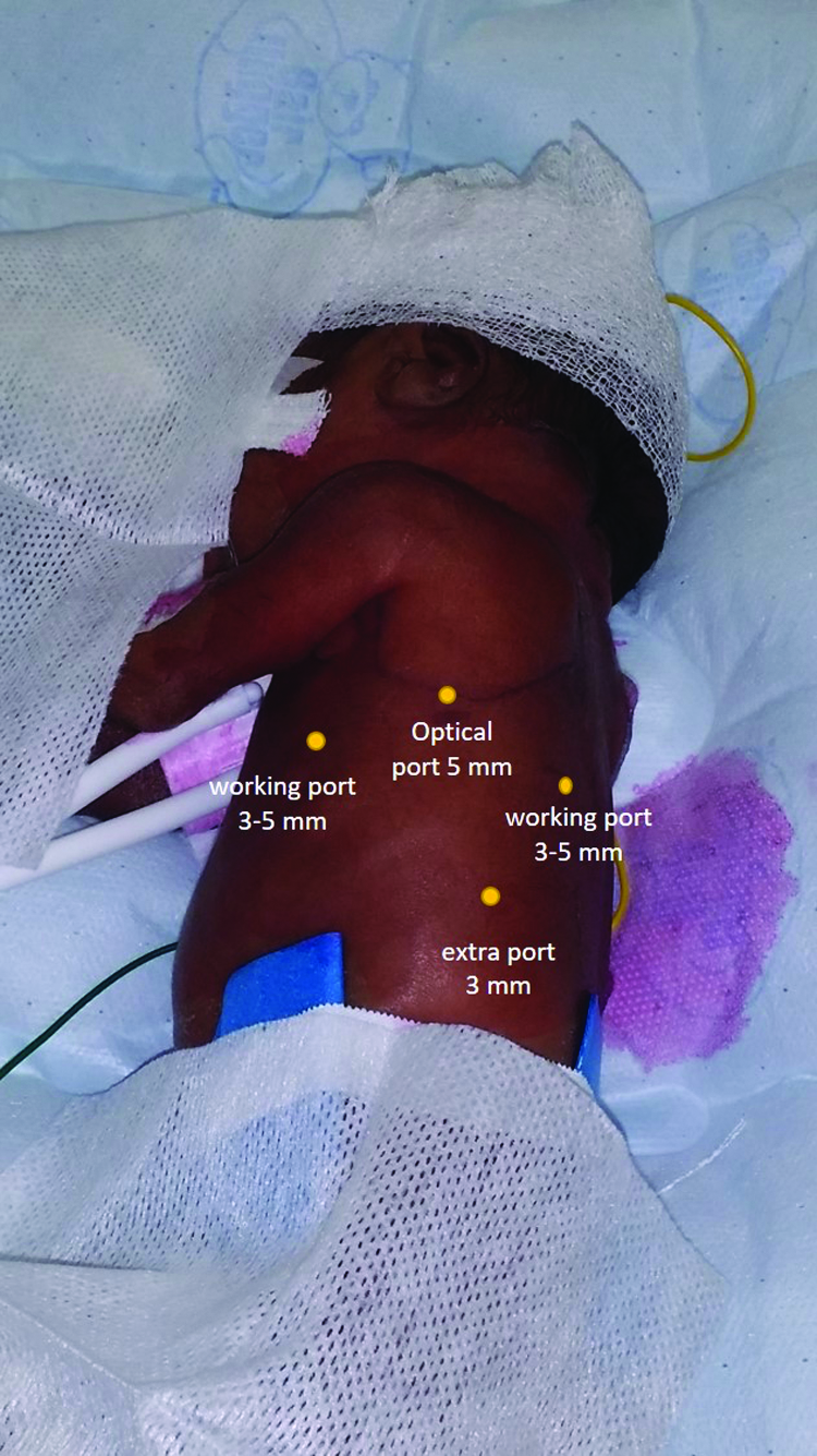
Ports placement for both persistent ductus arteriosus and vascular ring ligation.
Statistical analysis
We compared the patients with PDA and VR, operated on by thoracoscopy between 2008 and 2016 by a single operator, with two historical series of PDA and VR operated on by thoracotomy between 1997 and 2010, patients having been matched one to one for age and weight at surgery. We used the Student's t-test to compare the two surgical groups for both pathologies.
Results
Patients operated on by thoracoscopy by the same operator (AB) included 13 PDA (median age and weight at surgery: 34 days and 1800 g) and 11 VR (median age and weight at surgery: 8 months and 7000 g).
All PDA patients operated on by thoracoscopy were premature neonates (except one born at 37 WG), with a median gestational age at birth of 29.5 WG (min–max: 25–37) and a median weight at birth of 1255 g (min–max: 615–2520), and all were operated before 2 months of life. The smallest baby weighed 795 g, but was converted to open surgery because of the lack of working space.
The 22 VR patients presented various clinical signs at diagnosis, with no difference between the thoracoscopic group and the historical series operated on by thoracotomy. Results are presented in Table 1. One patient was diagnosed prenatally with an abnormal karyotype 47XXX. The anatomical description was assessed in all cases by contrast-enhanced computed tomography (CT) scanner: Complete double aortic arch (N = 12), right aortic arch with arterial ligament (Neuhauser anomaly) (N = 10). We retrieved a slight difference of age at surgery in the VR group, the rarity of the disease having prevented us to match the two groups for age and weight at surgery (Table 2).
Significant statistical difference, Student's t-test.
N/A, not applicable.
Operative time between the two surgical techniques was not different for both PDA and VR. Two cases of PDA and one case of VR required conversion for anesthetic reasons and lack of working space at the beginning of the learning curve. Residual shunt was not detected in any PDA patients on immediate postoperative cardiac ultrasound.
Recurrent nerve palsy systematically assessed by fibroscopy occurred in three cases of PDA, two cases by thoracotomy, and one by thoracoscopy. They were all transient and not retrieved at 6 months follow-up. Chylothorax occurred in two cases of VR both by thoracoscopy, resolving without reoperation. One case of VR operated by thoracotomy required reoperation for persistent tracheal compression. These small numbers prevented us from performing any further statistical analysis.
Regarding the immediate postoperative course of PDA, we failed to demonstrate a statistical trend neither for thoracic tube duration, nor for mechanical ventilation and/or switch from high-frequency oscillation to conventional ventilation for PDA. Results regarding the use of analgesia were not comparable since the protocol changed between these two periods with a trend to decrease the morphinic requirement. Results are presented in Tables 2 and 3.
Student's t-test.
N/A, not applicable; HFO, high-frequency oscillation.
Surgery-related mortality was null for both PDA and VR in both groups. Median follow-up was of 6 months.
Discussion
In this study, we provided a comparative study of both VR and PDA treatments by open or thoracoscopic surgery. Our results, despite the small number of patients, tended to show that the mini-invasive approach in experienced hands is as safe and effective than the open approach. It provides a better cosmetic result and a minimal risk for thoracic sequelae, which can occur after thoracotomy, especially in preterm babies.
Thoracoscopic surgery in neonates and infants for aortic anomalies are mostly performed around the world by cardiothoracic surgeons. Large series of PDA in children have already been published since many years by them, fewer regarding neonates,10–13 and small series of VR.3,4,14,15 However, pediatric thoracoscopic surgery is currently spreading throughout the general pediatric surgery community and tends to become the gold standard, especially in experimented teams, such as S. Rothenberg's department. 16 Indeed, this technique applied to aortic malformations requires an experienced pediatric laparoscopic surgeon, familiar with suturing, intracorporeal knots, and adequate using of clips. They have demonstrated that this technique, as surgeons become more comfortable with the approach, is safe and effective, minimizing physiologic and cosmetic adverse effects. The learning curve allowed them to use laparoscopy even in patients less than 4 kg in the last half of their series, with a minimum weight of 2 kg, and a mean weight of 8.5 kg. This is the main difference with our series, which included mostly premature babies, but with similar results in terms of success and conversion rate. They mentioned only 3 failures of PDA ligation among 78 patients, due to a torn ductus and 2 recurrences, respectively, treated by surgery and cardiac catheterization. We had no recurrence, but had to convert the smallest baby (795 g) due to lack of working space. Regarding VR treatment, they did not mention any complication, such as chylothorax, like us. We unfortunately did not manage to avoid this complication in two laparoscopic cases despite the magnification of the laparoscope, although both patients recovered without requiring surgery. Similarly, all our patients were clinically improved at short-term evaluation (6 months) by the VR surgery.
Due to the relative rarity on these cardiovascular malformations, and the skills required to perform laparoscopic surgery on these patients, very few comparative studies with open surgery are available in the literature.3,17 It is therefore difficult to prove to the community that it is worth to acquire these skills, in terms of potential risk-to-benefit ratio for the patient. Indeed, our statistical analysis is based on a very small series, but it stills appears that we did not have major complications, nor high rate of recurrence. Postoperative morphinic requirement has been difficult to compare in our study since the 20-year period have seen so many changes in the postoperative pain control in neonates. This is even more comprehensible in the PDA group, where no drain was left progressively over the period after the chest closure, in both open and thoracoscopic techniques. This also might have an effect on the duration of the mechanical ventilation requirement, although we failed to demonstrate any statistical result. In such premature babies with potential risk of pulmonary dysplasia, a long-term evaluation on a larger series is needed. The operative time, in experienced hands nowadays, is quite even, compared with older series, 3 and the complications tend to decrease compared with open surgery.
Thoracotomy in neonates and especially in preterm babies may present some immediate complication such as ribs fractures and late complications such as chest wall deformities and/or scoliosis. We think that in these particular surgical indications, thoracoscopic approach should be the preferable approach to minimize these sequelae and offer a better cosmetic result.
More reports are needed regarding the possibilities of robotic surgery in infants for PDA and VR treatments,18,19 and due to the lack of space within the chest cavity in these small infants, miniaturizing of surgical ports and instruments remains mandatory.
Conclusion
Our short-term results in thoracoscopic PDA closure and VR anomalies treatment in neonates and infants is comparable to our experience in open surgery. Thoracoscopy in neonates and premature babies provides a better cosmetic result and decreases the risk of chest wall deformities, which can occur after thoracotomy. A larger series is mandatory to confirm these preliminary results.
Footnotes
Disclosure Statement
No competing financial interests exist.
References
Supplementary Material
Please find the following supplemental material available below.
For Open Access articles published under a Creative Commons License, all supplemental material carries the same license as the article it is associated with.
For non-Open Access articles published, all supplemental material carries a non-exclusive license, and permission requests for re-use of supplemental material or any part of supplemental material shall be sent directly to the copyright owner as specified in the copyright notice associated with the article.