
Abstract
Background:
To evaluate the effectiveness and safety of endoscopic decompression with a transanal drainage tube (ED-TDT) in the treatment of neonatal Hirschsprung's disease (HD).
Materials and Methods:
Six newborns (4 boys and 2 girls; age at operation 9–29 days, median 24 days) were admitted to our hospital due to the delayed passage of meconium after birth, abdominal distension, and vomiting. HD was diagnosed by the retrograde enema radiography. All patients underwent ED-TDT for decompression of the dilated colon. During the operation, an endoscope was inserted through the anus to the ascending colon or ileocecal region, which was then used to insert a guidewire to the ascending colon. The endoscope was then removed, and a colorectal drainage tube was introduced over the guidewire. This drainage was placed until a pull-through procedure was performed.
Results:
The ED-TDT was successfully performed in all newborns. The intraoperative rectal biopsy confirmed the diagnosis of HD. The duration of the operation was 25–70 minutes (median 52.5 minutes). There were no perforations, bleeding, or other complications after surgery. After surgery, the abdominal distention was significantly relieved, and the babies could be fed with milk after 6 hours. These babies were discharged after 2–11 days (median 2.5 days) and underwent a pull-through procedure after 1–3 months (median 3 months).
Conclusions:
ED-TDT is a safe and effective treatment for neonatal HD before a pull-through procedure. It could be used as an alternative therapy to colostomy.
Introduction
Hirschsprung's disease (HD) often leads to abdominal distension, vomiting, poor feeding, and delayed passage of meconium, which in turn may result in failure to thrive, malnutrition, and enterocolitis in children. The transition zone shown by the barium enema is usually regarded as the classic imaging of HD, and is found between the contracted aganglionic zone and the dilated normal bowel. According to the transitional zone location, HD is usually classified into the short- and long-segment type. The aganglionic tract involves the rectum and the sigmoid colon in the short-segment HD. The long-segment disease extends toward the proximal end of the colon. Resection of the aganglionic colon and a pull-through procedure are the only effective treatments for HD; however, it still remains controversial whether or not a single-stage procedure is needed in newborns. Generally, rectal irrigation or enterostomy is the selected treatment to decompress the dilated colon and prevent enterocolitis in newborns with HD. For the short-segment HD, rectal irrigation may be effective in most newborns before a definitive procedure. However, for the long-segment HD or total colonic aganglionosis, the newborns with HD are usually treated by enterostomy. In this study, the endoscopic decompression with a transanal drainage tube (ED-TDT) was used as an alternative to enterostomy in treating neonatal HD before a pull-through procedure.
Materials and Methods
A total of 6 newborns (4 boys and 2 girls; age at operation 9–29 days, median 24 days) were admitted to our hospital between November 2016 and January 2020 due to the delayed passage of meconium after birth, abdominal distension, and vomiting. There were obvious abdominal distension, varicose veins, and high tension in the abdominal wall (Fig. 1A). The rectal examination demonstrated an explosive discharge of stool and gas. An abdominal X-ray examination showed intestinal obstruction and dilated colon. HD was diagnosed by using retrograde enema radiography (Fig. 2), revealing a long-segment HD in 4 patients and a short-segment HD in 2 patients. The traditional enemas were first used to resolve the abdominal distention and intestinal obstruction. However, the traditional enemas failed to decompress the colon in these 6 patients. Therefore, all babies underwent an ED-TDT to relieve the abdominal distention and intestinal obstruction. The detailed data are shown in Table 1. This study was according to the institutional research committee's ethical standards, and all the infants' guardians signed the informed consent before surgery.
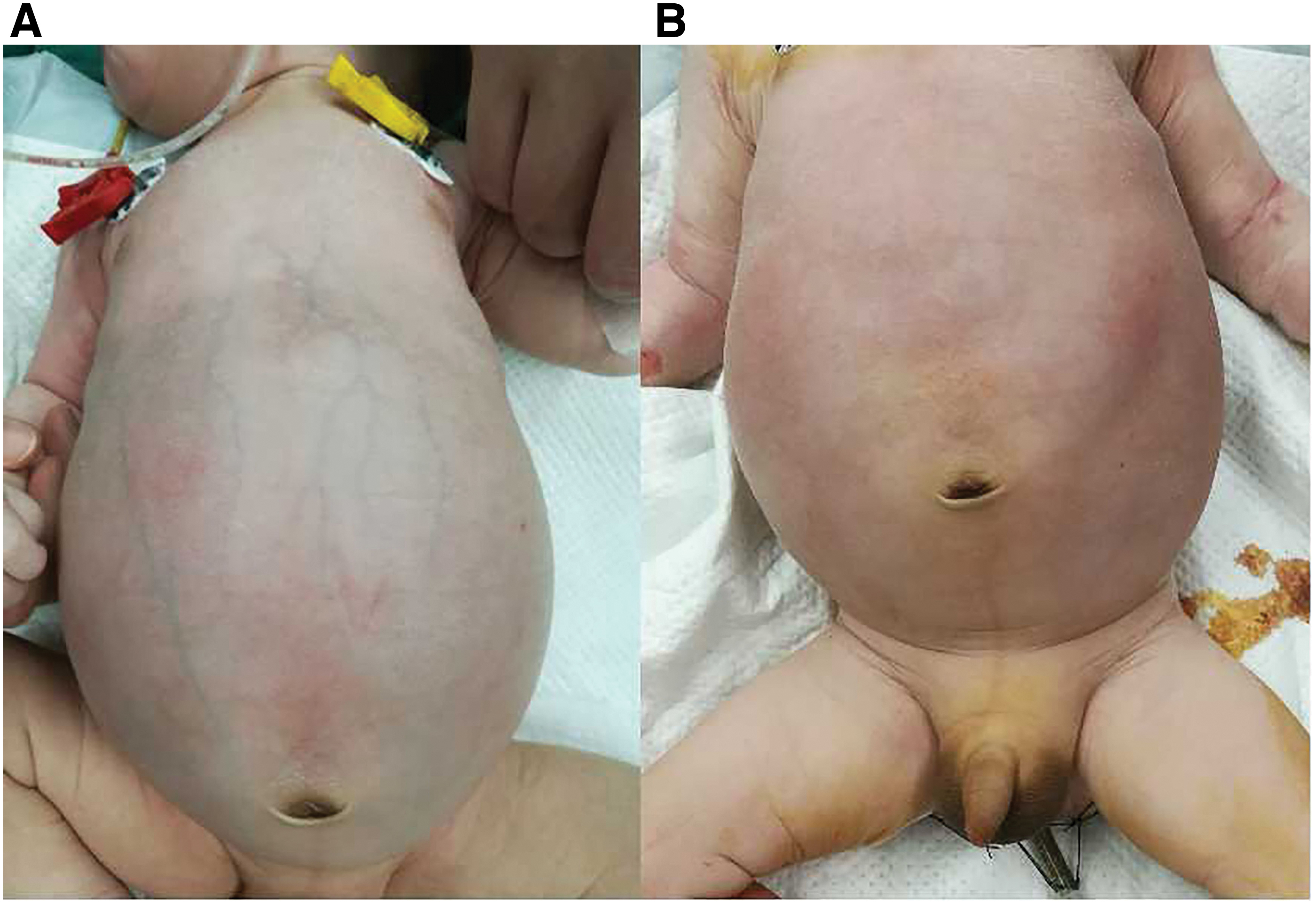
Obvious abdominal distension, varicose veins, and high tension of the abdominal wall observed before the placement of a drainage tube
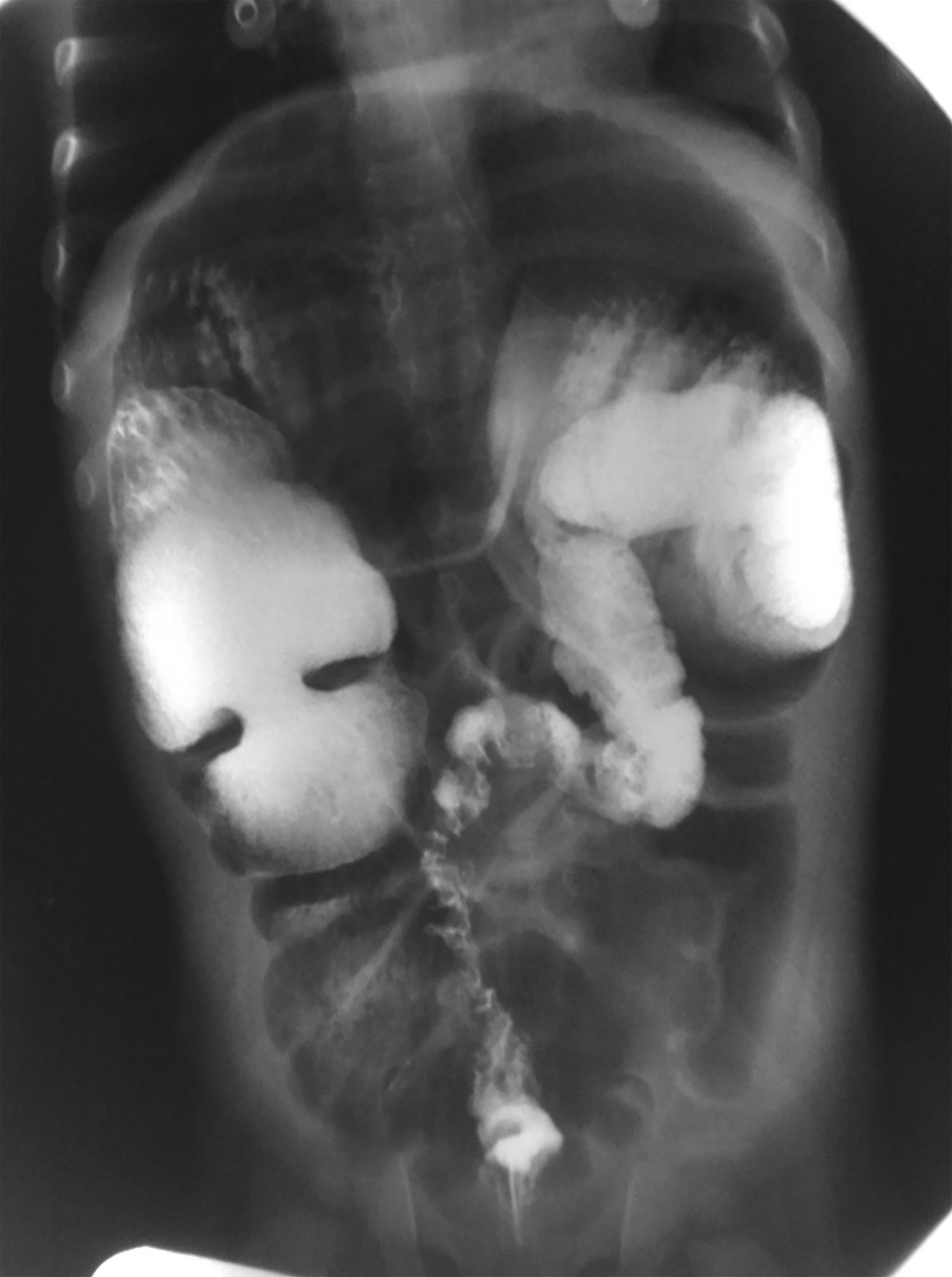
The preoperative enema radiography showed the contracted, transitional, and dilated zone of HD. HD, Hirschsprung's disease.
The General Conditions and Outcomes of Patients
The drainage tube fell off by accident after 1 month, and the baby suffered from abdominal distention and constipation again and had to undergo a pull-through procedure in advance. Pre: before the placement of the drainage tube; post: after the placement of the drainage tube, and before the pull-through procedure; final diagnosis of HD refers to the diagnosis of HD that was determined by pathology after the pull-through procedure; diagnosis of HD by radiography refers to the HD diagnosed by the retrograde enema radiography before the placement of the tube; long, long-segment HD; short, short-segment HD; total, total colonic aganglionosis.
HD, Hirschsprung's disease.
Surgical methods
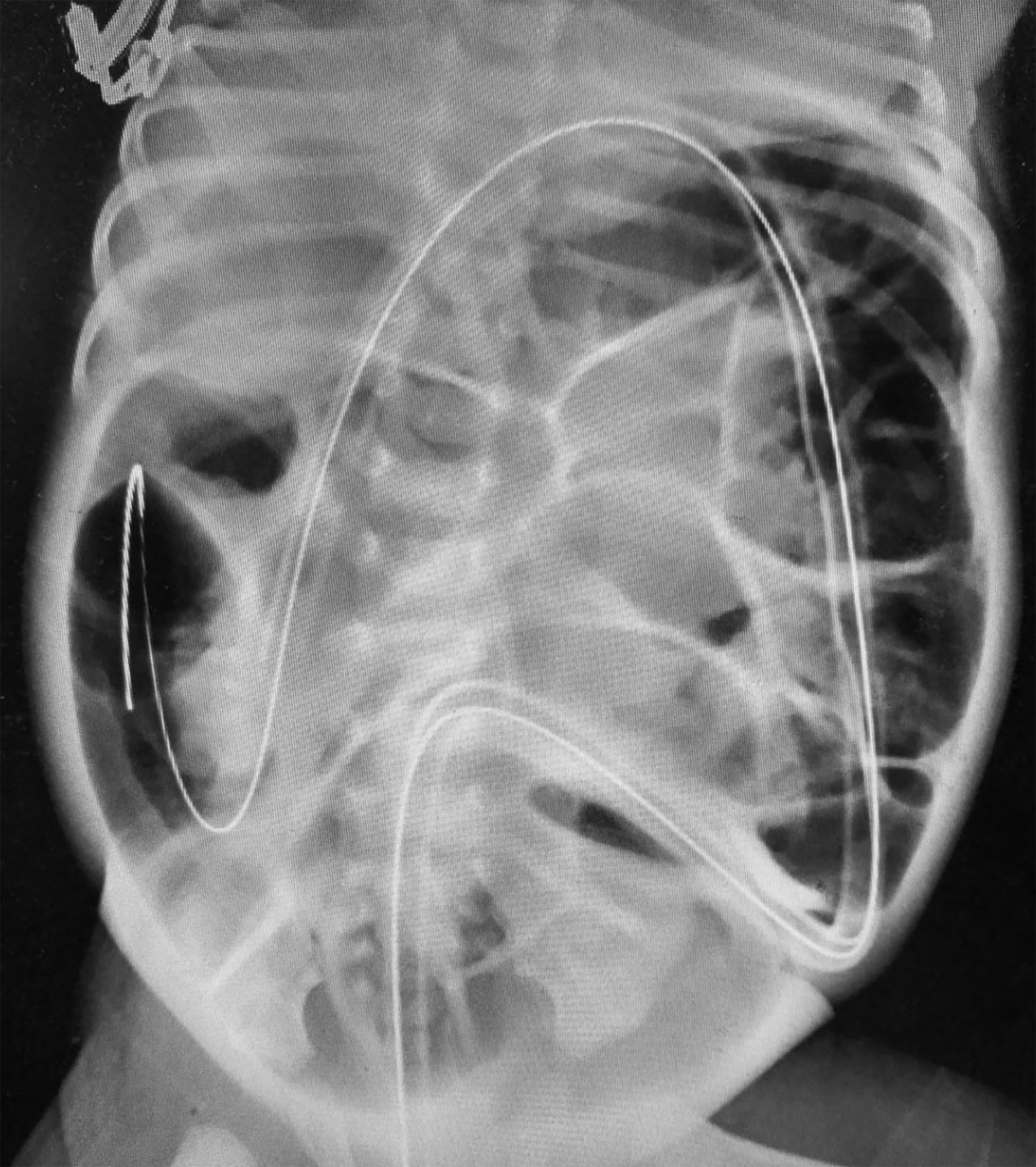
Glycerol enema was carried out before the procedure. An endoscope was inserted through the anus to the ileocecal region during the operation, which was then used to insert a 0.13 cm guidewire to the ascending colon (Fig. 3). The endoscope was removed, and a colorectal drainage tube (12F) was introduced over the guidewire, and its distal end was placed at the proximal end of the ascending colon (Fig. 4). An intraoperative abdominal X-ray examination confirmed that the distal end of the tube was placed at the proximal end of the ascending colon (Fig. 5). After removing the guidewire, the gas and stool in the bowel run out from the tube. Finally, the tube was fixed at the anus using a tape. A drainage bag was attached to the tube. A rectal biopsy was performed during the operation to identify the diagnosis of HD.

A 0.13 cm guidewire was inserted into the ascending colon through the endoscope.
A colorectal drainage tube was introduced over the guidewire, and its distal end was placed at the ascending colon.

The intraoperative abdominal X-ray examination confirmed that the tube's distal end was placed at the ascending colon.
After surgery, the tube was cleaned two times daily using 200 mL of saline for the prevention of blockage. The management of drainage tube was performed by the infants' guardians at home after discharge. This drainage tube was placed until a pull-through procedure was performed after 2–3 months.
Results
The outcomes are shown in Table 1. The ED-TDT was successfully performed in all newborns. The duration of the operation was 25–70 minutes (median 52.5 minutes); the length of the drainage tube in the colon was 30–38 cm (median 31.5 cm); the abdominal distention was significantly relieved (Fig. 1B), and the babies were fed with milk after 6 hours. These babies were discharged after 2–11 days (median 2.5 days).
The rectal biopsy revealed the absence of ganglion cells and the presence of hypertrophic nerve trunks, which confirmed the diagnosis of HD in all newborns. There were no perforations, bleeding, or other complications after surgery. Four patients underwent a pull-through procedure after 3 months, and 1 patient underwent a pull-through procedure after 2 months. The drainage tube fell off by accident after 1 month in 1 patient (Patient no. 1 in Table 1), who consequently suffered from abdominal distention and constipation and had to undergo a pull-through procedure in advance. The body weight was significantly increased after drainage. The HD types were finally diagnosed by the pathological biopsy after the pull-through procedure, resulting in two with a long-segment HD, three with a short-segment HD, and one with a total colonic aganglionosis.
Discussion
HD is the second most common cause of intestinal obstruction in newborns, 1 which may lead to intestinal perforation if not timely treated. 2 Approximately 60% of neonatal HD presents with vomiting, abdominal distension, and failure to pass meconium in the first 48 hours of life. HD-associated enterocolitis has been identified as a major reason for the high mortality in neonatal HD. Therefore, early diagnosis and colostomy can reduce both the mortality and the incidence of enterocolitis. However, although the barium enema's inaccuracy in the diagnosis of neonatal HD has been emphasized, some studies have reported diagnostic accuracy of about 76%, 87%, and 89% in this population.3–5 Therefore, the high diagnostic rate still relies on the rectal biopsy for making the definitive diagnosis. 6 In this study, all of the patients were diagnosed as HD by retrograde enema radiography, which was further confirmed by an intraoperative rectal biopsy. In addition, it still remains controversial whether an immediate colostomy is needed. Previous studies have suggested that irrigation is an effective therapy and that some infants may be too ill to undergo colostomy.7–9 Yet, other studies showed that a colostomy could reduce the mortality in newborn series.1,10 In our hospital, we usually train children's guardians to perform a day-to-day enema and anal dilation for the infants with a short-segment HD, after which a pull-through procedure is performed at 3 months. However, the colon's aforementioned management is usually not effective for the long-segment HD due to failure in the decompression of the bowel, where colostomy is needed to decompress the bowel and prevent enterocolitis.
It still remains controversial whether or not a single-stage pull-through procedure should be performed in infants <3 months. Carcassonne et al. 11 suggested that curative surgical treatment of HD (including total colonic HD) is satisfactory in patients <3 months and that preoperative decompression using a routine colostomy is not necessary. Ekema et al. 12 performed a one-stage transanal Soave operation in newborns with the short-segment disease, eliminating the need for an abdominal incision and colostomy. However, if the child suffered from HD-associated enterocolitis or a significantly dilated colon, a colostomy should be performed for several months until the child recovered; the pull-through procedure was usually performed 4–6 months after colostomy placement.13,14 In our study, all patients suffered from the dilated colon and intestinal obstruction. They were all supposed to be treated by a colostomy according to the aforementioned strategy. Nevertheless, colostomy has some disadvantages. First, a routine colostomy is considered as the main etiologic factor in secondary perineal dermatitis and late encopresis, even with normal manometric studies. It delays the acquisition of normal organic and psychological corticoanal connections at least until the age of 12 months. 11 Second, abdominal incision and a secondary anastomosis need to be performed to close the stoma, leading to more pain and trauma in children. Third, there may be the disused atrophy of the distal colon after colostomy, which increases the difficulty of the pull-through procedure and the time of recovering the colonic function after surgery. Fourth, there is a risk of stoma prolapse or retraction. To avoid these disadvantages of colostomy, we performed ED-TDT in this study.
Transanal drainage tube placement for relief of acute colorectal obstruction is commonly used in adult patients. The ED-TDT has been reported for the management of acute colorectal obstruction in adults15–17 as an effective and safe approach that can be used to avoid a colostomy. The tube could decompress as well as clean the dilated colon. However, most of the previous ED-TDT reports involved adult patients, and there are few reports in children. The colorectal obstruction in adults is usually caused by colorectal carcinoma, whereas the colonic dysfunction causes intestinal obstruction in children with HD. According to its function of decompression, ED-TDT may be suitable for HD treatment by decompressing the dilated colon and preventing enterocolitis. Therefore, we performed the ED-TDT in neonatal HD. According to a previous study, tube placement was complicated by perforation in 0%–7%, whereas other complications such as bleeding were not reported. 15 In this study, all procedures were successfully completed, and there were no complications.
To avoid the risk of perforation, there were some special procedures in the neonatal ED-TDT. First, a 9 mm soft endoscope was used due to the smaller neonatal colon lumen. Second, we made sure that the bowel's tension was not too high, as it may lead to perforation. Therefore, the volume of injected gas should be moderate, and the injection and suction of gas should be balanced. Third, the endoscope should be gently inserted and smoothly moved forward. Fourth, the insertion to the sigmoid, transverse, and ascending colon was relatively difficult. Therefore, an assistant pressed the abdomen to help with the insertion. Fifth, it was sometimes difficult to pass an endoscope for the contracted aganglionic segment of HD; thus we injected some saline to dilate the bowel. Compared with colostomy, there was no abdominal incision, diarrhea, and electrolyte disorder after the tube's placement. In addition, the cost of stoma care can be eliminated. Good bowel preparation is very important when a surgical procedure is performed in a patient with HD to decrease postoperative complications. Before a pull-through procedure, the HD patients usually undergo the rectal irrigation several times a day until the colon is clean. Therefore, the colonic tube placement is beneficial for preparing the bowel before a pull-through procedure due to its drainage for intestinal contents. Yet, in our study, the tube fell out after 1 month in 1 patient, and he had to undergo a pull-through procedure before the expected time (after 3 months). Therefore, the fixation and nursing of the tube are very important.
The limitation of this study is its retrospective nature; obviously, a larger prospective randomized trial is warranted to confirm the efficacy of this new strategy for the treatment of neonatal HD. In addition, the number of cases should be increased in future studies, and a comparative study between the ED-TDT and colostomy is warranted.
In conclusion, ED-TDT is a safe and effective treatment for neonatal HD before a pull-through procedure. It could be used as an alternative therapy to colostomy in HD.
Footnotes
Disclosure Statement
No competing financial interests exist.
Funding Information
This study has received funding from National Nature Science Foundation of China (Code: 81770595).