
Abstract
Introduction:
Epiphrenic diverticulum is a rare disease caused by mucosa and submucosa herniation through the muscular layers of the esophageal wall. This study presents a case of a patient with a symptomatic epiphrenic diverticulum treated with surgery under endoscopic assistance. A review of the literature on this unusual condition was conducted, focusing on the pathogenesis, presentation, and surgical challenges.
Methods:
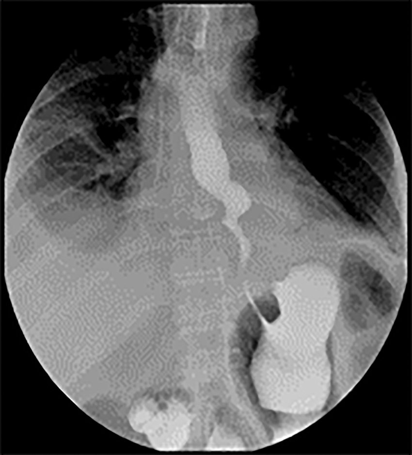
A 75-year-old woman who underwent an esophagogastroduodenoscopy (EGD) after experiencing worsening dysphagia for food and liquids. The EGD revealed a large epiphrenic diverticulum 36 cm from the upper incisor teeth. The presence of a diverticulum was verified by barium swallow, which revealed a 6 cm diameter epiphrenic diverticulum on the right side of the esophagus.
Results:
Patient underwent laparoscopic diverticulectomy associated with Heller's myotomy and anterior partial fundoplication. A gastroscope was placed intraoperatively to calibrate the esophagus to prevent stenosis during diverticulectomy, and it was also used to check the integrity of the esophageal wall. The patient tolerated clear liquids on postoperative day 2. Postoperative course was complicated by right pleural effusion and fever managed with antibiotics and pleural drainage.
Conclusion:
Epiphrenic diverticulectomy in conjunction with management of the underlying motor dysfunction and avoidance of gastroesophageal reflux disease is an effective procedure. Laparoscopy is considered the approach of choice for the majority of patients. Endoscopic assistance during surgery can help the identification of the diverticulum and verify the integrity of the staple line.
Introduction
Esophageal diverticula are rare with a prevalence of 0.06%–3.6% described on radiologic and endoscopic procedures. 1 Diverticula can occur in any portion of the esophagus: Rokitansky diverticula are generally found in the thoracic esophagus and are typically associated with tuberculous mediastinal lymph nodes. 2 Zenker diverticulum is a pharyngoesophageal junction diverticulum. Epiphrenic diverticula occur in the distal 10 cm of the esophagus, representing the 10% of the esophageal diverticula. 3 Epiphrenic diverticulum is a pulsion diverticulum; this condition is due to the herniation of mucosa and submucosa through the muscle layers of the esophageal wall. 4 Primary esophageal motility disorder frequently occurs in association with epiphrenic diverticula5–10 and achalasia represents the most described associated disorder (>60% of cases).11,12
Dysphagia, regurgitation of undigested food, 8 chest pain, and weight loss are the most common symptoms;8,13–15 these symptoms usually may be caused by the underlying esophageal dysmotility more than the diverticulum per se5,10,16 and the size of diverticulum do not correlate with them.11,13,17,18 Respiratory symptoms such as nocturnal cough, asthma, laryngitis, and pneumonia can be due to episodes of aspiration;8,18 in patients with esophageal achalasia, stasis and fermentation of the food can cause heartburn. 19 Severe complications as bleeding and perforation are rare, as well as malignant transformation.3,20,21 Finally, 40%–60% of the cases of epiphrenic diverticulum can be asymptomatic.11,22
Barium esophagogram and upper digestive endoscopy are the diagnostic procedures most frequently applied.13,22 Probably the barium swallow is the most important test to evaluate the symptoms but especially to plan the surgical intervention. Barium swallow test defines the size of the diverticulum, its shape, neck, location, and the distance from the gastroesophageal junction, and it can also identify any complications, although they are rare, as fistulae or underlying motility disorder.3,13 Epiphrenic diverticulum's size ranges from 1 to 14 cm, with a median size of about 4–7 cm.8,10,23 Most of cases (>70%) present epiphrenic diverticulum on the right side of the esophageal wall;8,23 although the largest part of patients has a single epiphrenic diverticulum, ∼15% of patients may have multiple diverticula.5,23 Upper digestive endoscopy is also essential to rule out any associated diseases. 11
Methods
A 75-year-old Caucasian female presented to our Institution for worsening dysphagia for solids and liquids that had continued for about 3 years. Her medical history included arterial hypertension and lymphoma treated with systemic chemotherapy with complete remission. Physical examination, cardiological, and laboratory test findings were otherwise unremarkable. Esophagogastroduodenoscopy (EGD) revealed a large epiphrenic diverticulum 36 cm from the upper incisor teeth. Barium swallow confirmed the presence of the epiphrenic diverticulum on the right side of the esophagus measuring 6 cm in the long-axis diameter (Fig. 1). Conventional manometry of the esophagus excluded underlying dysmotility disorders. Laparoscopic Heller myotomy with resection of the esophageal diverticulum and anterior partial fundoplication was scheduled.

Barium swallow shows a 60 mm epiphrenic diverticulum on the right wall of the lower esophagus.
Operative technique
With the patient in a modified lithotomy position, 3 12 mm trocars were inserted: 1 at the umbilical level and 2 at the epigastrium at the level of the left and right midclavicular lines. Two additional 5 mm trocars were placed, one at the left anterior axillary line at the level of the umbilicus and in the right subcostal position for the liver retractor. The procedure began with a dissection through the gastrohepatic ligament to the diaphragm's right crus. The lower esophagus was then dissected circumferentially in the loose areolar tissue till the diverticulum was found. The right chest pleural lining was detached from the diverticulum laterally, taking care not to get into the right hemithorax. When the diverticulum was fully free of its attachments, 8 cm of distal esophagus was dissected within the lower mediastinum.
After that, an endoscopy was performed until the diverticular aperture was clearly spotted. The endoscope's endoluminal insufflation aided in the completion of the diverticular dissection. To prevent the esophagus from being narrowed, the diverticulum was resected at its base using a 45-mm stapler and direct endoluminal visualization. The dissecting hook was used to carry out the myotomy. The myotomy had been performed 6 cm from the lower esophageal sphincter and 2 cm away from it, until the stomach oblique muscle fibers were reached. After that, the endoscope was reinserted, revealing a clearly visible gastroesophageal (GE) connection that was extensively patent. The myotomy was finished with no visible damage to the esophageal mucosa. The coagulating shears were used to divide the fundus's small gastric branches up to the left diaphragmatic crus.
After that, the fundus was passed anteriorly around the esophagus. The Dor fundoplication was then created by sewing the wrap's edges to the myotomy's margins with a 0 Poly (ethylene terephthalate) suture. The wrap was additionally superiorly fastened to the diaphragm's right crus. The esophagus and stomach were then irrigated with saline. There were no bubbles, indicating that the esophagus mucosa was not damaged. The entire procedure took 3 hours.
Results
On postoperative day 2 a barium swallow demonstrated that the esophagus was widely patent and had no leakage from either the diverticulectomy suture line or the myotomy site (Fig. 2). The patient then started a full liquid diet. Following the onset of fever on postoperative day 3, chest-CT was performed revealing right pleural effusion with basal dysventilation (Fig. 3). This complication was managed with antibiotics and thoracentesis followed by pleural catheter positioning. Furthermore, the patient underwent a EGD that showed the integrity of the suture line without leakages. The patient was then discharged on postoperative day 7. The patient is currently 24 months status post-Heller myotomy with no dysphagia or symptomatic reflux.
Postoperative contrast esophagography shows smooth flow of contrast medium to the stomach.
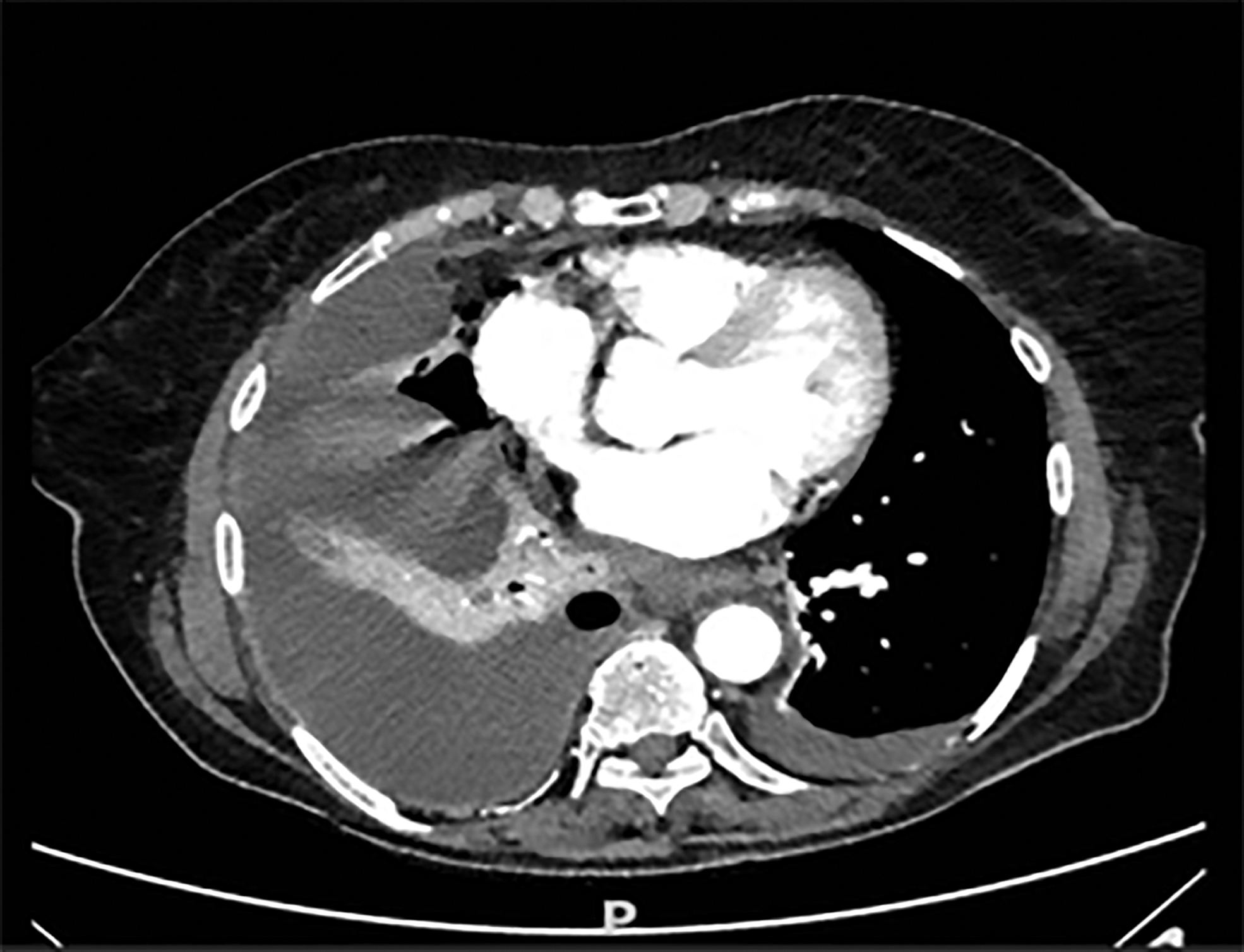
Contrast-enhanced computed tomography shows a right pleural effusion.
Discussion
Esophageal diverticula (ED) are caused by elevated intraluminal pressures, which are frequently triggered by achalasia or some underlying esophageal motility issue that results in “pulsion” physiology. Infact, the majority, if not all, ED patients have an esophageal motor dysfunction. Surgery is recommended for individuals with symptomatic epiphrenic diverticulum. Diverticulectomy, diverticuloplasty, and diverticulopexy are surgical treatments for an epiphrenic diverticulum, with diverticulectomy being the most commonly used. The most commonly adopted surgical technique is diverticulectomy with Heller's myotomy and partial fundoplication.13,14
When treating a symptomatic diverticulum, a cardiomyotomy is usually required, even if dysmotility was not found by esophageal manometry for various reasons: first, esophageal dysmotility may not be detected by conventional parameters and may be missed by inexperienced physiologists; second, esophageal dysmotility, rather than the diverticulum per se, may be responsible for the majority of symptoms, such as dysphagia and pulmonary symptoms; and third, the addition of a myotomy reduces the chance of leak due to lower intraluminal pressure.5,13,14,24 The first step of the surgical intervention is the identification and the mobilization of the Angle of His to create a retroesophageal window; 25 the esophagus is encircled, for example placing a Penrose drain around the esophagogastric junction to obtain a good traction to simplify the dissection. 26
When the ED is identified, it proceeds with the dissection of the pouch, obliterating the diverticular neck with a stapler jaws, keeping the line of suture parallel to the axis of the esophagus.27,28 Furthermore, to close the muscular layer, it may need two or three nonabsorbable stitches or also a continuous suture with a nonabsorbable one. 28
Intraoperative endoscopy is fundamental to guide the obliteration line of the stapler jaws to avoid any mucosal injuries, then to confirm the complete exclusion of the esophageal diverticulum and finally to verify the right patency of the lumen of the esophagus.27,28
The following step of the surgical intervention provides the performances of the Heller myotomy, starting from the cardia up to the inferior edge of the diverticular neck. Heller myotomy generally is performed on the opposite side of the esophageal diverticulum to allow the line of the suture over the staple line: this makes it possible to reduce the risk of a leak.11,17,25
The myotomy allows better transit of the bolus and prevents overpressure at the level of the staple line suture. Furthermore, we believe that intraoperative endoscopy is useful since it clearly outlines the extent of our GE junction and facilitates in dissection of the muscle fibers. Finally, as soon as the myotomy is concluded, the hiatus is closed by nonabsorbable stitches, when necessary.
At the end, anterior Dor fundoplication is performed to prevent gastroesophageal reflux.27,28 The fundoplication offers the additional benefit of reducing postoperative reflux while having little impact on recurrent dysphagia. Dor fundoplication was chosen because of the expected benefit of closing the exposed esophageal mucosa in the case of a leak. Furthermore, we identify esophageal mucosal leakage by saline insufflation at the myotomy site. Intraoperative repair of perforations is therefore possible. Several articles have discussed whether the transhiatal or thoracic approaches should be used.
Laparoscopy
The laparoscopic approach to epiphrenic diverticulum is now considered the preferred surgical therapy for the vast majority of patients. 4 This method has several advantages, including improved access for a better view of the esophagogastric junction, better and simpler performance of myotomy and fundoplication, and less pulmonary impact.14,17,25 Laparoscopic diverticulectomy with modified Heller myotomy and partial fundoplication is the treatment of choice in patients with an epiphrenic diverticulum at 5 cm or less from the gastroesophageal junction. 29
Thoracoscopy
Thoracic technique is recommended for patients with large or extremely high diverticula, or when the center has inadequate experience with abdominal/laparoscopic approaches. 4 The transthoracic approach not only makes it easier to dissect the upper part of the diverticulum and release tenacious adhesions but it also necessitates single lung ventilation, pleural drainage placement, and makes it difficult to perform a long myotomy and an antireflux procedure.17,24,27 Video-assisted thoracoscopic surgery (VATS) from the right side is favored, considering that allows a better exposure both for the right- and for the left-sided diverticula.9,30
Combined thoracoscopic and laparoscopic approach
Thoracoscopic diverticulectomy, myotomy from the diverticulectomy to the diaphragm, laparoscopic myotomy with partial fundoplication is an option in patients with epiphrenic diverticula distant more than 5 cm from the gastroesophageal junction. 29 The combined VATS and laparoscopic approach are performed when proximal location of the diverticulum and/or significant mediastinal adhesions do not allow a safe transhiatal resection. 9
Conclusions
Laparoscopic approach to epiphrenic diverticulum, today, is considered the surgical treatment of choice for the majority of patients. Intraoperative endoscopy has the potential to enhance surgical outcomes by allowing surgeons to address technical concerns before they become postoperative complications.
Footnotes
Authors' Contributions
Design of the work: N.T. and S.M. Drafting of the work and revising it critically: G.A. Final approval: N.T, S.M., and G.A. All authors agree on all aspects of the work.
Disclosure Statement
No competing financial interests exist.
Funding Information
No funding was received for this article.