
Abstract
Featured Article
Hagerling, R., et al. (2017). “VIPAR, a quantitative approach to 3D histopathology applied to lymphatic malformations.” JCI Insight 2(16).
BACKGROUND: Lack of investigatory and diagnostic tools has been a major contributing factor to the failure to mechanistically understand lymphedema and other lymphatic disorders in order to develop effective drug and surgical therapies. One difficulty has been understanding the true changes in lymph vessel pathology from standard 2D tissue sections. METHODS: VIPAR (volume information-based histopathological analysis by 3D reconstruction and data extraction), a light-sheet microscopy-based approach for the analysis of tissue biopsies, is based on digital reconstruction and visualization of microscopic image stacks. VIPAR allows semiautomated segmentation of the vasculature and subsequent nonbiased extraction of characteristic vessel shape and connectivity parameters. We applied VIPAR to analyze biopsies from healthy lymphedematous and lymphangiomatous skin. RESULTS: Digital 3D reconstruction provided a directly visually interpretable, comprehensive representation of the lymphatic and blood vessels in the analyzed tissue volumes. The most conspicuous features were disrupted lymphatic vessels in lymphedematous skin and a hyperplasia (4.36-fold lymphatic vessel volume increase) in the lymphangiomatous skin. Both abnormalities were detected by the connectivity analysis based on extracted vessel shape and structure data. The quantitative evaluation of extracted data revealed a significant reduction of lymphatic segment length (51.3% and 54.2%) and straightness (89.2% and 83.7%) for lymphedematous and lymphangiomatous skin, respectively. Blood vessel length was significantly increased in the lymphangiomatous sample (239.3%). CONCLUSION: VIPAR is a volume-based tissue reconstruction data extraction and analysis approach that successfully distinguished healthy from lymphedematous and lymphangiomatous skin. Its application is not limited to the vascular systems or skin. FUNDING: Max Planck Society, DFG (SFB 656), and Cells-in-Motion Cluster of Excellence EXC 1003.
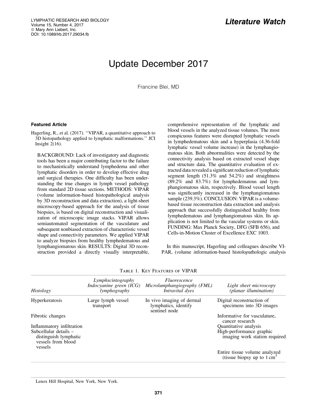
In this manuscript, Hagerling and colleagues describe VIPAR, (volume information-based histolopathologic analysis by 3D reconstruction and data extraction) a light-sheet microscopy- based technology which reconstructs tissue sections into 3-D images, to structurally and quantitatively analyze blood vessel and lymphedematous specimens. This technique is aimed at expanding the current diagnostic techniques, which are summarized below. Based on the data provided in this study, VIPAR, via computer-assisted 3D reconstruction of tissue samples, addresses the limitations of light microscopy and other techniques, which do not provide adequate information for analyzing lymphatic vessel structure. The key features of VIPAR are the 3D images, high resolution of structural details, and objective quantitative analysis of various tissue parameters (Table 1).
In this feasibility study, human skin biopsies were analyzed blood and lymphatic vessels from normal controls were compared with skin biopsies from one patient with WILD (warts, depressed cell-mediated immunity, primary lymphedema, anogenital dysplasia). Multiple images are presented, demonstrating the VIPAR technique's ability to provide enhanced data from skin biopsies. Quantitative data assessing vessels shape and connectivity/branching is also presented. Although this is a small study, the technology is intriguing, and will likely hold promise for future studies. Some issues that this article addresses are very timely—lymphedema is more common and can affect one's quality of life, and further research towards improved therapies are necessary.
Basic Science
Absinta, M., et al. (2017). “Human and nonhuman primate meninges harbor lymphatic vessels that can be visualized noninvasively by MRI.” Elife 6. [EPub Oct 03]
Asano, Y., et al. (2017). “Transplantation of artificial human lymphatic vascular tissues fabricated using a cell-accumulation technique and their engraftment in mouse tissue with vascular remodeling.” J Tissue Eng Regen Med. [EPub Sep 06]
Transplantation of engineered tissues with microvascular structure is advancing towards therapeutic application to improve the flow of blood and/or lymphatic fluids. In lymphatic disorders, transplantation of tissue-engineered lymphatic grafts can be an ideal treatment for draining excessive lymphatic fluid. In this study, we examined the transplantation of three-dimensional artificial human lymphatic network tissue (AHLT) fabricated by the cell accumulation technique into the subcutaneous tissue and fascia of mice. At 2 weeks after transplantation, the AHLT showed engraftment of artificial lymphatic vessels immunopositive for human CD31 and human podoplanin. Notably, we also observed the generation of blood vessel-like structure comprising endothelial cells immunopositive for human CD34 and mural-like cells immunopositive for human CD90 and alphaSMA, which were considered as myofibroblasts. In the fabrication of AHLT in vitro, the sporadic emergence of human CD34-positive / Prox-1-negative sites was observed, followed by the formation of blood vessel-like structure in the graft within 7 days after transplantation. The fine structure of engrafted AHLT observed by transmission electron microscopy showed that the engrafted artificial lymphatic vessels possess the specific structures of native lymphatic capillaries such as loose inter-endothelial connections and anchoring filaments. In contrast, blood vessel-like structure showed tight inter-endothelial connections, thick basement membranes, and layers of mural-like cells, which resemble small blood vessels. These results suggested the remodeling of artificial lymphatic network to form blood vessel-like structure associated with mural-like cells along with AHLT fabrication and engraftment.
Behringer, E. J., et al. (2017). “Calcium and electrical dynamics in lymphatic endothelium.” J Physiol. [EPub Oct 09]
Subsequent to a rise in intracellular Ca2+ ([Ca2+ ]i), hyperpolarization of the endothelium coordinates vascular smooth muscle relaxation along resistance arteries during blood flow control. In the lymphatic vasculature, collecting vessels generate rapid contractions coordinated along lymphangions to propel lymph, but the underlying signalling pathways are unknown. We tested the hypothesis that lymphatic endothelial cells (LECs) exhibit Ca2+ and electrical signalling properties that facilitate lymph propulsion. To study electrical and intracellular Ca2+ signalling dynamics in lymphatic endothelium, we excised collecting lymphatic vessels from the popliteal fossa of mice and removed their muscle cells to isolate intact LEC tubes (LECTs). Intracellular recording revealed a resting membrane potential of approximately −70 mV. Acetylcholine (ACh) increased [Ca2+ ]i with a time course similar to that observed in endothelium of resistance arteries (i.e. rapid initial peak with a sustained “plateau”). In striking contrast to the endothelium-derived hyperpolarization (EDH) characteristic of arteries, LECs depolarized (>15 mV) to either ACh or TRPV4 channel activation. This depolarization was facilitated by the absence of Ca2+ -activated K+ channels (KCa) as confirmed with PCR, persisted in the absence of extracellular Ca2+, was abolished by LaCl3 and was attenuated approximately 70% in LECTs from Trpv4-/- mice. Computational modelling of ion fluxes in LECs indicated that omitting K+ channels supports our experimental results. These findings reveal novel signalling events in LECs, which are devoid of the KCa activity abundant in arterial endothelium. Absence of EDH with effective depolarization of LECs may promote the rapid conduction of contraction waves along lymphatic muscle during lymph propulsion. This article is protected by copyright. All rights reserved.
Dieterich, L. C., et al. (2017). “Distinct transcriptional responses of lymphatic endothelial cells to VEGFR-3 and VEGFR-2 stimulation.” Sci Data 4: 170106.
Dora, K. A. and D. F. van Helden (2017). “Endothelial tubes - another window into lymphatic function.” J Physiol. [EPub Oct 24]
Dubey, L. K., et al. (2017). “Interactions between fibroblastic reticular cells and B cells promote mesenteric lymph node lymphangiogenesis.” Nat Commun 8(1): 367.
Lymphatic growth (lymphangiogenesis) within lymph nodes functions to promote dendritic cell entry and effector lymphocyte egress in response to infection or inflammation. Here we demonstrate a crucial role for lymphotoxin-beta receptor (LTbetaR) signaling to fibroblastic reticular cells (FRCs) by lymphotoxin-expressing B cells in driving mesenteric lymph node lymphangiogenesis following helminth infection. LTbetaR ligation on fibroblastic reticular cells leads to the production of B-cell-activating factor (BAFF), which synergized with interleukin-4 (IL-4) to promote the production of the lymphangiogenic factors, vascular endothelial growth factors (VEGF)-A and VEGF-C, by B cells. In addition, the BAFF-IL-4 synergy augments expression of lymphotoxin by antigen-activated B cells, promoting further B cell-fibroblastic reticular cell interactions. These results underlie the importance of lymphotoxin-dependent B cell-FRC cross talk in driving the expansion of lymphatic networks that function to promote and maintain immune responsiveness.The growth of lymph nodes in response to infection requires lymphangiogenesis. Dubey et al. show that the mesenteric lymph node lymphangiogenesis upon helminth infection depends on the signaling loop between the B and fibroblastic reticular cells (FRCs), whereby the FRCs respond to lymphotoxin secreted by B cells by releasing B cell activating factor.
Gambino, T. J., et al. (2017). “A three-dimensional lymphatic endothelial cell tube formation assay to identify novel kinases involved in lymphatic vessel remodeling.” Assay Drug Dev Technol 15(1): 30–43.
The lymphatic system is a series of vessels that transport cells and excess fluid from tissues to the blood vascular system. Normally quiescent, the lymphatics can grow or remodel in response to developmental, immunological, or cells pathological stimuli. Lymphatic vessels comprise lymphatic endothelial cells (LECs) that can respond to external growth factors by undergoing proliferation, migration, adhesion, and tube and lumen formation into new vessel structures, a process known as lymphangiogenesis. To understand the key gene and signaling pathways necessary for lymphangiogenesis and lymphatic vessel remodeling, we have developed a three-dimensional LEC tube formation assay to explore the role of kinase signaling in these processes. The collagen-overlay-based assay was used with primary human adult dermal LECs to investigate a library of 60 tyrosine kinase (TK) and TK-like genes by siRNA knockdown. Nine candidate genes were identified and characterized for their ability to modify key parameters of lymphatic tube formation, including tube length, area, thickness, branching, and number of blind-ended sacs. Four genes-ZAP70, IRAK4, RIPK1, and RIPK2-were identified as high-confidence hits after tertiary deconvolution screens and demonstrate the utility of the assay to define LEC genes critical for the formation of tube structures. This assay facilitates the identification of potential molecular targets for novel drugs designed to modulate the remodeling of lymphatics that is important for the metastatic spread of cancer and other pathologies.
Han, L., et al. (2017). “Interleukin-33 promotes inflammation-induced lymphangiogenesis via ST2/TRAF6-mediated Akt/eNOS/NO signalling pathway.” Sci Rep 7(1): 10602.
The interplay between inflammation and lymphangiogenesis is mediated by various cytokines. However, most of these molecules and their associated mechanism are yet to be defined. Here, we explored the role of IL-33 in modulating inflammation-induced lymphangiogenesis (ILA) and its underlying mechanisms using an ILA mouse model and a lymphatic endothelial cell (LEC) line. Our results show that IL-33 promoted the proliferation, migration and tube formation of LECs and ILA in vivo. The pro-lymphangiogenic activity of IL-33 was abolished by ST2 blockage. In mechanisms, IL-33 induced the phosphorylation of Akt/eNOS to produce NO in LECs. The IL-33-induced Akt/eNOS activation was suppressed by the PI3K-specific-inhibitor wortmannin, and NO-production was inhibited by both wortmannin and the NO synthase-inhibitor NMA. Knock-down of ST2 or TRAF6 suppressed Akt/eNOS phosphorylation and NO production. The reduction of NO treated with wortmannin or NMA abolished the promoting effects of IL-33 on the chemotactic motility and tube formation of HDLECs. In vivo, IL-33-induced ILA was also impaired in eNOS-/- mice. In conclusion, our study is the first to show that IL-33 promotes inflammation-induced lymphangiogenesis via a ST2/TRAF6-mediated Akt/eNOS/NO signalling pathway. This findings may provide us more opportunities to treat inflammation and lymphangiogenesis associated diseases.
Huang, L. H., et al. (2017). “Cardiac lymphatic vessels, transport, and healing of the infarcted heart.” JACC Basic Transl Sci 2(4): 477–483.
Huang, S. S., et al. (2017). “Development of the LYVE-1 gene with an acidic-amino-acid-rich (AAAR) domain in evolution is associated with acquisition of lymph nodes and efficient adaptive immunity.” J Cell Physiol. [Epub Aug 19]
CRSBP-1 (mammalian LYVE-1) is a membrane glycoprotein highly expressed in lymphatic endothelial cells (LECs). It has multiple ligands, including hyaluronic acid (HA) and growth factors/cytokines (e.g., PDGF-BB and VEGF-A) containing CRS motifs (clusters of basic amino-acid residues). The ligand binding activities are mediated by Link module and acidic-amino-acid-rich (AAAR) domains, respectively. These CRSBP-1/LYVE-1 ligands have been shown to induce opening of lymphatic intercellular junctions in LEC monolayers and in lymphatic vessels in wild-type mice. We hypothesize that CRSBP-1/LYVE-1 ligands, particularly CRS-containing growth factors/cytokines, are secreted by immune and cancer cells for lymphatic entry during adaptive immune responses and lymphatic metastasis. We have looked into the origin of the Link module and AAAR domain of LYVE-1 in evolution and its association with the development of lymph nodes and efficient adaptive immunity. Lymph nodes represent the only major recent innovation of the adaptive immune systems in evolution particularly to mammals and bird. Here we demonstrate that the development of the LYVE-1 gene with the AAAR domain in evolution is associated with acquisition of lymph nodes and adaptive immunity. LYVE-1 from other species, which have no lymph nodes, lack the AAAR domain and efficient adaptive immunity. Synthetic CRSBP-1 ligands PDGF and VEGF peptides, which contain the CRS motifs of PDGF-BB and VEGF-A, respectively, specifically bind to CRSBP-1 but do not interact with either PDGFbetaR or VEGFR2. These peptides function as adjuvants by enhancing adaptive immunity of pseudorabies virus (PRV) vaccine in pigs. These results support the notion that LYVE-1 is involved in adaptive immunity in mammals.
Janardhan, H. P., et al. (2017). “Hdac3 regulates lymphovenous and lymphatic valve formation.” J Clin Invest 127(11): 4193–4206.
Lymphedema, the most common lymphatic anomaly, involves defective lymphatic valve development; yet the epigenetic modifiers underlying lymphatic valve morphogenesis remain elusive. Here, we showed that during mouse development, the histone-modifying enzyme histone deacetylase 3 (Hdac3) regulates the formation of both lymphovenous valves, which maintain the separation of the blood and lymphatic vascular systems, and the lymphatic valves. Endothelium-specific ablation of Hdac3 in mice led to blood-filled lymphatic vessels, edema, defective lymphovenous valve morphogenesis, improper lymphatic drainage, defective lymphatic valve maturation, and complete lethality. Hdac3-deficient lymphovenous valves and lymphatic vessels exhibited reduced expression of the transcription factor Gata2 and its target genes. In response to oscillatory shear stress, the transcription factors Tal1, Gata2, and Ets1/2 physically interacted with and recruited Hdac3 to the evolutionarily conserved E-box-GATA-ETS composite element of a Gata2 intragenic enhancer. In turn, Hdac3 recruited histone acetyltransferase Ep300 to form an enhanceosome complex that promoted Gata2 expression. Together, these results identify Hdac3 as a key epigenetic modifier that maintains blood-lymph separation and integrates both extrinsic forces and intrinsic cues to regulate lymphatic valve development.
Jha, S. K., et al. (2017). “Efficient activation of the lymphangiogenic growth factor VEGF-C requires the C-terminal domain of VEGF-C and the N-terminal domain of CCBE1.” Sci Rep 7(1): 4916.
The collagen- and calcium-binding EGF domains 1 (CCBE1) protein is necessary for lymphangiogenesis. Its C-terminal collagen-like domain was shown to be required for the activation of the major lymphangiogenic growth factor VEGF-C (Vascular Endothelial Growth Factor-C) along with the ADAMTS3 (A Disintegrin And Metalloproteinase with Thrombospondin Motifs-3) protease. However, it remained unclear how the N-terminal domain of CCBE1 contributed to lymphangiogenic signaling. Here, we show that efficient activation of VEGF-C requires its C-terminal domain both in vitro and in a transgenic mouse model. The N-terminal EGF-like domain of CCBE1 increased VEGFR-3 signaling by colocalizing pro-VEGF-C with its activating protease to the lymphatic endothelial cell surface. When the ADAMTS3 amounts were limited, proteolytic activation of pro-VEGF-C was supported by the N-terminal domain of CCBE1, but not by its C-terminal domain. A single amino acid substitution in ADAMTS3, identified from a lymphedema patient, was associated with abnormal CCBE1 localization. These results show that CCBE1 promotes VEGFR-3 signaling and lymphangiogenesis by different mechanisms, which are mediated independently by the two domains of CCBE1: by enhancing the cleavage activity of ADAMTS3 and by facilitating the colocalization of VEGF-C and ADAMTS3. These new insights should be valuable in developing new strategies to therapeutically target VEGF-C/VEGFR-3-induced lymphangiogenesis.
Jiang, X., et al. (2017). “Lymphatic dysfunction, leukotrienes, and lymphedema.” Annu Rev Physiol. [EPub Oct 13]
The lymphatic system is essential for the maintenance of tissue fluid homeostasis, gastrointestinal lipid absorption, and immune trafficking. Whereas lymphatic regeneration occurs physiologically in wound healing and tissue repair, pathological lymphangiogenesis has been implicated in a number of chronic diseases such as lymphedema, atherosclerosis, and cancer. Insight into the regulatory mechanisms of lymphangiogenesis and the manner in which uncontrolled inflammation promotes lymphatic dysfunction is urgently needed to guide the development of novel therapeutics: These would be designed to reverse lymphatic dysfunction, either primary or acquired. Recent investigation has demonstrated the mechanistic role of leukotriene B4 (LTB4) in the molecular pathogenesis of lymphedema. LTB4, a product of the innate immune response, is a constituent of the eicosanoid inflammatory mediator family of molecules that promote both physiological and pathological inflammation. Here we provide an overview of lymphatic development, the pathophysiology of lymphedema, and the role of leukotrienes in lymphedema pathogenesis. Expected final online publication date for the Annual Review of Physiology Volume 80 is February 10, 2018. Please see http://www.annualreviews.org/page/journal/pubdates for revised estimates.
Kong, L. L., et al. (2017). “The optimum marker for the detection of lymphatic vessels.” Mol Clin Oncol 7(4): 515–520.
Podoplanin, lymphatic vessel endothelial hyaluronic acid receptor-1, prospero-related homeobox-1 and vascular endothelial growth factor receptor 3 have been demonstrated to have crucial roles in the development of the lymphatic system and lymphangiogenesis process by combining with their corresponding receptors. Thus, the four markers have been widely used in labelling lymphatic vessels for the detection of lymphangiogenesis and lymphatic vessel invasion. Numerous authors have aimed to identify the roles of these four markers in the lymphatic system and the mechanisms have been partly clarified at the molecular level. The aim of the present review was to comprehensively clarify the characteristics and latent action modes of the four markers in order to determine which is the best one for the detection of lymphangiogenesis and lymphatic vessel invasion.
Lin, Y. C., et al. (2017). “Arf6 in lymphatic endothelial cells regulates lymphangiogenesis by controlling directional cell migration.” Sci Rep 7(1): 11431.
The small GTPase Arf6 plays pivotal roles in a wide variety of cellular events such as endocytosis, exocytosis, and actin cytoskeleton reorganization. However, the physiological functions of Arf6 at the whole animal level have not yet been thoroughly understood. Here, we show that Arf6 regulates developmental and tumor lymphangiogenesis in mice. Lymphatic endothelial cell (LEC)-specific Arf6 conditional knockout (LEC-Arf6 cKO) mouse embryos exhibit severe skin edema and impairment in the formation of lymphatic vessel network at the mid-gestation stage. Knockdown of Arf6 in human LECs inhibits in vitro capillary tube formation and directed cell migration induced by vascular endothelial growth factor-C (VEGF-C) by inhibiting VEGF-C-induced internalization of beta1 integrin. Finally, we found that LEC-Arf6 cKO mice transplanted with B16 melanoma cells attenuated tumor lymphangiogenesis and progression. Collectively, these results demonstrate that Arf6 in LECs plays a crucial role in physiological and pathological lymphangiogenesis.
Lokmic, Z. (2017). “Utilising lymphatic cell markers to visualise human lymphatic abnormalities.” J Biophotonics. [EPub Sep 04]
The in vivo visualisation of the human lymphatic system is limited by the mode of delivery of tracing agents, depth of field and size of the area examined, and specificity of the cell markers used to distinguish lymphatic endothelium from the blood vessels and the surrounding tissues. These limitations are particularly problematic when imaging human lymphatic abnormalities. Firstly, limited understanding of the lymphatic disease aetiology exists with respect to genetic causes and phenotypic presentations. Secondly, the ability of a tracer to reach the entire lymphatic network within the diseased tissue is suboptimal. Thirdly, what is known about the expression of lymphatic endothelial cell markers, such as podoplanin, LYVE-1, PROX-1 and VEGFR-3 in rodent lymphatic vessels and healthy human lymphatic endothelial cells may not necessarily apply in the human lymphatic disease settings. The aim of this review is to highlight challenges in visualising lymphatic vessels in human lymphatic abnormalities with respect to distribution patterns of the cellular markers currently employed to visualise abnormal human lymphatic vessels in experimental settings. Integrating these limitations into current technological designs aimed at improving the diagnostic visualisation of the human lymphatic vasculature in vivo is likely to improve the current ability to image human lymphatic diseases.
Ma, W. and G. Oliver (2017). “Lymphatic endothelial cell plasticity in development and disease.” Physiology (Bethesda) 32(6): 444–452.
The lymphatic vasculature is crucial for maintaining tissue-fluid homeostasis, providing immune surveillance and mediating lipid absorption. The lymphatic vasculature is tightly associated with the blood vasculature, although it exhibits distinct morphological and functional features. Endothelial cells (ECs) lineage fate specification is determined during embryonic development; however, accumulating evidence suggests that differentiated ECs exhibit remarkable heterogeneity and plasticity. In this review, we provide an overview of the molecular mechanisms promoting lymphatic cell fate specification in the mammalian embryo. We also summarize available data suggesting that lymphatic EC fate is reprogrammable in normal and pathological settings. We further discuss the possible advantages of cell fate manipulation to treat certain disorders associated with lymphatic dysfunction.
Myllyla, T., et al. (2017). “Assessment of the dynamics of human glymphatic system by near-infrared spectroscopy (NIRS).” J Biophotonics. [EPub Aug 12]
Fluctuations in brain water content has attracted increasing interest, particularly as regards studies of the glymphatic system, which is connected with the complex organisation of dural lymphatic vessels, responsible for cleaning tissue. Disturbances of glymphatic circulation are associated with several brain disorders, including dementia. This paper introduces an approach to non-invasive measurement of water dynamics in the human brain utilizing near-infrared spectroscopy (NIRS). We demonstrate the possibility to sense dynamic variations of water content between the skull and grey matter, for instance, in the subarachnoid space. Measured fluctuations in water content, especially in the cerebrospinal fluid (CSF), are assumed to be correlated with the dynamics of glymphatic circulation. The sampling volume for the NIRS optode was estimated by MC-modelling for the wavelengths of 660 nm, 740 nm, 830 nm and 980 nm. In addition, using combinations of these wavelengths, this paper presents calculation models for quantifying water and hemodynamics. The presented NIRS technique allows long-term functional brain monitoring, including sleeping time. Furthermore, it is used in combination with different magnetic neuroimaging techniques, particularly Magnetic Resonance Encephalography (MREG). Using the combined setup, we report preliminary results on the interaction between CSF and blood oxygen level-dependent (BOLD) fluctuations.
Park, H. J., et al. (2017). “Interleukin-17A negatively regulates lymphangiogenesis in T helper 17 cell-mediated inflammation.” Mucosal Immunol. [EPub Sep 20]
During inflammation lymphatic vessels (LVs) are enlarged and their density is increased to facilitate the migration of activated immune cells and antigens. However, after antigen clearance, the expanded LVs shrink to maintain homeostasis. Here we show that interleukin (IL)-17A, secreted from T helper type 17 (TH17) cells, is a negative regulator of lymphangiogenesis during the resolution phase of TH17-mediated immune responses. Moreover, IL-17A suppresses the expression of major lymphatic markers in lymphatic endothelial cells and decreases in vitro LV formation. To investigate the role of IL-17A in vivo, we utilized a cholera toxin-mediated inflammation model and identified inflammation and resolution phases based on the numbers of recruited immune cells. IL-17A, markedly produced by TH17 cells even after the peak of inflammation, was found to participate in the negative regulation of LV formation. Moreover, blockade of IL-17A resulted in not only increased density of LVs in tissues but also their enhanced function. Taken together, these findings improve the current understanding of the relationship between LVs and inflammatory cytokines in pathologic conditions.Mucosal Immunology advance online publication 20 September 2017; doi:10.1038/mi.2017.76.
Pujol, F., et al. (2017). “Dachsous1-Fat4 signaling controls endothelial cell polarization during lymphatic valve morphogenesis-brief report.” Arterioscler Thromb Vasc Biol 37(9): 1732–1735.
OBJECTIVE: The purpose of this study was to investigate the role of Fat4 and Dachsous1 signaling in the lymphatic vasculature. APPROACH AND RESULTS: Phenotypic analysis of the lymphatic vasculature was performed in mice lacking functional Fat4 or Dachsous1. The overall architecture of lymphatic vasculature is unaltered, yet both genes are specifically required for lymphatic valve morphogenesis. Valve endothelial cells (Prox1high [prospero homeobox protein 1] cells) are disoriented and failed to form proper valve leaflets. Using Lifeact-GFP (green fluorescent protein) mice, we revealed that valve endothelial cells display prominent actin polymerization. Finally, we showed the polarized recruitment of Dachsous1 to membrane protrusions and cellular junctions of valve endothelial cells in vivo and in vitro. CONCLUSIONS: Our data demonstrate that Fat4 and Dachsous1 are critical regulators of valve morphogenesis. This study highlights that valve defects may contribute to lymphedema in Hennekam syndrome caused by Fat4 mutations.
Qiu, X., et al. (2017). “Hybrid nanoclusters for near-infrared to near-infrared upconverted persistent luminescence bioimaging.” ACS Appl Mater Interfaces 9(38): 32583–32590.
Persistent luminescence (PL) bioimaging provides an optimal method of eliminating autofluorescence for a higher resolution and sensitivity because of the absence of excitation light. However, ultraviolet light is still necessary in common energy charging processes, which limits its reactivation in vivo because of its low penetration depth. In the present study, we introduce a type of hybrid nanocluster (UCPL-NC) composed of upconversion nanoparticles, beta-NaYbF4:Tm@NaYF4, and persistent nanoparticles, Zn1.1Ga1.8Ge0.1O4:0.5%Cr, which can be activated by a 980 nm laser and exhibits an afterglow at 700 nm to realize near-infrared (NIR) to NIR UCPL bioimaging. The PL of the UCPL-NCs can be reactivated even when covered with a 10 mm pork. We demonstrate that these polyethylene glycol-modified phospholipid-functionalized UCPL-NCs can be reactivated in vivo and applied in the PL lymphatic imaging on small animals.
Roman, B. L. and A. P. Hinck (2017). “ALK1 signaling in development and disease: New paradigms.” Cell Mol Life Sci 74(24): 4539–4560.
Activin A receptor like type 1 (ALK1) is a transmembrane serine/threonine receptor kinase in the transforming growth factor-beta receptor family that is expressed on endothelial cells. Defects in ALK1 signaling cause the autosomal dominant vascular disorder, hereditary hemorrhagic telangiectasia (HHT), which is characterized by development of direct connections between arteries and veins, or arteriovenous malformations (AVMs). Although previous studies have implicated ALK1 in various aspects of sprouting angiogenesis, including tip/stalk cell selection, migration, and proliferation, recent work suggests an intriguing role for ALK1 in transducing a flow-based signal that governs directed endothelial cell migration within patent, perfused vessels. In this review, we present an updated view of the mechanism of ALK1 signaling, put forth a unified hypothesis to explain the cellular missteps that lead to AVMs associated with ALK1 deficiency, and discuss emerging roles for ALK1 signaling in diseases beyond HHT.
Ruiz, S., et al. (2017). “Tacrolimus rescues the signaling and gene expression signature of endothelial ALK1 loss-of-function and improves HHT vascular pathology.” Hum Mol Genet. [EPub Sep 14]
Hereditary hemorrhagic telangiectasia (HHT) is a highly debilitating and life-threatening genetic vascular disorder arising from endothelial cell (EC) proliferation and hypervascularization, for which no cure exists. Because HHT is caused by loss-of-function mutations in BMP9-ALK1-Smad1/5/8 signaling, interventions aimed at activating this pathway are of therapeutic value. We interrogated the whole-transcriptome in human umbilical vein ECs (HUVECs) and found that ALK1 signaling inhibition was associated with a specific pro-angiogenic gene expression signature, which included a significant elevation of DLL4 expression. By screening the NIH clinical collections of FDA-approved drugs, we identified tacrolimus (FK-506) as the most potent activator of ALK1 signaling in BMP9-challenged C2C12 reporter cells. In HUVECs, tacrolimus activated Smad1/5/8 and opposed the pro-angiogenic gene expression signature associated with ALK1 loss-of-function, by notably reducing Dll4 expression. In these cells, tacrolimus also inhibited Akt and p38 stimulation by VEGF, a major driver of angiogenesis. In the BMP9/10-immunodepleted postnatal retina-a mouse model of HHT vascular pathology-tacrolimus activated endothelial Smad1/5/8 and prevented the Dll4 overexpression and hypervascularization associated with this model. Finally, tacrolimus stimulated Smad1/5/8 signaling in C2C12 cells expressing BMP9-unresponsive ALK1 HHT mutants and in HHT patient blood outgrowth ECs (BOECs). Tacrolimus repurposing has therefore therapeutic potential in HHT.
Singh, A. P., et al. (2017). “A role for BRG1 in the regulation of genes required for development of the lymphatic system.” Oncotarget 8(33): 54925–54938.
Lymphatic vasculature is an important part of the cardiovascular system with multiple functions, including regulation of the return of interstitial fluid (lymph) to the bloodstream, immune responses, and fat absorption. Consequently, lymphatic vasculature defects are involved in many pathological processes, including tumor metastasis and lymphedema. BRG1 is an important player in the developmental window when the lymphatic system is initiated. In the current study, we used tamoxifen inducible Rosa26CreERT2-BRG1floxed/floxed mice that allowed temporal analysis of the impact of BRG1 inactivation in the embryo. The BRG1floxed/floxed/Cre-TM embryos exhibited edema and hemorrhage at embryonic day-13 and began to die. BRG1 deficient embryos had abnormal lymphatic sac linings with fewer LYVE1 positive lymphatic endothelial cells. Indeed, loss of BRG1 attenuated expression of a subset of lymphatic genes in-vivo. Furthermore, BRG1 binds at the promoters of COUP-TFII and LYVE1, suggesting that BRG1 modulates expression of these genes in the developing embryos. Conversely, re-expression of BRG1 in cells lacking endogenous BRG1 resulted in induction of lymphatic gene expression in-vitro, suggesting that BRG1 was both required and sufficient for lymphatic gene expression. These studies provide important insights into intrinsic regulation of BRG1-mediated lymphatic-gene expression, and further an understanding of lymphatic gene dysregulation in lymphedema and other disease conditions.
Song, L., et al. (2017). “Nucleoside/nucleotide reverse transcriptase inhibitors attenuate angiogenesis and lymphangiogenesis by impairing receptor tyrosine kinases signalling in endothelial cells.” Br J Pharmacol. [EPub Sep 14]
BACKGROUND AND PURPOSE: Cardiovascular disease associated with antiretroviral therapy (ART) has become a major clinical challenge for HIV-positive patients. However, the role of ART in blood vessel growth is largely unknown. Here, we examined an integral component of ART, nucleoside/nucleotide reverse transcriptase inhibitors (NRTIs) and investigated their effects on key microvascular functions, including angiogenesis and lymphangiogenesis. EXPERIMENTAL APPROACH: The angiogenesis/lymphangiogenesis capability of endothelial cells (ECs) was evaluated using migration, proliferation and tube formation assays in vitro, and mouse ear and Matrigel plug assays in vivo. Expressions of signalling molecules and mitochondrial antioxidant catalases were determined using Western blotting. Receptor tyrosine kinase (RTK) internalization and endocytosis were examined using flow cytometry and confocal immunofluorescence microscopy respectively. Mitochondrial DNA copy number and ROS were determined using quantitative real-time PCR and MitoSOX staining respectively. KEY RESULTS: Pharmaceutical doses of NRTIs [azidothymidine (AZT), tenofovir disoproxil fumarate (TDF) and lamivudine (3TC)] inhibited angiogenesis and lymphangiogenesis both in vivo and in vitro by affecting the proliferation and migration of ECs. Correspondingly, NRTIs selectively attenuated the activation and transduction of endothelial RTK signals, VEGFR2 and FGFR1 pathways, in vascular ECs and the VEGFR3 pathway in lymphatic ECs. Both TDF and 3TC restrained RTKs' endocytosis into early endosomes but not internalization, while AZT blocked the protein maturation of RTKs. Excessive ROS levels were detected in NRTI-treated ECs, and the MnSOD mimic MnTMPyP alleviated the angiogenic/lymphangiogenic defects induced by NRTIs. CONCLUSIONS AND IMPLICATIONS: NRTIs negatively regulate angiogenesis and lymphangiogenesis by inducing mitochondrial oxidative stress and subsequently impairing RTK signalling in ECs.
Sun, B. L., et al. (2017). “Lymphatic drainage system of the brain: A novel target for intervention of neurological diseases.” Prog Neurobiol. [Sep 14]
The belief that the vertebrate brain functions normally without classical lymphatic drainage vessels has been held for many decades. On the contrary, new findings show that functional lymphatic drainage does exist in the brain. The brain lymphatic drainage system is composed of basement membrane-based perivascular pathway, a brain-wide glymphatic pathway, and cerebrospinal fluid (CSF) drainage routes including sinus-associated meningeal lymphatic vessels and olfactory/cervical lymphatic routes. The brain lymphatic systems function physiological as a route of drainage for interstitial fluid (ISF) from brain parenchyma to nearby lymph nodes. Brain lymphatic drainage helps maintain water and ion balance of the ISF, waste clearance, and reabsorption of macromolecular solutes. A second physiological function includes communication with the immune system modulating immune surveillance and responses of the brain. These physiological functions are influenced by aging, genetic phenotypes, sleep-wake cycle, and body posture. The impairment and dysfunction of the brain lymphatic system has crucial roles in age-related changes of brain function and the pathogenesis of neurovascular, neurodegenerative, and neuroinflammatory diseases, as well as brain injury and tumors. In this review, we summarize the key component elements (regions, cells, and water transporters) of the brain lymphatic system and their regulators as potential therapeutic targets in the treatment of neurologic diseases and their resulting complications. Finally, we highlight the clinical importance of ependymal route-based targeted gene therapy and intranasal drug administration in the brain by taking advantage of the unique role played by brain lymphatic pathways in the regulation of CSF flow and ISF/CSF exchange.
Thompson, R. L., et al. (2017). “Design principles for lymphatic drainage of fluid and solutes from collagen scaffolds.” J Biomed Mater Res A. [EPub Sep 06]
In vivo, tissues are drained of excess fluid and macromolecules by the lymphatic vascular system. How to engineer artificial lymphatics that can provide equivalent drainage in biomaterials remains an open question. This study elucidates design principles for engineered lymphatics, by comparing the rates of removal of fluid and solute through type I collagen gels that contain lymphatic vessels or unseeded channels, or through gels without channels. Surprisingly, no difference was found between the fluid drainage rates for gels that contained vessels or bare channels. Moreover, solute drainage rates were greater in collagen gels that contained lymphatic vessels than in those that had bare channels. The enhancement of solute drainage by lymphatic endothelium was more pronounced in longer scaffolds and with smaller solutes. Whole-scaffold imaging revealed that endothelialization aided in solute drainage by impeding solute reflux into the gel without hindering solute entry into the vessel lumen. These results were reproduced by computational models of drainage with a flow-dependent endothelial hydraulic conductivity. This study shows that endothelialization of bare channels does not impede the drainage of fluid from collagen gels and can increase the drainage of macromolecules by preventing solute transport back into the scaffold. (c) 2017 Wiley Periodicals, Inc. J Biomed Mater Res Part A, 2017.
Vaahtomeri, K., et al. (2017). “Lymphangiogenesis guidance by paracrine and pericellular factors.” Genes Dev 31(16): 1615–1634.
Volek, E., et al. (2017). “Evaluation of lymphatic vessel dilatations by anterior segment swept-source optical coherence tomography: Case report.” BMC Ophthalmol 17(1): 194.
BACKGROUND: Conjunctival lymphangiectasia is a rare condition presumably caused by the obstruction of lymphatic channels or by an abnormal connection between conjunctival lymphatic and blood vessels. Diagnosis is based on clinical appearance and histology. We report a case of conjunctival lymphangiectasia in which anterior segment optical coherence tomography (OCT) was used to assist the diagnosis and the planning of the biopsy location. CASE PRESENTATION: A 31-year-old woman was referred with repeated episodes of conjunctival “hemorrhages” and chemosis with extended recovery periods over the last months. Other symptoms were dryness, redness, burning sensation and itching. Photo documentation, anterior segment OCT, ultrasound, computer tomography (CT) and magnetic resonance imaging (MRI) of the brain were performed. MRI revealed dilated atypical Virchow-Robin space (VRS). Conjunctival biopsy was taken and the location of the biopsy was selected based on OCT findings. Based on the clinical appearance we suspected the case to be conjunctival lymphangiectasia or lymphangioma. Histology and immunhistochemistry confirmed the diagnosis of conjunctival lymphangiectasia. CONCLUSIONS: Anterior segment OCT is a non-invasive tool, useful in the evaluation of conjunctival lesions and planning surgery.
Wang, Y., et al. (2017). “Smooth muscle cell recruitment to lymphatic vessels requires PDGFB and impacts vessel size but not identity.” Development 144(19): 3590–3601.
Tissue fluid drains through blind-ended lymphatic capillaries, via smooth muscle cell (SMC)-covered collecting vessels into venous circulation. Both defective SMC recruitment to collecting vessels and ectopic recruitment to lymphatic capillaries are thought to contribute to vessel failure, leading to lymphedema. However, mechanisms controlling lymphatic SMC recruitment and its role in vessel maturation are unknown. Here, we demonstrate that platelet-derived growth factor B (PDGFB) regulates lymphatic SMC recruitment in multiple vascular beds. PDGFB is selectively expressed by lymphatic endothelial cells (LECs) of collecting vessels. LEC-specific deletion of Pdgfb prevented SMC recruitment causing dilation and failure of pulsatile contraction of collecting vessels. However, vessel remodelling and identity were unaffected. Unexpectedly, Pdgfb overexpression in LECs did not induce SMC recruitment to capillaries. This was explained by the demonstrated requirement of PDGFB extracellular matrix (ECM) retention for lymphatic SMC recruitment, and the low presence of PDGFB-binding ECM components around lymphatic capillaries. These results demonstrate the requirement of LEC-autonomous PDGFB expression and retention for SMC recruitment to lymphatic vessels, and suggest an ECM-controlled checkpoint that prevents SMC investment of capillaries, which is a common feature in lymphedematous skin.
Watson, D. J., et al. (2017). “Integrated geometric and mechanical analysis of an image-based lymphatic valve.” J Biomech. [EPub Oct 07]
Lymphatic valves facilitate the lymphatic system's role in maintaining fluid homeostasis. Malformed valves are found in several forms of primary lymphoedema, resulting in incurable swelling of the tissues and immune dysfunction. Their experimental study is complicated by their small size and operation in low pressure and low Reynolds number environments. Mathematical models of these structures can give insight and complement experimentation. In this work, we present the first valve geometry reconstructed from confocal imagery and used in the construction of a subject-specific model in a closing mode. A framework is proposed whereby an image is converted into a valve model. An FEA study was performed to identify the significance of the shear modulus, the consequences of smoothing the leaflet surface and the effect of wall motion on valve behaviour. Smoothing is inherent to any analysis from imagery. The nature of the image, segmentation and meshing all cause attenuation of high-frequency features. Smoothing not only causes loss of surface area but also the loss of high-frequency geometric features which may reduce stiffness. This work aimed to consider these effects and inform studies by taking a manual reconstruction and through manifold harmonic analysis, attenuating higher frequency features to replicate lower resolution images or lower degree-of-freedom reconstructions. In conclusion, two metrics were considered: trans-valvular pressure required to close the valve, DeltaPc, and the retrograde volume displacement after closure. The higher DeltaPc, the greater the volume of lymph that will pass through the valve during closure. Retrograde volume displacement after closure gives a metric of compliance of the valve and for the quality of the valve seal. In the case of the image-specific reconstructed valve, removing features with a wavelength longer than four mum caused changes in DeltaPc. Varying the shear modulus from 10kPa to 60kPa caused a 3.85-fold increase in the retrograde volume displaced. The inclusion of a non-rigid wall caused DeltaPc to increase from 1.56 to 2.52cmH2O.
Williams, S. P., et al. (2017). “Genome-wide functional analysis reveals central signaling regulators of lymphatic endothelial cell migration and remodeling.” Sci Signal 10(499).
Lymphatic vessels constitute a specialized vasculature that is involved in development, cancer, obesity, and immune regulation. The migration of lymphatic endothelial cells (LECs) is critical for vessel growth (lymphangiogenesis) and vessel remodeling, processes that modify the lymphatic network in response to developmental or pathological demands. Using the publicly accessible results of our genome-wide siRNA screen, we characterized the migratome of primary human LECs and identified individual genes and signaling pathways that regulate LEC migration. We compared our data set with mRNA differential expression data from endothelial and stromal cells derived from two in vivo models of lymphatic vessel remodeling, viral infection and contact hypersensitivity-induced inflammation, which identified genes selectively involved in regulating LEC migration and remodeling. We also characterized the top candidates in the LEC migratome in primary blood vascular endothelial cells to identify genes with functions common to lymphatic and blood vascular endothelium. On the basis of these analyses, we showed that LGALS1, which encodes the glycan-binding protein Galectin-1, promoted lymphatic vascular growth in vitro and in vivo and contributed to maintenance of the lymphatic endothelial phenotype. Our results provide insight into the signaling networks that control lymphangiogenesis and lymphatic remodeling and potentially identify therapeutic targets and biomarkers in disease specific to lymphatic or blood vessels.
Yan, H., et al. (2017). “CD146 is required for VEGF-C-induced lymphatic sprouting during lymphangiogenesis.” Sci Rep 7(1): 7442.
VEGF-C is essential for lymphangiogenesis during development and tumor progression. VEGFR-3 is the well-known cognate receptor of VEGF-C to regulate lymphatic migration and proliferation, but the receptor of VEGF-C in regulating lymphatic sprouting, the initiating step of lymphangiogenesis, still remains elusive. Here we use both in vitro and in vivo methods to demonstrate CD146 as a receptor of VEGF-C to regulate lymphangiogenesis, especially at the sprouting step. Mechanistically, CD146 selectively activates the downstream p38 kinase, upon VEGF-C stimulation, to regulate lymphatic sprouting. Moreover, CD146 can also activate ERK to mediate VEGF-C regulation of the subsequent proliferation and migration of lymphatic endothelial cells. In zebrafish embryos, knockdown or dysfunction of CD146 results in similar developmental defects in lymphatic sprouting, capillary network, parachordal lymphangioblast (PL), and thoracic duct (TD) similar to down-regulation of VEGF-C. Altogether, our data reveals a critical role of CD146 to mediate VEGF-C signaling pathway in lymphangiogenesis.
Yang, Y., et al. (2017). “Construction of tissue-engineered lymphatic vessel using human adipose derived stem cells differentiated lymphatic endothelial like cells and decellularized arterial scaffold: A preliminary study.” Biotechnol Appl Biochem. [EPub Oct 05]
We have previously demonstrated that human adipose-derived stem cells (hADSCs) can be differentiated into lymphatic endothelial like cells. The purpose of this study was to investigate the feasibility of utilizing the induced lymphatic endothelial like cells and decellularized arterial scaffold to construct the tissue-engineered lymphatic vessel. The hADSCs were isolated from adipose tissue in healthy adults and were characterized the multilineage differentiation potential. Decellularized arterial scaffold was prepared using the Triton x-100 method. ADSCs were differentiated into lymphatic-like endothelial cells, and the induced cells were then seeded onto the decellularized arterial scaffold to engineer the lymphatic vessel. The histological analyses were performed to examine the endothelialized construct. The decellularized arterial scaffold was successfully obtained and was able to maintain its vessel morphology. The isolated ADSCs can be differentiated into osteocytes and adipocytes. After seeding onto the scaffold, the seeded cells attached and grew well on the decellularized arterial scaffold. Our preliminary results demonstrated that the induced lymphatic endothelial like cells combined with decellularized arterial scaffold could be utilized to successfully engineer the lymphatic vessel. Our findings may be helpful for the development of tissue-engineering of the lymphatic graft.
Yu, P., et al. (2017). “FGF-dependent metabolic control of vascular development.” Nature 545(7653): 224–228.
Zhong, W., et al. (2017). “Prox1-GFP/Flt1-DsRed transgenic mice: An animal model for simultaneous live imaging of angiogenesis and lymphangiogenesis.” Angiogenesis 20(4): 581–598.
The roles of angiogenesis in development, health, and disease have been studied extensively; however, the studies related to lymphatic system are limited due to the difficulty in observing colorless lymphatic vessels. But recently, with the improved technique, the relative importance of the lymphatic system is just being revealed. We bred transgenic mice in which lymphatic endothelial cells express GFP (Prox1-GFP) with mice in which vascular endothelial cells express DsRed (Flt1-DsRed) to generate Prox1-GFP/Flt1-DsRed (PGFD) mice. The inherent fluorescence of blood and lymphatic vessels allows for direct visualization of blood and lymphatic vessels in various organs via confocal and two-photon microscopy and the formation, branching, and regression of both vessel types in the same live mouse cornea throughout an experimental time course. PGFD mice were bred with CDh5CreERT2 and VEGFR2lox knockout mice to examine specific knockouts. These studies showed a novel role for vascular endothelial cell VEGFR2 in regulating VEGFC-induced corneal lymphangiogenesis. Conditional deletion of vascular endothelial VEGFR2 abolished VEGFA- and VEGFC-induced corneal lymphangiogenesis. These results demonstrate the potential use of the PGFD mouse as a powerful animal model for studying angiogenesis and lymphangiogenesis.
Clinical
Alvarez-Chinchilla, P., et al. (2017). “Vulvar lymphedema and refractory VIN-III heralding GATA2 deficiency syndrome.” Eur J Obstet Gynecol Reprod Biol 218: 138–140.
Brambila-Tapia, A. J. L., et al. (2017). “GATA2 null mutation associated with incomplete penetrance in a family with Emberger syndrome.” Hematology 22(8): 467–471.
INTRODUCTION: GATA2 mutations are associated with several conditions, including Emberger syndrome which is the association of primary lymphedema with hematological anomalies and an increased risk for myelodysplasia and leukemia. OBJECTIVE: To describe a family with Emberger syndrome with incomplete penetrance. METHODS: A DNA sequencing of GATA2 gene was performed in the parents and offspring (five individuals in total). RESULTS: The family consisted of 5 individuals with a GATA2 null mutation (c.130G>T, p.Glu44*); three of them were affected (two of which were deceased) while two remained unaffected at the age of 40 and 13 years old. The three affected siblings (two boys and one girl) presented with lymphedema of the lower limbs, recurrent warts, epistaxis and recurrent infections. Two died due to hematological abnormalities (AML and pancytopenia). In contrast, the two other family members who carry the same mutation (the mother and one brother) have not presented any symptoms and their blood tests remain normal. DISCUSSION: Incomplete penetrance may indicate that GATA2 haploinsufficiency is not enough to produce the phenotype of Emberger syndrome. It could be useful to perform whole exome or genome sequencing, in cases where incomplete penetrance or high variable expressivity is described, in order to probably identify specific gene interactions that drastically modify the phenotype. In addition, skewed gene expression by an epigenetic mechanism of gene regulation should also be considered.
Brittain, J. M., et al. (2017). “Stewart-Treves Syndrome on the lower extremity associated to idiopathic chronic lymphedema visualized on FDG PET/CT.” Clin Nucl Med 42(12): e519–e522.
Angiosarcomas are highly malignant and rare tumors of vascular or lymphatic endothelial cell origin with a poor prognosis. Lymphangiosarcoma associated with chronic lymphedema is known as Stewart-Treves syndrome. Stewart-Treves syndrome is primarily described in patients with lymphedema of an upper extremity occurring after breast cancer surgery including radical axillary lymph node dissection and subsequent radiotherapy. It is rarely described in the presence of idiopathic chronic lymphedema of the lower extremities. We present a case of lymphangiosarcoma visualized on F-FDG PET/CT, where Stewart-Treves syndrome is secondary to probably a combination of idiopathic chronic lymphedema of the lower extremities and systemic immunosuppressive treatment.
Brouillard, P., et al. (2017). “Loss of ADAMTS3 activity causes Hennekam lymphangiectasia-lymphedema syndrome 3.” Hum Mol Genet 26(21): 4095–4104.
Primary lymphedema is due to developmental and/or functional defects in the lymphatic system. It may affect any part of the body, with predominance for the lower extremities. Twenty-seven genes have already been linked to primary lymphedema, either isolated, or as part of a syndrome. The proteins that they encode are involved in VEGFR3 receptor signaling. They account for about one third of all primary lymphedema cases, underscoring the existence of additional genetic factors. We used whole-exome sequencing to investigate the underlying cause in a non-consanguineous family with two children affected by lymphedema, lymphangiectasia and distinct facial features. We discovered bi-allelic missense mutations in ADAMTS3. Both were predicted to be highly damaging. These amino acid substitutions affect well-conserved residues in the prodomain and in the peptidase domain of ADAMTS3. In vitro, the mutant proteins were abnormally processed and sequestered within cells, which abolished proteolytic activation of pro-VEGFC. VEGFC processing is also affected by CCBE1 mutations that cause the Hennekam lymphangiectasia-lymphedema syndrome syndrome type1. Our data identifies ADAMTS3 as a novel gene that can be mutated in individuals affected by the Hennekam syndrome. These patients have distinctive facial features similar to those with mutations in CCBE1. Our results corroborate the recent in vitro and murine data that suggest a close functional interaction between ADAMTS3 and CCBE1 in triggering VEGFR3 signaling, a cornerstone for the differentiation and function of lymphatic endothelial cells.
Chauhan, N., et al. (2017). “Improving the efficacy of a prophylactic vaccine formulation against lymphatic filariasis.” Parasitol Res. [EPub Aug 21]
Mass drug administration (MDA) is the current strategy for interrupting the transmission of lymphatic filariasis (LF) infection and control of the disease in endemic areas. However, subject non-compliance has resulted in the presence of several “transmission hotspots” in the endemic regions threatening the reemergence of LF. This situation is further complicated by the fact that the drugs used in MDA are not effective against adult LF worms, a major concern for the control strategy. Thus, there is clearly a need for an effective and sustainable approach to control LF. Prophylactic vaccine combined with targeted treatment of infected patients and vector control is suggested as a more sustainable strategy to eliminate LF infection from endemic regions. A multivalent vaccine (rBmHAT) developed in our laboratory conferred about 90% protection in rodents. However, when we tested the rBmHAT vaccine along with alum in rhesus macaques, only about 40% protection was achieved and the immune response obtained was Th2 biased. In an attempt to improve the vaccine, in this study, we tested two vaccine antigens (rBmHAT and rBmHAX) along with two adjuvant formulations [alum + GLA (AL019) and mannosylated chitosan (MCA)] in a mouse model. Our results show that rBmHAT is a better vaccine antigen than rBmHAX. Combination of rBmHAT with AL019 or MCA adjuvants gave 94 and 88% protection, respectively, against challenge infections. Immunized animals developed antigen-specific memory T cells that secreted significant levels of IL-4, IFN-gamma, and IL-17 suggesting the generation of a balanced Th1/Th2 responses following immunization. A major advantage of MCA adjuvant is that the vaccine booster doses can be administered orally. These studies thus showed that rBmHAT is a better vaccine antigen and can be given in combination with AL019 or MCA adjuvant to obtain excellent results.
Chavhan, G. B., et al. (2017). “MR lymphangiography in children: Technique and potential applications.” Radiographics 37(6): 1775–1790.
Dayan, J. H., et al. (2017). “Lymphedema: Pathogenesis and novel therapies.” Annu Rev Med. [EPub Sep 06]
Lymphedema affects up to 1 in 6 patients who undergo treatment for a solid tumor in the United States. Its prevalence has increased as more effective oncologic therapies have improved patient survival, but there remains no definitive cure. Recent research has elucidated new details in the pathogenesis of the disease and has demonstrated that it is fundamentally an immunologic progress that ultimately results in inflammation, fibroadipose deposition, impaired lymphangiogenesis, and dysfunctional lymphatic pumping. These findings have allowed for the development of novel medical and surgical therapies that may potentially alter the standard of care for a disease that has largely been treated by compression. This review seeks to provide an overview of the emerging therapies and how they can be utilized for effective management of lymphedema. Expected final online publication date for the Annual Review of Medicine Volume 69 is January 29, 2018. Please see http://www.annualreviews.org/page/journal/pubdates for revised estimates.
DiBardino, D. M., et al. (2017). “Blue bronchoscopy: Confirmation of aberrant pulmonary lymphatic perfusion during lymphangiography.” J Bronchology Interv Pulmonol 24(4): e51-e52.
Forbrich, A., et al. (2017). “Photoacoustic imaging of lymphatic pumping.” J Biomed Opt 22(10): 1–6.
Gentileschi, S., et al. (2017). “Lymphatic and sensory function of the upper limb after brachioplasty in post-bariatric massive weight loss patients.” Aesthet Surg J 37(9): 1022–1031.
Hassanein, A. H., et al. (2017). “Diagnostic accuracy of lymphoscintigraphy for lymphedema and analysis of false-negative tests.” Plast Reconstr Surg Glob Open 5(7): e1396.
Hayashi, A., et al. (2017). “Effective and efficient lymphaticovenular anastomosis using preoperative ultrasound detection technique of lymphatic vessels in lower extremity lymphedema.” J Surg Oncol. [EPub Oct 23]
Hosokawa, Y., et al. (2017). “Yellow nail syndrome with dramatic improvement of nail manifestations after endoscopic sinus surgery.” Clin Med Insights Ear Nose Throat 10: 1179550617718184.
Itkin, M. (2017). “Magnetic resonance lymphangiography and lymphatic embolization in the treatment of pulmonary complication of lymphatic malformation.” Semin Intervent Radiol 34(3): 294–300.
Kar, S. K., et al. (2017). “Lymphatic pathology in asymptomatic and symptomatic children with Wuchereria bancrofti infection in children from Odisha, India and its reversal with DEC and albendazole treatment.” PLoS Negl Trop Dis 11(10): e0005631.
Kreutzer, C. and G. Kreutzer (2017). “The lymphatic system: The Achilles Heel of the Fontan-Kreutzer Circulation.” World J Pediatr Congenit Heart Surg 8(5): 613–623.
Landau, M. J., et al. (2017). ““Vectra 3D imaging for quantitative volumetric analysis of the upper limb: A feasibility study for tracking outcomes of lymphedema treatment”.” Plast Reconstr Surg. [EPub Sep 14]
Latour-Alvarez, I., et al. (2017). “Skin tumors arising on chronic lymphedema.” Actas Dermosifiliogr. [EPub Aug 21]
Santecchia, L., et al. (2017). “Interosseous membrane release for long-standing upper limb lymphedema: A procedure often neglected.” Plast Reconstr Surg Glob Open 5(8): e1452.
Scaglioni, M. F., et al. (2017). “Systematic review of lymphovenous anastomosis (LVA) for the treatment of lymphedema.” Microsurgery. [EPub Oct 03]
Sosin, M., et al. (2017). “Lymphatic reconstitution and regeneration after face transplantation.” Ann Plast Surg 79(5): 505–508.
Spier, A. B., et al. (2017). “A novel case of lymphangiomatosis in HIV.” Exp Mol Pathol 102(2): 251–254.
Surampalli, V., et al. (2017). “Primary intestinal lymphangiectasia (Waldmann's Disease) presenting with chylous effusions in a 15-year-old.” J Clin Diagn Res 11(8): OD17-OD18.
Veldtman, G. R., et al. (2017). “Cardiovascular adaptation to the Fontan circulation.” Congenit Heart Dis. [EPub Aug 10]
Visconti, G. and M. Salgarello (2017). “Venule valve graft in lymphatic supermicrosurgery: A novel strategy for managing blood backflow.” Microsurgery. [EPub Sep 12]
Yang, X., et al. (2017). “Ultrasound 2D strain measurement for arm lymphedema using deformable registration: A feasibility study.” PLoS One 12(8): e0181250.
Zaleska, M. T. and W. L. Olszewski (2017). “Imaging lymphatics in human normal and lymphedema limbs-Usefulness of various modalities for evaluation of lymph and edema fluid flow pathways and dynamics.” J Biophotonics.[EPub Aug 31]
Oncology
Habenicht, L. M., et al. (2017). “Tumor regulation of lymph node lymphatic sinus growth and lymph flow in mice and in humans.” Yale J Biol Med 90(3): 403–415.
He, B., et al. (2017). “IR-780-loaded polymeric micelles enhance the efficacy of photothermal therapy in treating breast cancer lymphatic metastasis in mice.” Acta Pharmacol Sin. [EPub Aug 10]
Cancer metastasis is responsible for over 90% of breast cancer-related deaths, and inhibiting lymph node metastasis is an option to treat metastatic disease. Herein, we report the use of IR-780-loaded polymeric micelles (IPMs) for effective photothermal therapy (PTT) of breast cancer lymphatic metastasis. The IPMs were nanometer-sized micelles with a mean diameter of 25.6 nm and had good stability in simulated physiological solutions. Under 808-nm laser irradiation, IPMs exhibited high heat-generating capability in both in vitro and in vivo experiments. After intravenous injection, IPMs specifically accumulated in the tumor and metastatic lymph nodes and penetrated into these tissues. Moreover, a single IPMs treatment plus laser irradiation significantly inhibited primary tumor growth and suppressed lymphatic metastasis by 88.2%. Therefore, IPMs are an encouraging platform for PTT applications in treatment of metastatic breast cancer.
Ishii, N., et al. (2017). “Lymphedema of the lower extremities due to refractory malignant lymphoma treated by lymphaticovenous anastomosis.” Plast Reconstr Surg Glob Open 5(8): e1475.
Keeley, V. (2017). “Advances in understanding and management of lymphoedema (cancer, primary).” Curr Opin Support Palliat Care 11(4): 355–360.
Muchowicz, A., et al. (2017). “Inhibition of lymphangiogenesis impairs antitumour effects of photodynamic therapy and checkpoint inhibitors in mice.” Eur J Cancer 83: 19–27.
Syed, S. N., et al. (2017). “S1P provokes tumor lymphangiogenesis via macrophage-derived mediators such as IL-1beta or Lipocalin-2.” Mediators Inflamm 2017: 7510496.
A pleiotropic signaling lipid, sphingosine-1-phosphate (S1P), has been implicated in various pathophysiological processes supporting tumor growth and metastasis. However, there are only a few descriptive studies suggesting a role of S1P in tumor lymphangiogenesis, which is critical for tumor growth and dissemination. Corroborating own data, the literature suggests that apoptotic tumor cell-derived S1P alters the phenotype of tumor-associated macrophages (TAMs) to gain protumor functions. However, mechanistically, the role of TAM-induced lymphangiogenesis has only been poorly described, mostly linked to the production of lymphangiogenic factors such as vascular endothelial growth factor C (VEGF-C) and VEGF-D, or transdifferentiation into lymphatic endothelial cells. Recent findings highlight a rather underappreciated role of S1P in tumor lymphangiogenesis, referring to the production of interleukin-1beta (IL-1beta) and lipocalin-2 (LCN2) by a tumor-promoting macrophage phenotype. In this review, we aim to provide to the readers with the current understanding of the molecular mechanism how apoptotic cell-derived S1P triggers TAMs to promote lymphangiogenesis.
Zhou, X. M., et al. (2017). “Bone marrow derived mesenchymal stem cells involve in the lymphangiogenesis of lung cancer and jinfukang inhibits the involvement in vivo.” J Cancer 8(10): 1786–1794.
Lymphangiogenesis plays an important role in cancer metastasis. Bone marrow-derived mesenchymal stem cells (BMMSCs) migrate to the site of tumorigenesis and in turn promote the metastasis. However, whether BMMSCs involve in the lymphangiogenesis of lung cancer is unclear. Jinfukang has clinically been used for the treatment of non small cell lung cancer (NSCLC) in China. In this study, to investigate the involvement of BMMSCs in lymphangiogenesis in lung cancer, and evaluate the inhibitory effect of Jinfukang on the lymphangiogenesis, chimeric mice were prepared by transplanting bone marrow from green fluorescent protein (GFP) transgenic mice (C57BL/6-EGFP) into irradiated C57BL/6 mice. Then, the chimeric mice were injected subcutaneously with freshly prepared Lewis lung carcinoma cell suspension to make lung tumor model, and the model mice were further orally administrated with Jinfukang once per day for 3 weeks. Four weeks after the bone marrow transplantation, GFP-positive cells primarily existed in bone marrow of acceptor mice, and three more weeks after, Lewis lung carcinoma cells formed a tumor mass in chimeric mice. Observation of GFP-positive cells revealed that BMMSCs transferred into the lung tumor. Immunofluorescent analyses of lymphatic vessel endothelial hyaluronan receptor-1 (LYVE-1), a lymphatic endothelium marker, demonstrated a part of lymphatic endothelial cells in lung cancer were derived from BMMSCs, and those lymphatic endothelial cells contributed to the lung tumor lymphangiogenesis. Furthermore, Jinfukang treatment resulted in a significant reduction of the average weight of the tumor mass in chimeric mice, and displayed a significant lower number of LYVE-1 positive cells. The present results suggest that BMMSCs transfer to tumor, differentiate into lymphatic endothelial cells, and involve in the lymphangiogenesis in lung cancer of mice. Jinfukang inhibits the lung tumor mass via suppression of the BMMSCs transformation and lung tumor lymphangiogenesis. Our findings might provide the potential for the cancer therapies.
Vascular Anomalies
Amyere, M., et al. (2017). “Germline loss-of-function mutations in EPHB4 cause a second form of capillary malformation-arteriovenous malformation (CM-AVM2) deregulating RAS-MAPK signaling.” Circulation 136(11): 1037–1048.
BACKGROUND: Most arteriovenous malformations (AVMs) are localized and occur sporadically. However, they also can be multifocal in autosomal-dominant disorders, such as hereditary hemorrhagic telangiectasia and capillary malformation (CM)-AVM. Previously, we identified RASA1 mutations in 50% of patients with CM-AVM. Herein we studied non-RASA1 patients to further elucidate the pathogenicity of CMs and AVMs. METHODS: We conducted a genome-wide linkage study on a CM-AVM family. Whole-exome sequencing was also performed on 9 unrelated CM-AVM families. We identified a candidate gene and screened it in a large series of patients. The influence of several missense variants on protein function was also studied in vitro. RESULTS: We found evidence for linkage in 2 loci. Whole-exome sequencing data unraveled 4 distinct damaging variants in EPHB4 in 5 families that cosegregated with CM-AVM. Overall, screening of EPHB4 detected 47 distinct mutations in 54 index patients: 27 led to a premature stop codon or splice-site alteration, suggesting loss of function. The other 20 are nonsynonymous variants that result in amino acid substitutions. In vitro expression of several mutations confirmed loss of function of EPHB4. The clinical features included multifocal CMs, telangiectasias, and AVMs. CONCLUSIONS: We found EPHB4 mutations in patients with multifocal CMs associated with AVMs. The phenotype, CM-AVM2, mimics RASA1-related CM-AVM1 and also hereditary hemorrhagic telangiectasia. RASA1-encoded p120RASGAP is a direct effector of EPHB4. Our data highlight the pathogenetic importance of this interaction and indicts EPHB4-RAS-ERK signaling pathway as a major cause for AVMs.
Baluk, P., et al. (2017). “Rapamycin reversal of VEGF-C-driven lymphatic anomalies in the respiratory tract.” JCI Insight 2(16).
Burrows, P. E. (2017). “Angioarchitecture of hereditary arteriovenous malformations.” Semin Intervent Radiol 34(3): 250–257.
Cavalli, G. (2017). “SMAD4 gene mutation and risk of aortic dilation: Lessons from hereditary hemorrhagic telangiectasia.” Int J Cardiol 245: 145–146.
Chen, J., et al. (2017). “Mechanisms of action of microRNAs in infantile hemangioma tissue and vascular endothelial cells in different periods.” Med Sci Monit 23: 4214–4224.
Cunha, S. I., et al. (2017). “Deregulated TGF-beta/BMP signaling in vascular malformations.” Circ Res 121(8): 981–999.
Correct organization of the vascular tree requires the balanced activities of several signaling pathways that regulate tubulogenesis and vascular branching, elongation, and pruning. When this balance is lost, the vessels can be malformed and fragile, and they can lose arteriovenous differentiation. In this review, we concentrate on the transforming growth factor (TGF)-beta/bone morphogenetic protein (BMP) pathway, which is one of the most important and complex signaling systems in vascular development. Inactivation of these pathways can lead to altered vascular organization in the embryo. In addition, many vascular malformations are related to deregulation of TGF-beta/BMP signaling. Here, we focus on two of the most studied vascular malformations that are induced by deregulation of TGF-beta/BMP signaling: hereditary hemorrhagic telangiectasia (HHT) and cerebral cavernous malformation (CCM). The first of these is related to loss-of-function mutation of the TGF-beta/BMP receptor complex and the second to increased signaling sensitivity to TGF-beta/BMP. In this review, we discuss the potential therapeutic targets against these vascular malformations identified so far, as well as their basis in general mechanisms of vascular development and stability.
Lapinski, P. E., et al. (2017). “Somatic second hit mutation of RASA1 in vascular endothelial cells in capillary malformation-arteriovenous malformation.” Eur J Med Genet. [EPub Oct 9]
Ma, J., et al. (2017). “Diagnosis and surgical treatment of cervical macrocystic lymphatic malformations in infants.” Exp Ther Med 14(2): 1293–1298.
Malic, C. C., et al. (2017). “Lymphatic malformation architecture: Implications for treatment with OK-432.” J Craniofac Surg 28(7): 1721–1724.
Martins, L., et al. (2017). “Computational analysis for GNAQ mutations: New insights on the molecular etiology of Sturge-Weber syndrome.” J Mol Graph Model 76: 429–440.
Somatic activating mutations in the GNAQ have been recently associated with several congenital genetic disorders and tumors; however, the molecular mechanism/etiology that leads to GNAQ somatic mosaic mutation are unknown. Here, we reported a case of Sturge-Weber Syndrome (SWS) manifesting cutaneous vascular malformations (hemifacial Port-wine stain), cerebral and ocular vascular abnormalities (including epilepsy and glaucoma) and harboring a c.548G>A (p.R183Q) somatic mosaic mutation in GNAQ. Computational modeling studies were performed to assistant with the comprehension of the functional impact of p.R183Q and p.Q209L mutations in GNAQ, which encodes a G protein subunit alpha q (Galphaq). The p.R183Q mutation was predicted to abolish hydrogen bonds between R183 residue and GDP molecule, destabilizing the inactive GDP-bound conformation of the Galphaq mutants. Furthermore, replacement of R183 by Q183 residue was predicted to promote conformation changes in protein surface features affecting the switch I region, a key region that undergoes conformational changes triggered by receptor binding during signal transduction. In addition, replacement of Q209 by L209 residue was predicted to affect the molecular interaction between Galphaq and Gbeta subunit, impairing formation of the inactive heterotrimeric complex. These findings, in association with PPI network analysis, indicate that p.R183Q and p.Q209L mutations result in the over-activation of different downstream effectors, which in turn will determine the distinct cell responses and phenotype. These findings bring new insights on molecular etiology of vascular malformations associated to SWS and on different mechanisms underlying hyperactivation of downstream pathways to Galphaq.
Mu, W., et al. (2017). “Characterization of pulmonary arteriovenous malformations in ACVRL1 versus ENG mutation carriers in hereditary hemorrhagic telangiectasia.” Genet Med. [EPub Oct 19]
PurposePulmonary arteriovenous malformations (pAVMs) are major contributors to morbidity and mortality in hereditary hemorrhagic telangiectasia (HHT). Mutations in ENG and ACVRL1 underlie the vast majority of clinically diagnosed cases. The aims of this study were to characterize and compare the clinical and morphologic features of pAVMs between these two genotype groups.MethodsSixty-six patients with HHT and affected family members were included. Genotype, phenotypic data, and imaging were obtained from medical records. Morphologic features of pAVMs were analyzed using computed tomography angiography. HHT symptoms, pAVM imaging characteristics, frequency of procedural intervention, and HHT severity scores were compared between ENG and ACVRL1 genotype groups.ResultsENG mutation carriers were more likely than ACVRL1 mutation carriers to have pAVMs (P < 0.001) or multiple lesions (P = 0.03), and to undergo procedural intervention (P = 0.02). Additionally, pAVMs in ENG carriers were more likely to exhibit bilateral lung involvement and growth over time, although this did not reach statistical significance. The HHT severity score was significantly higher in ENG than in ACVRL1 (P = 0.02). ConclusionThe propensity and multiplicity of ENG-associated pAVMs may contribute to the higher disease severity in this genotype, as reflected by the HHT severity score and the frequency of interventional procedures.GENETICS in MEDICINE advance online publication, 19 October 2017; doi:10.1038/gim.2017.160.
Nitschke, M., et al. (2017). “Retrograde lymph flow leads to chylothorax in transgenic mice with lymphatic malformations.” Am J Pathol 187(9): 1984–1997.
Chylous pleural effusion (chylothorax) frequently accompanies lymphatic vessel malformations and other conditions with lymphatic defects. Although retrograde flow of chyle from the thoracic duct is considered a potential mechanism underlying chylothorax in patients and mouse models, the path chyle takes to reach the thoracic cavity is unclear. Herein, we use a novel transgenic mouse model, where doxycycline-induced overexpression of vascular endothelial growth factor (VEGF)-C was driven by the adipocyte-specific promoter adiponectin (ADN), to determine how chylothorax forms. Surprisingly, 100% of adult ADN-VEGF-C mice developed chylothorax within 7 days. Rapid, consistent appearance of chylothorax enabled us to examine the step-by-step development in otherwise normal adult mice. Dynamic imaging with a fluorescent tracer revealed that lymph in the thoracic duct of these mice could enter the thoracic cavity by retrograde flow into enlarged paravertebral lymphatics and subpleural lymphatic plexuses that had incompetent lymphatic valves. Pleural mesothelium overlying the lymphatic plexuses underwent exfoliation that increased during doxycycline exposure. Together, the findings indicate that chylothorax in ADN-VEGF-C mice results from retrograde flow of chyle from the thoracic duct into lymphatic tributaries with defective valves. Chyle extravasates from these plexuses and enters the thoracic cavity through exfoliated regions of the pleural mesothelium.
Seiffert, A., et al. (2017). “Incidence, treatment patterns, and health care costs of infantile hemangioma: Results of a retrospective German database analysis.” Pediatr Dermatol 34(4): 450–457.
Soblet, J., et al. (2017). “Blue Rubber Bleb Nevus (BRBN) Syndrome is caused by somatic TEK (TIE2) mutations.” J Invest Dermatol 137(1): 207–216.
Blue rubber bleb nevus syndrome (Bean syndrome) is a rare, severe disorder of unknown cause, characterized by numerous cutaneous and internal venous malformations; gastrointestinal lesions are pathognomonic. We discovered somatic mutations in TEK, the gene encoding TIE2, in 15 of 17 individuals with blue rubber bleb nevus syndrome. Somatic mutations were also identified in five of six individuals with sporadically occurring multifocal venous malformations. In contrast to common unifocal venous malformation, which is most often caused by the somatic L914F TIE2 mutation, multifocal forms are predominantly caused by double (cis) mutations, that is, two somatic mutations on the same allele of the gene. Mutations are identical in all lesions from a given individual. T1105N-T1106P is recurrent in blue rubber bleb nevus, whereas Y897C-R915C is recurrent in sporadically occurring multifocal venous malformation: both cause ligand-independent activation of TIE2, and increase survival, invasion, and colony formation when expressed in human umbilical vein endothelial cells.
Tan, E. M. S., et al. (2017). “Embryonic stem cell-like subpopulations in venous malformation.” Front Med (Lausanne) 4: 162.
BACKGROUND: Venous malformation (VM) consists of a network of ectatic anomalous thin-walled venous channels. A role for an activating TIE2 mutation in the development of the dilated luminal vessels in VM, and its proposed involvement of embryonic stem cells (ESCs), led us to investigate the expression of ESC markers in subcutaneous VM (SCVM) and intramuscular VM (IMVM). METHODS: Formalin-fixed paraffin-embedded sections of SCVM from seven patients and IMVM samples from seven patients were analyzed for the expression of Nanog, pSTAT3, OCT4, SOX2, SALL4, and CD44, using 3,3'-diaminobenzidine (DAB) immunohistochemical (IHC) staining. All these samples did not express lymphatic marker D2-40. NanoString mRNA analysis and RT-PCR were performed on snap-frozen samples of SCVM (n = 3) and IMVM (n = 3) from the respective original cohorts of patients included in DAB IHC staining. To confirm co-expression of two proteins, immunofluorescent (IF) IHC staining on two representative samples of IMVM and SCVM samples from the original cohorts of patients included for DAB IHC staining was performed. RESULTS: DAB IHC staining demonstrated expression of all of the above ESC markers in both SCVM and IMVM samples. IF IHC staining showed that these markers were localized to the endothelium within these lesions and that Nanog, pSTAT3, SOX2, and CD44 were also expressed by cells outside of the endothelium. NanoString mRNA analysis confirmed transcription activation of pSTAT3, OCT4, and CD44. RT-qPCR confirmed transcription activation of Nanog, SOX2, and SALL4. CONCLUSION: Our findings support the presence of two ESC-like subpopulations, one within and one outside of the endothelium, of both SCVM and IMVM. Given that the endothelial ESC-like subpopulation expresses the more primitive marker, OCT4, it is exciting to speculate that they give rise to the non-endothelial subpopulation.
Vorselaars, V. M. M., et al. (2017). “SMAD4 gene mutation increases the risk of aortic dilation in patients with hereditary haemorrhagic telangiectasia.” Int J Cardiol 245: 114–118.
BACKGROUND: Mutations in the genes ENG, ACVRL1 and SMAD4 that are part of the transforming growth factor-beta signalling pathway cause hereditary haemorrhagic telangiectasia (HHT). Mutations in non-HHT genes within this same pathway have been found to associate with aortic dilation. Therefore, we investigated the presence of aortic dilation in a large cohort of HHT patients as compared to non-HHT controls. METHODS: Chest computed tomography of consecutive HHT patients (ENG, ACVRL1 and SMAD4 mutation carriers) and non-HHT controls were reviewed. Aortic root dilation was defined as a z-score >1.96. Ascending and descending aorta dimensions were corrected for age, gender and body surface area. RESULTS: In total 178 subjects (57.3% female, mean age 43.9+/-14.9years) were included (32 SMAD4, 47 ENG, 50 ACVRL1 mutation carriers and 49 non-HHT controls). Aortopathy was present in a total of 42 subjects (24% of total). Aortic root dilatation was found in 31% of SMAD4, 2% of ENG, 6% of ACVRL1 mutation carriers, and 4% in non-HHT controls (p < 0.001). The aortic root diameter was 36.3+/-5.2mm in SMAD4 versus 32.7+/-3.9mm in the non-SMAD4 group (p = 0.001). SMAD4 was an independent predictor for increased aortic root (beta-coefficient 3.5, p < 0.001) and ascending aorta diameter (beta-coefficient 1.6, p = 0.04). CONCLUSIONS: SMAD4 gene mutation in HHT patients is independently associated with a higher risk of aortic root and ascending aortic dilation as compared to other HHT patients and non-HHT controls.
Wu, K. Q., et al. (2017). “M1 macrophage-induced endothelial-to-mesenchymal transition promotes infantile hemangioma regression.” Am J Pathol 187(9): 2102–2111.
Infantile hemangiomas are benign tumors of vascular endothelial cells (ECs), characterized by three distinct stages: proliferating phase, involuting phase, and involuted phase. The mechanisms that trigger involution of hemangioma into fibro-fatty tissue remain unknown. We report a novel mechanism by which M1-polarized macrophages induce endothelial-to-mesenchymal transition (EndMT) and promote hemangioma regression. M1- but not M2-polarized macrophages induced EndMT in ECs. Tumor necrosis factor-alpha and, to a lesser extent, IL-1beta and interferon-gamma were the most potent cytokines produced by the M1 macrophages that induce in vitro EndMT. Western blot analysis and gene expression profiling showed that ECs treated with M1 macrophages, tumor necrosis factor-alpha, or IL-1beta decreased the expression of endothelial markers, whereas mesenchymal markers increased concomitantly. Immunohistochemical staining of patient samples revealed that a significant perivascular infiltration of M1, but not M2, macrophages coincides with endothelial expression of the critical EndMT transcription factors Snail/Slug in involuting hemangiomas. Most strikingly, M1 macrophage-treated ECs isolated from patient hemangiomas (HemECs) but not untreated HemECs readily differentiated into adipocytes on adipogenic induction. Thus, in vitro EndMT and adipogenesis of HemECs have, in part, recapitulated the natural history of hemangioma regression. In conclusion, our findings indicate that EndMT induced by M1 macrophages promotes infantile hemangioma regression and may lead to novel therapeutic treatments for this vascular tumor.
Yang, J. G., et al. (2017). “Lymphotoxins promote the progression of human lymphatic malformation by enhancing lymphatic endothelial cell proliferation.” Am J Pathol 187(11): 2602–2615.
Formation of inflammation-related tertiary lymphoid organs promotes human lymphatic malformation (LM) development. However, the role of lymphotoxins (LTs) and LT-related inducible ligand, the crucial mediators for tertiary lymphoid organ formation, is undetermined in LMs. Herein, we show that LTs and LT-related inducible ligand promote LM development by enhancing lymphatic endothelial cell (LEC) proliferation via activating NF-kappaB pathways. The expression of LTs and their receptors was increased in LMs, especially the infected ones, when compared with normal skins. Nuclear translocation of p65, p52, and RelB in the LECs of LMs indicated the activation of classic and alternative NF-kappaB pathways. Pearson's correlation and cluster analysis suggested the close relationship between LEC proliferation and NF-kappaB activation. Moreover, in vitro data demonstrated LTs accelerated the proliferation of human dermal LECs (HdLECs) through activation of NF-kappaB. In addition, lipopolysaccharide (LPS) up-regulated LT receptor expression in HdLECs, leading to increased sensitivity to LTs. Suppression of LT receptors hampered LPS-enhanced HdLEC proliferation, indicating the crucial role of LT pathways in inflammatory lymphangiogenesis. Besides, evidence from the LM rat models demonstrated LTalpha and LPS enhanced LEC proliferation, therefore promoting LM development. Blocking LT pathways by neutralizing antibodies against LTalpha and lymphotoxin beta receptor may decelerate the growth of the disease. In summary, our present study demonstrated activation of LT signaling pathways in LECs contributed to the progression of LMs.
Yang, L., et al. (2017). “The expression and function of miR-424 in infantile skin hemangioma and its mechanism.” Sci Rep 7(1): 11846.
Infantile hemangioma is the most common benign tumor in infants. Many studies have confirmed that basic fibroblast growth factor (bFGF) and its key receptor FGFR1 are highly expressed in hemangioma. Moreover, several miRNAs can regulate angiogenesis. In this regard, miR-424 often plays a role as tumor suppressor gene. This study was designed to investigate the mechanism of miR-424 in infantile skin hemangioma. Our results showed low expression of miR-424 in infantile skin hemangioma tissues, and that miR-424 overexpression downregulated FGFR1 expression in hemangioma-derived endothelial cells, while miR-424 inhibition upregulated FGFR1 expression. Luciferase reporter analysis confirmed that FGFR1 was a target gene of miR-424. CCK-8, flow cytometry, transwell migration and tube formation assays demonstrated that miR-424 overexpression inhibited cell proliferation, migration and tube formation, at least in part by blocking the bFGF/FGFR1 pathway. In contrast, miR-424 inhibition significantly enhanced these functions. Furthermore, miR-424 overexpression significantly inhibited ERK1/2 phosphorylation, whereas miR-424 inhibition enhanced ERK1/2 phosphorylation. In conclusion, miR-424 could suppress the bFGF/FGFR1 pathway, thereby inhibit ERK1/2 phosphorylation, and thus inhibit cell proliferation, migration and tube formation capabilities and the development of infantile skin hemangioma.
Zhang, L., et al. (2017). “Estrogen-mediated hemangioma-derived stem cells through estrogen receptor-alpha for infantile hemangioma.” Cancer Manag Res 9: 279–286.
BACKGROUND: Infantile hemangiomas (IHs) are the most common benign vascular tumor of infancy. They occur more frequently in female infants. The cause of hemangioma is currently unknown; however, current studies suggested the importance of estrogen (E2) signaling in hemangioma proliferation. METHODS: Hemangioma-derived stem cells (HemSCs) were cultured with estrogen for 48–72 h; the cell viability and proliferation were evaluated with the messenger RNA (mRNA) and protein expression levels of fibroblast growth factor 2 (FGF2), vascular endothelial growth factor-A (VEGF-A) and estrogen receptor-alpha (ER-alpha), by application of several in vitro assays, such as methyl thiazolyl tetrazolium (MTT), reverse transcriptase-polymerase chain reaction (RT-PCR), real-time PCR, enzyme-linked immunosorbent assay (ELISA) and Western blotting. Also, the cell population's response to external estrogen was investigated by in vivo experiments. HemSCs and human umbilical vein endothelial cells (HUVECs) were mixed and injected subcutaneously into 20 flank of BALB/c-nu mice, which were randomly divided into 5 groups based on different E2 treatment doses (0, 0.01, 0.1 and 1 mg, respectively), 0.1 mg dimethyl sulfoxide (DMSO) as control. Each group of mice were treated intramuscularly every week, then 2 and 4 weeks later, the subcutaneous implants were harvested and evaluated the tumor tissues with microvessel density (MVD) assay and immunohistochemistry. RESULTS: The study demonstrated that application of E2 increased the expression of FGF2, VEGF-A, and ER-alpha in HemSCs with the optimal concentration from 10-9 to 10-5 M. Two-week treatment of E2 promoted expression of VEGF-A and FGF2 in HemSCs culture. Morphological, histological and immunohistological improvements were observed in vivo using murine IH model in which HemSCs and HUVECs were implanted into BALB/c-nu mice that were post-injected with E2. In the grafts, mean MVD was markedly increased. CONCLUSION: The results suggested that E2 promotes angiogenesis via combination with ER-alpha to up-regulate the expression of VEGF-A in HemSCs, promoting proliferation of IHs. These findings provide critical insight into the potential mechanisms of E2 action on IHs.