
Abstract
Programmed death-1 (PD-1) is a transmembrane protein that shares homology with the B7/CD28 family of T cell signaling molecules. PD-1 interacts with its ligands PD-L1 and/or PD-L2 and provides a negative regulatory signal to CD4 and CD8 T cells that results ultimately in a phenotype termed T cell exhaustion. Here we expressed and purified mouse PD-1 protein and developed a monoclonal antibody (MAb) against mouse PD-1 by immunizing BALB/c mice with a specific region of the extracellular domains of PD-1 as antigen, which was expressed in Escherichia coli. A stable hybridoma cell line was established by animal immunization, cell fusion, and hybridoma screening. The MAb was then prepared from mouse ascites after inoculating the hybridoma cells. Different methods were used to analyze the characterization of the MAb, including ELISA, Western blotting, flow cytometry, and RT-PCR techniques. The results showed that the PD-1 MAb can bind to the PD-1 protein and promote lymphocyte proliferation. This PD-1 MAb will be a valuable tool for further investigation of programmed death-1 functions.
Introduction
P
Materials and Methods
Cell culture
Lymphocytes, mouse myeloma cell line SP2/0 cells, and mouse fibroblast cells L929 were cultured in RPMI-1640 medium supplemented with 10% fetal bovine serum (FBS), 100 U penicillin, and 100 μg/mL streptomycin at 37°C and 5% CO2.
Expression and purification of recombinant sPD-1 protein
The construction of pGEX-4T1-sPD-1 plasmid has previously been described.(6) Escherichia coli BL21 (DE3) cells were transformed with pGEX-4T1-sPD-1. The recombinant protein sPD-1 was induced at 30°C for 12 h under 0.1 mM IPTG. The E. coli cells were then harvested, re-suspended in lysis buffer (PBS containing 0.5%Triton X-100 and 1 mM PMSF), and disrupted by sonication. The recombinant protein was purified by glutathione beads as recommended by the manufacturer. Protein concentrations were determined using a Bradford Assay kit and purified proteins were stored at −20°C.
PD-1 protein identification
sPD-1 antigen was lysed in lysis buffer (4% SDS, 20% glycerol, 0.12 M Tris [pH 6.8], 1% bromophenol blue, and 1% 2-mercaptoethanol; Amresco, Solon, OH). The lysates were immediately boiled for 5 min and equal amounts of proteins were subjected to 10% SDS-PAGE and electro-blotted using Towbin transfer buffer (25 mM Tris, 192 mM glycine) onto a PVDF membrane (Millipore, Billerica, MA) with an electro-blotting apparatus (Bio-Rad, Hercules, CA) at 100 mA for 120 min. Non-specific binding was blocked with 5% non-fat milk in TBST (8.8 g NaCl, 2.42 g Tris, 500 μL Tween-20 in 1 L [pH 7.4]) overnight at 4°C and rinsed twice with TBST. The membrane was then incubated with anti-PD-1 antibody (Abcam, Cambridge, United Kingdom), diluted in a blocking solution at room temperature for 1 h, followed by HRP-labeled goat anti-mouse IgG for 1 h. The bands were visualized using the ECL system.
Antigen preparation and generation of monoclonal antibodies
Recombinant mouse extracellular domain His-tagged PD-1 protein was produced in E. coli purified using Ni-NTA magnetic beads (GE Healthcare, Piscataway, NJ) and injected into mice. After a high titer of antibodies was produced in the mouse, the hybridoma was prepared through B lymphocyte hybridoma fusion, hybridoma cloning, and subcloning. Positive clones were screened by ELISA and verified using Western blot analysis. Antibodies were purified from positive hybridomas by protein A/G chromatography and stored in PBS with 50% glycerol, 0.1% BSA.
PD-1 MAb sensitivity
One hundred μL/well (1.0 μg/mL) sPD-1 antigen in 0.05 M carbonate buffer (pH 7.2) were adsorbed on the surface of 96-well flexible microplates (Nunc, Roskilde, Denmark) by overnight incubation at 4°C. To avoid unspecific binding, the plates were blocked with 1% bovine serum albumin (BSA) in PBST. Hybridoma supernatants were incubated for 30 min at 37°C and then washed with PBST three times. The plates were incubated for 30 min at room temperature with HRP-labeled goat anti-mouse IgG (diluted 1:3000). After washing with PBST three times, ELISA were developed using 100 μL of Zymed TMB substrate (Invitrogen, Carlsbad, CA), stopped with 50 μL of 2 M H2SO4 and read at 450 nm.
PD-1 MAb specificity
The lymphocytes were isolated and resuspended in 0.1% FBS-PBS and then stimulated by ConA (0.04, 0.2, 1 μg/mL). After 48 h, the lymphocytes were harvested and incubated with PD-1 MAb 4D10 for 30 min and then washed with PBS three times. They were stained with anti-mouse IgG-FITC-conjugated MAb (eBioscience, San Diego, CA) for 30 min, and after washing three times, the cells were resuspended in 500 μL of the same buffer, then analyzed by FACS to determine the levels of surface expression of the PD-1.
Lymphocyte proliferation
Mouse lymphocytes were isolated and resuspended in 0.1% FBS-PBS, then stained with 1 μmol/L 5-(-6)-carboxyfluorescein diacetate, succinimidyl ester (CFSE) (Molecular Probes, Eugene, OR) for 15 min at 37°C.(7) The staining was quenched with 10% FBS-PBS at 37°C for 5 min. Excess CFSE was removed by repeated PBS washing. Mouse lymphocytes were co-cultured with mouse fibroblast cells L929, which were treated with CDDP (4 μg/mL) for 24 h and fixed with 2% paraformaldehyde, and added with different amounts of the antibody 4D10. After 48 h, cells were harvested and subjected to FACS to determine the cell proliferation.
RNA extraction and RT-PCR
The total RNA of the cultured mouse lymphocytes was extracted using SS First-Strand cDNA synthesis kit (Invitrogen) following the manufacturer's instructions. GAPDH served as an endogenous reference for mouse lymphocyte mRNA normalization. The PCR reaction was carried out in a total reaction volume of 25 μL. Primers used for PCR were: GAPDH: forward 5′-GGTTGTCTCCTGCGACTTCAA-3′, reverse 5′-CCACCCTGTTGCTGTAGCC-3′;IL-2: forward 5′-AACTCACCAGGATGCTCACATTTA-3′, reverse 5′-TCCCTGGGTCTTAAGTGAAAGTTT-3′; IFN-γ: forward 5′-GGAGGAACTGGCAAAAGGATGG-3′, reverse 5′-TGTTGCTGATGGCCTGATTGTC-3′.
Results and Discussion
Expression and purification of recombinant protein sPD-1
In order to obtain the PD-1 antigen, we constructed pGEX-4T1-sPD-1, expressed and purified sPD-1 protein. Figure 1 showed the SDS-PAGE results of purified 42 kDa GST-sPD-1(1A), which was fused with a GST to a PD-1 extracellular region. The protein was identified by Western blot using a PD-1 antibody (Fig. 1B).

Expression and identification of mouse recombinant sPD-1 in prokaryotic cells. (
Sensitivity and specificity determination of anti-PD-1 MAb
Sensitivity of anti-PD-1 MAb 4D10, which was selected by screening assay, was determined by ELISA. The results showed that linearity of PD-1 MAb was confirmed with serial dilution from 1000 to 64,000 fold. The R2 is 0.984 (Fig. 2A). In order to identify MAb 4D10 specificity, over-expressed PD-1 in lymphocytes was prepared with ConA stimulation. After 0.04–1 μg/mL of ConA stimulation, PD-1 expressions on the lymphocytes were 31.2%, 43.8%, and 54.6%, respectively, with PD-1 MAb 4D10 (Fig. 2B).
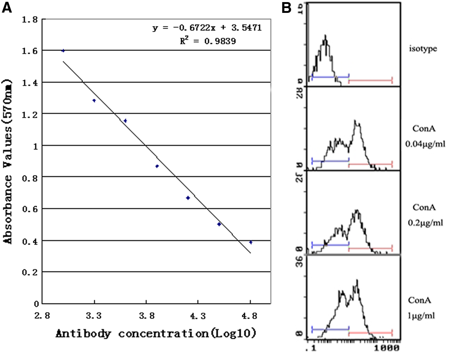
Sensitivity and specificity determination of anti-PD-1 MAb 4D10. (
PD-1 MAb promotes lymphocyte proliferation
Mouse fibroblast cells L929 expressed PD-L1 under cis-Dichlorodiamine platinum (II) (CDDP, 4 μg/mL) induction.(7,8) PD-L1 can bind to PD-1, which was widely expressed on activated T and B lymphocytes. The negative regulatory signals can be transmitted in the PD-1 expressed lymphocytes, where activities were inhibited, and the cytokine IL-2, IFN-γ secretion were decreased.(9) Here we prepared mouse lymphocytes and co-cultured with PD-L1 over-expressed mouse fibroblast cells L929, which were treated with CDDP. The results showed that the co-cultured lymphocyte proliferation was inhibited compared with lymphocytes alone (Fig. 3A). MAb 4D10 were obviously reversed in the inhibition in a dose-dependent manner when the MAb was added into the co-cultured lymphocytes. These results verify that MAb 4D10 can effectively block PD-1 and PD-L1 interaction. Meanwhile we harvested the co-cultured lymphocytes and the cytokines IL-2 and INF-γ were detected with RT-PCR. The results showed that the IL-2 and INF-γ expressions were increased by MAb 4D10 (Fig. 3B).
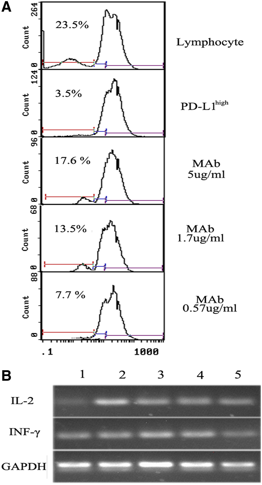
PD-1 MAb 4D10 promote lymphocyte proliferation. (
In summary, our results demonstrate that mouse PD-1 monoclonal antibody 4D10 emerged from quality control testing as an antibody that works best for flow cytometry and ELISA. Furthermore, this antibody specifically binds to PD-1 on the activated lymphocytes, blocks the PD-1 inhibitory signaling pathway, and promotes lymphocyte proliferation.
Footnotes
Author Disclosure Statement
The authors have no financial interests to disclose.