
Abstract
Bluetongue virus (BTV) is a member of the genus Orbivirus, within the family Reoviridae. The VP7 protein of BTV is used for developing group-specific serological assays. To prepare monoclonal antibody (MAb) against VP7 of the 25th serotype BTV, the RNA S7 encoding VP7 was cloned into prokaryotic expression vectors pET-28a (+) and pGEX-6P-1 to generate recombinant plasmids. The recombinant protein VP7 was expressed in Escherichia coli BL21 (DE3), respectively. The results of SDS-PAGE revealed that the VP7 was expressed and the molecular mass of recombinant fusion protein pET-28a (+)/VP7 and pGEX-6P-1/VP7 was approximately 44 kDa and 64 kDa, respectively. The Western blot analysis indicated that the recombinant VP7 possessed good immunoreactivity. After purification, pET-28a (+)/VP7 was used to immunize BALB/c mice, while pGEX-6P-1/VP7 was used to screen for well-to-well MAb-secreting hybridomas. The hybridoma cell line 3H7 against recombinant VP7 that secreted MAbs was obtained. The isotype of 3H7 was identified as IgG1. The purification of recombinant VP7 protein and the monoclonal antibody will have potential applications on competitive ELISA format for BT-specific serum detection method.
Introduction
B
BTV is a non-enveloped double-capsid virus that encodes seven structural proteins (VP1-VP7) and several non-structural proteins (NS1, NS2, NS3/3a, and NS4) from ten double-stranded RNA (dsRNA) segments of the genome.(8,9) VP7 encoded by the dsRNA segment 7 is a component of the core of BTV virion.(10) VP7 forming the core-surface layer consists of 349 amino acids and is about 36% of core protein.(11,12) The VP7 protein of BTV is a preferred choice for developing group-specific serological assays due to its highly conserved sequence and antigenicity between any BTV strains.(13) The economic losses from BT have had great significance in recent years, not only due to animal infection but also due to restrictions imposed by the International Animal Health Organization on animal trade and animal movement from where the virus is endemic.(14–18) Therefore, it is essential to establish cost-effective diagnostic methods for the detecting of BTV.
In this study, we successfully prepared and purified recombinant protein pGEX-6P-1/VP7 and pET-28a (+)/VP7 that could react to BTV-4 sheep positive serum. The results demonstrated that pET-28a (+)/VP7 had good antigenicity. At the same time, we prepared monoclonal antibodies against recombinant VP7 to establish a competitive ELISA method for detecting BT.
Materials and Methods
Reagents, antibodies, vectors, and kits
Commercial enzymes including T4 DNA ligase, restriction enzymes (BamH I and XhoI) were purchased from TaKaRa (Dalian, China). E. coli BL21 (DE3) was used for fusion protein expression. The vector of pET-28a (+) and pGEX-6P-1 were stored by our laboratory. The recombinant protein pCold-TF/VP7, pET-28a (+) /VP7(8), and three segments of BTV-25-VP2 (1-1206bp, 1009-1962bp, and 1813-2880bp) were expressed successfully in E. coli BL21 (DE3). Mouse anti-histidine (His) MAb (MA1-21315) was purchased from Zhongshan (Beijing, China). DNA ligation kits (6022Q) and isopropyl b-D-thiogalactopyranoside (IPTG) were purchased from TaKaRa Biotechnology. BHK21, serotype 8 bluetongue virus and BTV4 positive serum was provided by Dr. Donglai Wu (Harbin Veterinary Research Institute, CAAS).
Cell lines and cell culture
Mouse myeloma cells (SP2/0) were cultured in RPMI-1640 medium (HyClone, Thermo Fisher Scientific, Waltham, MA) supplemented with 20% fetal bovine serum (FBS, Invitrogen, Carlsbad, CA). Cells were cultured at 37°C/5% CO2 in a humidified environment.
Construction of recombinant plasmids
According to RNA sample of BTV-25 on GenBank (EU839843), the sequence of BTV-25 VP7 with restriction enzymes (BamH I and XhoI) was synthesized by Shenggong, Shanghai. The gene product digested with BamH I and XhoI was cloned into the pET-28a (+) and pGEX-6P-1 to construct prokaryotic expression vectors. The resulting recombinant plasmids were evaluated by enzyme digestion and sequencing and named pET-28a (+)/VP7 and pGEX-6P-1/VP7.
Expression and purification of recombinant protein
Recombination expression plasmids were transformed into E. coli BL21 (DE3). After induction with 1.0 mM IPTG at 37°C for 4 h, the recombination protein pET-28a (+)/VP7 was expressed at a high level in the cells as inclusion bodies. At the same time, recombinant expression vector pGEX-6P-1/VP7 was also expressed in E. coli BL21 (DE3). The condition of induction was 28°C for 6 h. The recombinant protein pET-28a (+)/VP7 were purified from their inclusion bodies by gel-cutting purification. The recombinant protein pGEX-6P-1/VP7 was purified from their soluble bodies by GST. The concentrations of the purified recombinant protein were determined.
Western blot analysis of recombinant protein
Bacterial lysates and purified proteins were boiled with 2×SDS–PAGE loading buffer for 10 min and then separated on 12% SDS–PAGE gels. The gels were stained with Coomassie Brilliant Blue G-250 (Bio-Rad, Hercules, CA). The boiled samples were fractionated by electrophoresis on 12% SDS–PAGE gels and then transferred onto nitrocellulose membranes. After blocking with 5% (w/v) non-fat dry milk in PBS at 4°C overnight, the membranes were incubated with anti-His and BTV-4 sheep positive serum, followed by incubation with an HRP-conjugated goat anti-mouse IgG (Zhongshan) and HRP-conjugated rabbit anti-sheep IgG (Zhongshan). After rinsing with PBST, images were acquired by DAB.
Immunization of mice and preparation of anti-BTV VP7 MAbs
Six-week-old BALB/c mice were purchased from Harbin Veterinary Research Institute (Harbin, China). The animal study was approved by the Animal Ethics Committee of Northeast Agricultural University (approval no. 1155-NCET-005). Each mouse was inoculated with 50 μg purified recombinant protein pET-28a (+)/VP7 and equal amount of complete Freund's adjuvant (CFA) on day 0. On days 14 and 28, mice were boosted with 50 μg purified recombinant protein pET-28a (+)/VP7 and equal amount of incomplete Freund's adjuvant (IFA). The anti-VP7 protein serum titer of immunized mice was detected using indirect ELISA on day 21 after immunization. Based on the indirect ELISA results, the spleen cells from the best-immunized mice were used for fusion with SP2/0 myeloma cells and generation of hybridomas conventionally. Indirect ELISA was adopted to test antibody production of the hybridomas with the blocking antigen pGEX-6P-1/VP7 using post-incubation serum as positive control and pre-incubation serum as negative control. The positive hybridomas were identified and cloned at least three times.
Preliminary characterization of monoclonal antibody
MAbs were isotyped using a mouse MAb isotyping kit (Sigma, Beijing, China). The hybridomas secreting monoclonal antibodies serially were passaged for 3 months; cell supernatant of monoclonal antibodies by indirect ELISA method was detected.
Western blot analysis of monoclonal antibody
The boiled protein pET-28a (+)/VP7, recombinant VP7 of the serotype 8 BTV, pGEX-6P-1/VP7, pCold-TF/VP7, pMAl-5x/VP2A, pMAl-5x/VP2B, and pMAl-5x/VP2C were separated on 12% SDS–PAGE gels and then transferred onto nitrocellulose membranes. After blocking with 5% (w/v) non-fat dry milk in PBS at 4°C overnight, the membranes were incubated with the monoclonal antibody 3H7, followed by incubation with an HRP-conjugated goat anti-mouse IgG (Zhongshan). After being washed with PBST, images were acquired by DAB.
Indirect immunofluorescence assays
BHK21 cells were cultured on glass coverslips in 24-well plates, and 80% confluence cell monolayers were infected with BTV. SP2/0 supernatant was utilized as negative control. After three washings with PBS, the cells were fixed with 4% (w/v) paraformaldehyde in PBS for 30 min followed by incubation with the undiluted MAb at 37°C for 1 h. Then the cells were incubated with fluorescein isothiocyanate (FITC)-labeled goat anti-mouse IgG (1:350 dilution in 1% BSA; Zhongshan) for 1 h in the dark, after washing with PBS.
Results
Construction of pET-28a (+)/VP7 and pGEX-6P-1/VP7 expression vector
The VP7 gene with restriction enzymes (BamH I and XhoI) was composed of 1050bp. The product was directionally inserted into pET-28a (+) and pGEX-6P-1. The recombinant plasmid was identified by restriction enzyme digestion and sequencing.
Expression and purification of recombinant protein
The recombinant bacteria induced by 1.0 mM IPTG were separated on 12% SDS–PAGE gels. SDS-PAGE revealed that the molecular weight of the recombinant protein pET-28a (+)/VP7 and pGEX-6P-1/VP7 was 44 kDa and 64 kDa, which corresponded to the molecular weight of the expected fusion protein (Fig. 1). The levels of protein expression are 26.8% and 27.3% of total cell protein. The concentrations of purified recombinant protein pET-28a (+)/VP7 and pGEX-6P-1/VP7 were 0.8 mg/mL and 1.0 mg/mL.

Expression analysis of recombinant protein. Lane M, unstained protein ladder; lane 1, recombinant bacteria pET-28a (+)/VP7 without IPTG induction; lane 2, recombinant bacteria induced pET-28a (+)/VP7 by IPTG; lane 3, expression protein of recombinant bacteria pGEX-6P-1/VP7 without IPTG induction; lane 4, recombinant bacteria pGEX-6P-1/VP7 with IPTG induction; lane 5, recombinant bacteria pET-28a (+)/VP7 without IPTG induction; lane 6, recombinant bacteria pET-28a (+)/VP7 induced by IPTG. Arrows indicate position of VP7 protein.
Western blot analysis of recombinant protein
To identify the antigen of the recombinant pET-28a (+)/VP7 and pGEX-6P-1-VP7, Western blot analysis was performed and the results suggested that pET-28a (+)/VP7 and pGEX-6P-1/VP7 were recognized by BTV-4 positive serum and anti-His antibody, which demonstrates that the fusion protein possessed good immunoreactivity and good reactivity (Figs. 2, 3).
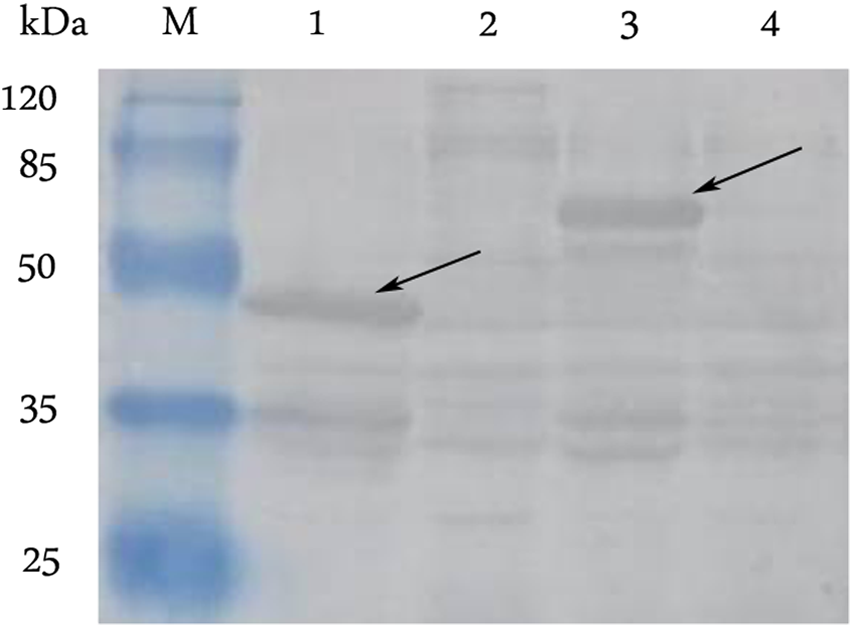
Western blot analysis of recombinant protein. Lane M, pre-stained protein ladder; lane 1, recombinant protein of pET-28a (+)/VP7 induced by IPTG; lane 2, recombinant protein of pET-28a (+)/VP7 without IPTG induction; lane 3, expression protein of pGEX-6P-1/VP7 induced by IPTG; lane 4, recombinant protein of pGEX-6P-1/VP7 without IPTG induction. Arrows indicate position of VP7 protein.
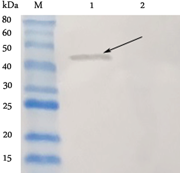
Western blot analysis of recombinant protein. Lane M, pre-stained protein ladder; lane 1, recombinant protein of pET-28a (+)/VP7 induced by IPTG; lane 2, expression protein of pET-28a (+)/VP7 without IPTG induction. Arrow indicates position of VP7 protein.
Titer determination of immunized mouse
An indirect ELISA test was performed to analyze the titer of antiserum against pET-28a (+)/VP7 using the recombinant pET-28a (+)/VP7 as coating antigens. The results revealed that the immunized mice acquired specific antibodies against the recombinant pET-28a (+)/VP7. Among them, the titer of the first mouse reached up to 1:12,800 for cell fusion.
Preliminary characterization of monoclonal antibody
MAb was isotyped using a mouse MAb isotyping kit (Sigma). The results of MAb isotyping indicated that 3H7 was identified as IgG1. The hybridoma-secreting monoclonal antibody serially was passaged for 3 months and frozen; cell supernatant of monoclonal antibody by indirect ELISA method was detected. The results indicate that 3H7 can stably secrete monoclonal antibody against recombinant protein VP7.
Western blot analysis of monoclonal antibody
Western blot results demonstrated that the monoclonal antibody 3H7 can react to pET-28a (+)/VP7, pET-28a (+)/VP7(8), pGEX-6P-1/VP7, and pCold-TF/VP7. However, 3H7 had no reaction with the recombinant protein pMAl-5x/VP2A, pMAl-5x /VP2B, and pMAl-5x /VP2C (Fig. 4), which revealed the specificity of the monoclonal antibody.
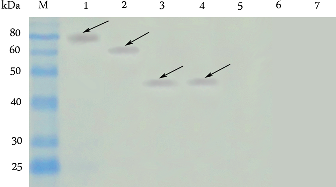
Western blot analysis of monoclonal antibody. Lane M, pre-stained protein ladder; lane 1, protein from pCold-TF/VP7; lane 2, protein from pGEX-6P-1/VP7; lane 3, protein from pET-28a (+)/VP7(8); lane 4, protein from pET-28a (+)/VP7; lane 5, protein from pMAl-5x/VP2A; lane 6, protein from pMAl-5x/VP2B; lane 7, protein from pMAl-5x/VP2C.
Reactivity of MAb 3H7
Immunofluorescence assays confirmed that MAb 3H7 reacted with BTV (Fig. 5). However, the supernatant of SP2/0 did not react with BTV. The MAb reacted with BTV exclusively.
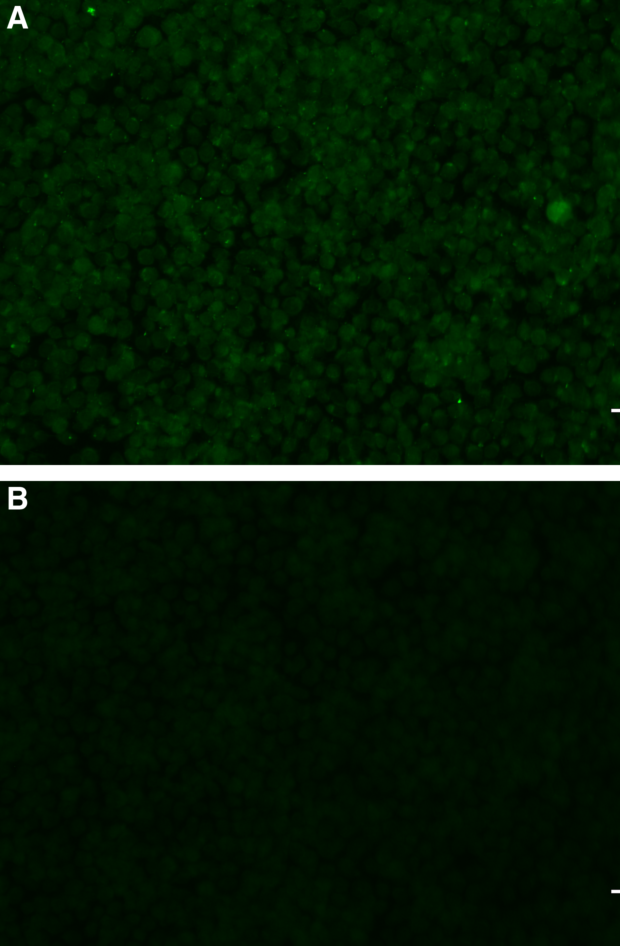
BHK21 cells were infected with BTV and MAb 3H7 or SP2/0 cell culture supernatant was used as primary antibody, respectively, followed by incubation of FITC-conjugated secondary antibody. A representative comparison is provided. (
Discussion
Bluetongue was once endemic in Africa, the Middle East, and India. However, due to global warming, there is a propensity for great BT outbreaks all over the world.(19,20) BT is economically significant because of direct losses due to the disease and indirect losses due to bans on livestock movement.(21) As one of the world's largest livestock producers and consumers, China imports a large number of livestock from abroad every year. To prevent transmission of BTV new serotype into new regions through the international trade of live animals and animal products, strict quarantine measures may be implemented on livestock entry.(22,23) The appearance of the 25th BT serotype has set an alarm for the prevention of BT. Although there is no outbreak of serotype 25 BTV within China, the danger of a new serotype BT cannot be ignored. Furthermore, sensitive, specific, high-throughput, and cost-effective methods for screening livestock sera are urgently required for China.(24) Additionally, TOV-specific serologic tools that use immunologically dominant epitopes of VP7 as recombinant ELISA antigens for detection of antibodies to TOV should be developed. These tools would facilitate epidemiologic studies to determine actual and retrospective sero-prevalences of TOV in goats and other domestic and wild ruminants in Switzerland and even throughout the world, in particular in countries that have long claimed to be free of BTV infections.
VP7 of BTV, which comprises approximately 36% of the total content of the virion, is the major sero-group-specific protein.(25) VP7 in particular is a highly immuno-dominant antigen, which is detected in the majority of BTV serogroup-specific serological assays and can be used to identify any bluetongue virus, helping to define the virus species.(26) Preparation of recombinant antigens in a bacterial system is simpler and more economical than in other systems.(27) Maintenance and culture of bacteria do not require strict observance of sterile conditions, nor does it require costly medium and serum as needed in mammalian or insect cell cultures.(28) Thus, we chose a bacterial system for expression and preparation of large quantities of highly purified viral sero-specific antigen VP7.
The positive sheep serum and anti-His antibody were utilized to test the reactivity of the recombinant VP7 protein pET-28a (+)/VP7 and pGEX-6P-1/VP7. To retain the antigenic and immunogenic properties of pET-28a (+)/VP7 and pGEX-6P-1/VP7 and overcome protein refolding issues, various induction conditions including IPTG concentration (0.2–2 mM), induction temperature (18, 28, and 37°C), and induction time (4–24 h) were optimized. Ultimately, induction of the bacteria with 1 mM IPTG for 4 h at 37°C was determined as the optimal conditions for pET-28a (+)/VP7 and 1 mM IPTG at 28°C for 6 h for pGEX-6P-1/VP7. The label of pET-28a (+) is 6 kDa, which impact on the specificity of the product antibody is insignificant, so we chose pET-28a (+)/VP7 as immunization antigen. Because pGEX-6P-1/VP7 is soluble and purified easily and highly, we chose pGEX-6P-1/VP7 as detecting antigen. The cell line of a monoclonal antibody (MAb) was generated using the hybridoma technique. The isotyping, stability, and reaction of MAbs were detected. The cell line 3H7 shows great advantage of specificity and stability, which generated large amounts of antibody.
Our results demonstrate that the recombinant VP7 of BTV-25 reacts well to BTV-4 and that the protein possesses good immunoreactivity. In addition, the protein could be prepared inexpensively in large quantities. Using this protein, a rapid and cheap assay (e.g., competitive ELISA) could be developed for detection of BTV group-specific antibodies, which may be useful for sero-epidemiology or sero-monitoring of BT. The protein may also be used as antigen in c-ELISA format.
The preparation of anti-VP7 MAb may be utilized in detecting BTV. Furthermore, the recombinant protein and monoclonal antibodies were used to establish the competitive ELISA for detecting BTV. The protein pGEX-6P-1/VP7 as coating antigen that proved to react to BTV-4 positive sheep serum and the monoclonal antibody were applied for cELISA. To determine whether the cELISA can be popularized for detecting BTV, further studies of assay development and its validation are required. Thus, the recombinant protein VP7 and its monoclonal antibody may lay the foundation for the establishment of a detection method.
Footnotes
Acknowledgments
The authors thank Dr. Donglai Wu (Harbin Veterinary Research Institute, CAAS) for providing BTV-4 positive sheep serum. This study was supported by the National Key Technology R&D Program of China (grant no. 2013BAD12B01).
Author Disclosure Statement
The authors have no financial conflicts of interest.