
Abstract
Human papillomavirus (HPV), a major pathogen of human cervical cancer, contains a full-length L1 gene encoding its surface capsid protein. One group of potential vaccine candidates against this virus in Iranian patients is based on surface protein components such as HPV31 L1 protein that can make virus-like particles (VLPs). The high immunity response stimulation of this effecter VLP was observed in host, suggesting that the individual characteristics of a particular effecter may require empirical testing for vaccination. In the present study, we decided to clone and express HPV31 L1 protein to investigate its use as a subunit vaccine and furthermore to insert the gene into an Escherichia coli background so as to analyze production of this recombinant protein. We report the presentation of HPV31 in 100 cervical lesion tissue samples based on polymerase chain reaction (PCR). Type of lesion, age, and other characteristics were reviewed and confirmed by a pathologist. The sequence from L1 genes of HPV was selected using special primers. The gene encoding the major capsid protein L1 was used for subcloning in pTG19-T and pET-32a plasmid. The recombinant protein expression was confirmed by RT-PCR using L1 primers and detected by absorption sodium dodecyl sulfate–polyacrylamide gel electrophoresis (SDS-PAGE) and immunoblot testing. The results presented here offer new insights into the in vivo response of HPV31 in Iranian patients and European models. On the other hand, the use of recombinant L1 protein for Iranian patient protection as well as vaccination studies will permit testing of this antigen protection rate and open the way to the discovery of protein biomarkers for monitoring clinical and subclinical cervical cancers.
Introduction
H
HPVs may lead to the development of cervical intraepithelial neoplasia (CIN), penile intraepithelial neoplasia, vulvar intraepithelial neoplasia, and anal intraepithelial neoplasia.(5) Most HPV infections are recognized rapidly by the immune system and do not progress to cervical cancer. Vaccination is used as a highly effective method for preventing cervical cancer in humans. Much effort has therefore been invested in the design of vaccines, which can protect high-risk groups from HPV. Although three vaccines are available to prevent the HPV, a recombinant vaccine based on surface protein is the most effective vaccine. The capsid antigens of HPV31 may be key to the organism's ability to avoid the host immune response. The immunoreactivity and suitability of L1 as a diagnostic antigen have been demonstrated.(6,7)
The L1 major capsid protein of HPV is ORF protein that makes virus-like particles (VLPs). This particle appears similar to the empty virion that was identified by electron microscopy for all investigated HPV strains. The L1 proteins of HPV strains differed by up to 15 amino acids. The late genes, L1 and L2, are transcribed, translated in the host epithelium, and encapsidate the new viral genomes.(8–12) Scientists worked on the expression of the L1 major capsid protein of six HPV16 strains in insect cells by recombinant baculoviruses.(13–19)
As a result, the capsid protein of HPV31 of Iranian patients as a recombinant antigen was expressed in Escherichia coli. We evaluated the capacity of the recombinant L1 as a recombinant capsid protein in E. coli. HPV tissue samples were obtained from the Khatam hospital, Tehran, for the detection of L1 gene sequences of Iranian patients for recombinant vaccine.
Materials and Methods
Preparation and staining of samples
One hundred archival tissue samples of cervical lesions obtained from Khatam laboratory hospital of Iran were collected and selected based on patient age and lesion type and then stored at 4°C. After paraffin removing, the samples were stained by hematoxylin and eosin to observe the nucleus and cytoplasm of the cells. After staining and microscopic analyzing, the paraffin in 44 samples was removed by xylol. In this step, sliced samples were incubated in microtubes, including 1 mL xylol. After vortexing, tubes were incubated in a heater for 15 minutes, followed by centrifuging at 15,000 rpm for 1 minute. One milliliter ethanol was added to the microtubes and centrifuge was repeated. The tissue cells were obtained for DNA extraction.
DNA extraction
Samples cells were harvested, followed by suspension in lysis buffer. The tubes were incubated at 56°C for 4 hours. For extraction of DNA, 300 μL precipitation buffer was used. In this step, microtubes were centrifuged at 15,000 rpm for 10 minutes. After centrifugation, washing buffer was added into the microtubes and we followed the kit (CinaGene) process for each sample. The genomic DNA was prepared and observed in agarose 0/8% (w/v) by the electrophoresis technique. For electrophoresis, 5 μL of samples was used to characterize and perform polymerase chain reaction (PCR).
PCR amplification of the β-actin gene for diagnosis
The HPV consensus L1 region and HPV genotypes along with the β-globin gene as an internal control were detected by multiplex PCR.(14) The PCR primers are described in Table 1. Final volume of 25 μL: master mix 12/5 μL, 1 μL of primers, and 3/5 μL of genomic DNA (as a template). Taq DNA polymerase was used with the following amplification program: reaction predenature step at 94°C, 35 cycles, including a denature step at 94°C for 30 seconds, annealing at 55°C for 45 seconds, polymerization at 72°C for 45 seconds, and an extension step at 72°C for 5 minutes.
HPV, human papillomavirus.
Amplification of full L1 gene by PCR reaction
HPV type 31L1 gene sequences from GenBank were used to design forward and reverse primers for whole gene PCR. Forward primer contains a restriction site for the EcoRI with 5′-AA GAA TTC ATG TGC CTG TAT ACA CGG GTC C-3′ sequences, and reverse primers have a restriction site for XhoI with 5′-ATC TCG AGT TCC TTC CTG GCA CGT ACACG-3′ sequences to clone the gene in other vectors. Master mix that includes 1× PCR buffer, MgCl2 1.5 mM, dNTP 5 mM primers 0/5 mM, Taq DNA polymerase 1U, and DNA ∼100 ng was prepared. PCR was carried out with an annealing temperature of 58°C and the PCR product was analyzed by 1.2% agarose gel electrophoresis.
Cloning and restriction endonuclease digestion
Cloning plasmid was amplified by transformation into E. coli DH5α. With the aim of constructing a plasmid encoding L1, pTG19-T plasmid was digested by BamH1 enzyme and the separated L1 gene was observed by electrophoresis. The resulting double-stranded L1 gene that contained XhoI and BamH1 overhangs was ligated into pET-32a-L1 using T4 ligase to obtain a vector, including the L1 gene.
The construct was screened by X-Gal/isopropyl-b-D-thiogalactopyranoside (IPTG), restriction enzyme digestion, and electrophoresis. For pET-32a-L1 cloning, double digests were performed. After using the first enzyme (BamH1), it was separated from digested plasmid DNA by heating at 65°C for 25 minutes. The second enzyme (XhoI) cut another side of the gene.
Expression of the L1 gene in expression plasmid
The production of fusion protein was induced by addition of glucose 1% and amounts of IPTG. After RNA extraction from recombinant cells by kit (CinaGene), reverse transcription polymerase chain reaction (RT-PCR) amplified the cDNA by extracted RNA and avian myeloblastosis virus (AMV) reverse transcriptase.
Immunoblotting
The recombinant cells were harvested and subjected to analysis. The total protein profiles of the induced bacterial cultures were obtained in Coomassie blue and electrophoresis on 12% sodium dodecyl sulfate (SDS) gel. Total antigen from recombinant E. coli was mixed with an equal volume of sample buffer. Samples were blotted onto nitrocellulose membranes at 37°C for 1 hour. This membrane was incubated with His-Tag antibody at 80 rpm for 2 hours. After washing with TBS1X, the bonds of protein were observed.
Result
Staining results
The late phase of growth in tissue samples and signs such as large nuclear, high mitosis, and keratinization that were observed are cancer signs. The microscopic results showed that lesions in the tissue samples of 136 patients included metaplasia (50 cases) 50%, CIN1 (18 cases) 18%, CIN 2/3 (8 cases) 8%, and squamous cell cancer (24 cases) 24%.
HPV typing, identification of the L1 gene in samples and cloning
Extraction of DNA of HPV was confirmed by PCR and electrophoresis. Among 100 obtained samples from the hospital, remarkable variability was shown: 12 HPV16, 9 HPV18, 3 HPV31, and 1 HPV35.
MY11 and MY09 consensus primers for the L1 gene amplified 450 bp fragments. The Oligo7 program designed the primers for complete sequence (1700 bp) of the gene encoding the major capsid protein L1 of the high-risk HPV type 31 in Iranian patients.
Cloning the L1 gene HPV in E. coli
pTG-19 plasmid was partially digested with BamH1, so the gene fragment was cloned. After screening of ligation by electrophoresis, the 1700 bp fragment produced by PCR with designed primers was cloned into pET 32a-L1. The screen of cloning of the L1 gene into expression plasmid was performed and confirmed by RT-PCR with MY09/11 primers. The results of amplification and expression are shown in Figures 1 and 2.
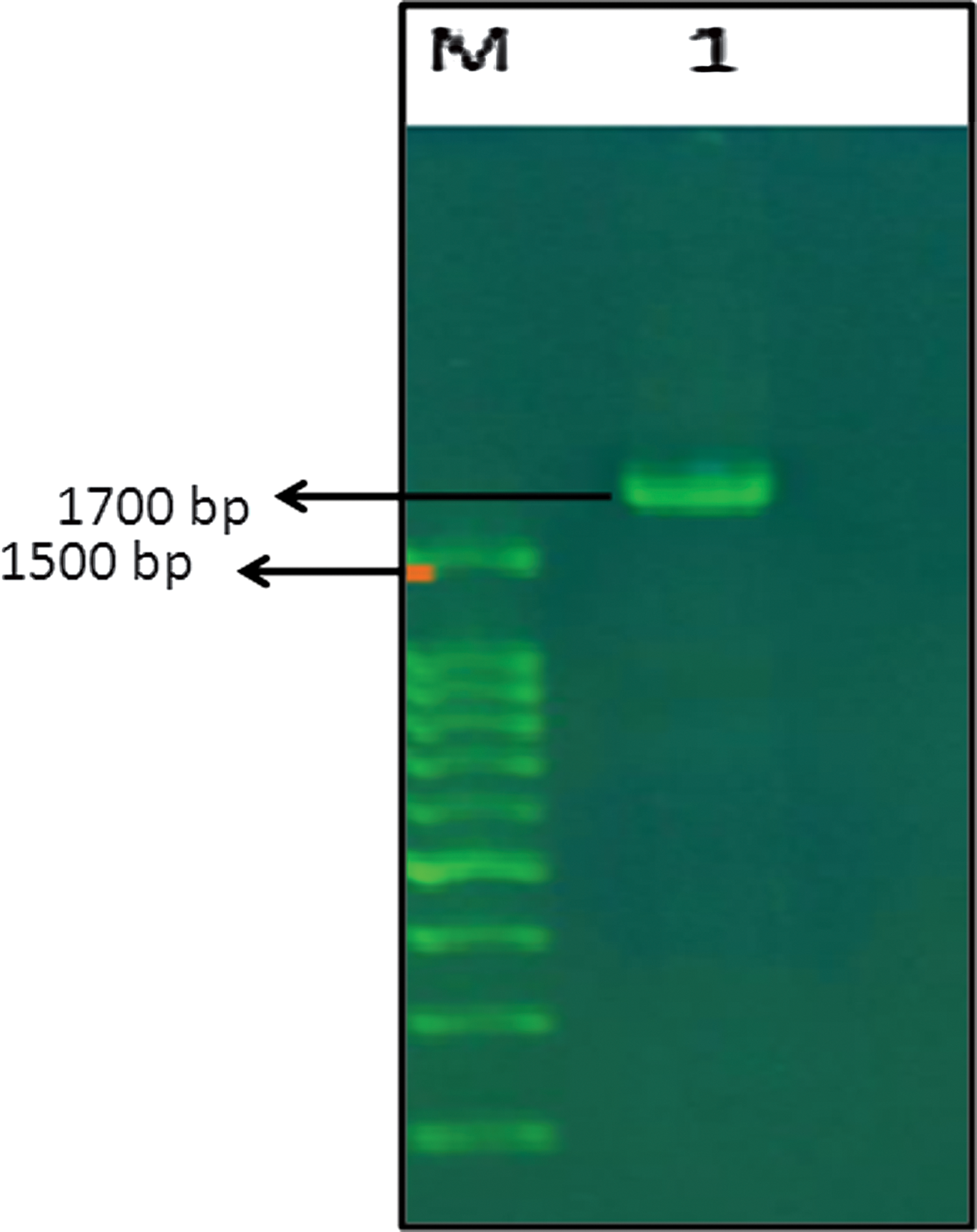
Amplified gene fragment of HPV31 L1. Lane m, marker; Lane 1, L1 gene.
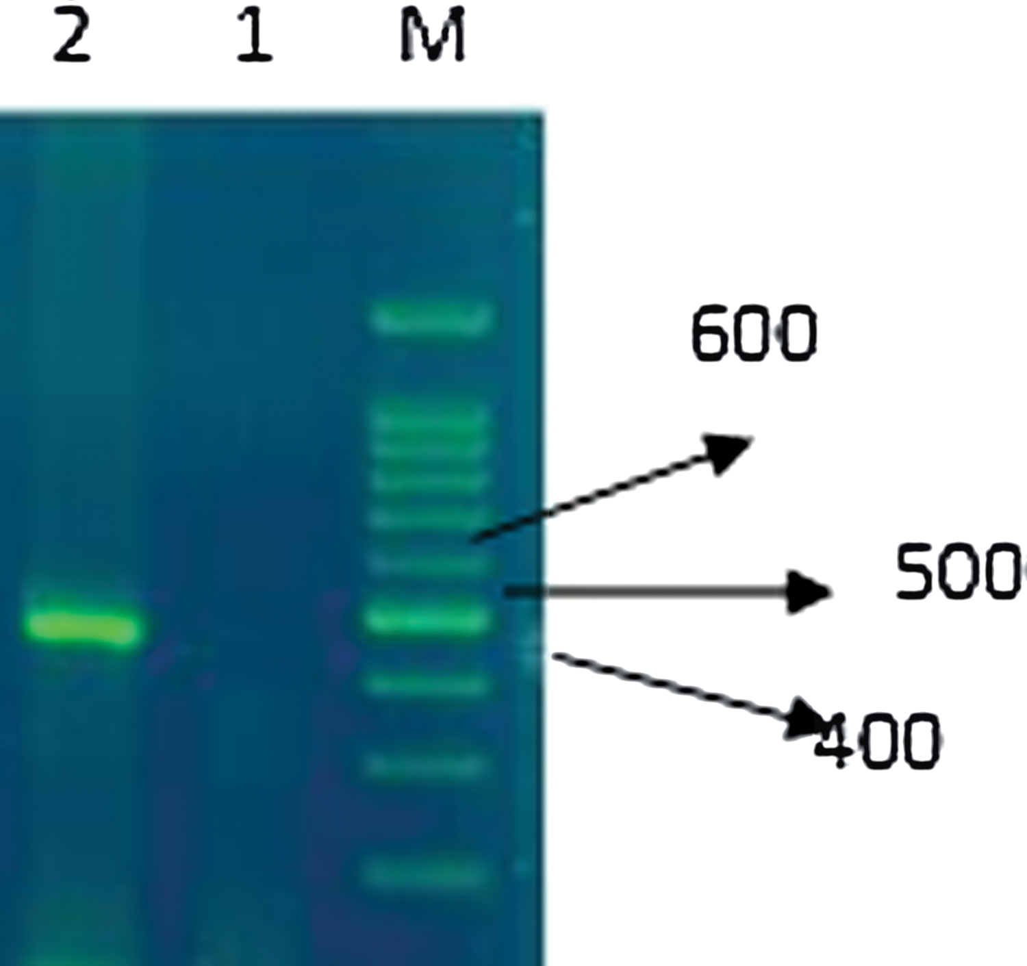
RT-PCR results. Lane m, marker; Lane 1, cDNA of transformed cells without inducer; Lane 2, cDNA of transformed cell with inducer. RT-PCR, reverse transcription polymerase chain reaction.
Expression of the L1 gene
One protein with 57 kDa was found to be increased in positive samples. Therefore, protein expression directed by this inserted DNA was analyzed in detail with SDS-polyacrylamide gel electrophoresis (SDS-PAGE). SDS-PAGE protocol was performed with the aim of characterizing, both qualitatively and quantitatively, the differences in upper phase and pellet of expression liquid. Figure 3 summarizes the obtained results.
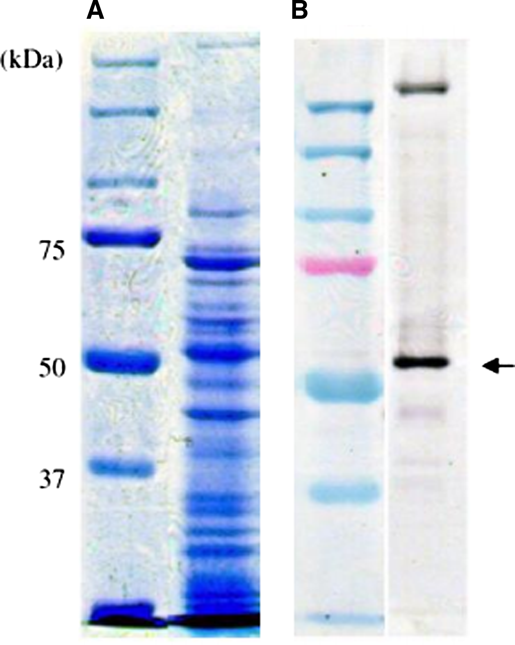
Comparing the expression of the same protein among different SDS-PAGE and immunoblotting M markers.
Discussion
HPV can cause cervical cancer and other cancers, including cancer of the vulva, vagina, penis, or anus. It can also cause cancer in the back of the throat, including the base of the tongue and tonsils. This disease occurs sporadically in world countries such as Iran. Although about 14–13 types of HPVs are responsible for cervical cancer, 16 and 18 types in the world, HPV type 31 accounts for about 12% of all HPV infections in Iran.(18–24) Most genital HPV infections are cleared from the body within 12–24 months.(19,20) Just a little (5%–10%) lead to persistent infection.(21)
Studies have shown that 79% of women worldwide are infected with HPV at some point in their lives.(22,23) Prospective studies in America and Britain suggest that there is more incidence of HPV in young women between 20 and 14 years of age.(24,25)
In this study, the frequency of lesions in patients with this form of metaplasia was found to be metaplasia (50 cases) 50%, CIN1 (18 cases) 18%, CIN 2/3 (8 cases) 8%, and squamous cell cancer (24 cases) 24%. Zhao et al. reported that the capsid protein HPV expresses L1 when found independently in eukaryotic cells by recombinant DNA technology and has the ability of spontaneous arrangements for VLPs.(26) This discovery led to the development of HPV vaccines based on proprietary VLP types. VLP vaccines are prophylactic vaccines, which can prevent the onset of HPV infection and therapeutic vaccines.(27)
When these vaccines are given in three doses, the antibody titer that is induced is several times higher than what is seen in natural infection. The antibody peak response is in the seventh month (1 month after the third dose) and 150–157 times higher than the natural infection-induced antibody response to the HPV.(28–30)
Considering the prevalence of HPV31 in Iran as well as the effectiveness of vaccines based on VLPs, the aim of this study was to determine the isolation and expression of HPV31 VLP-L1 nanoparticles in E. coli bacteria to prepare for production of the vaccine. In addition, the method used to clone the HPV31-L1 gene expression in bacteria is inexpensive and can be used for the investigation and production of a vaccine to prevent cervical cancer, using viral nanoparticles.
Footnotes
Author Disclosure Statement
No competing financial interests exist.