
Abstract
Podoplanin (PDPN)/Aggrus is a type-I transmembrane sialoglycoprotein, which possesses a platelet aggregation-stimulating (PLAG) domain. The O-glycosylation on Thr52 of human PDPN (hPDPN) is critical for the interaction of hPDPN with C-type lectin-like receptor-2 (CLEC-2), resulting in platelet aggregation. Many anti-hPDPN monoclonal antibodies (MAbs) against PLAG domains and non-PLAG domains have been established; however, mouse anti-PLAG2/3 MAb, the epitope of which is consistent with rat anti-PLAG2/3 MAb NZ-1, has not been established. NZ-1 inhibits the hPDPN-CLEC-2 interaction and is also useful for anti-PA tag MAb. We recently established CasMab technology to produce MAbs against membranous proteins. Herein, we produced a novel anti-hPDPN MAb, LpMab-13, which binds to PLAG2/3 domains. LpMab-13 recognized endogenous hPDPN of cancer cells, including glioblastoma, oral cancer, lung cancer, and malignant mesothelioma, and normal cells such as lymphatic endothelial cells and podocytes of kidney in Western blot, flow cytometry, and immunohistochemistry. LpMab-13 recognized glycan-deficient hPDPN in flow cytometry, indicating that the interaction between LpMab-13 and hPDPN is independent of its glycosylation. The minimum epitope of LpMab-13 was identified as Ala42–Asp49 of hPDPN using Western blot and flow cytometry. The combination of different epitope-possessing MAbs could be advantageous for the hPDPN-targeting diagnosis and therapy.
Introduction
P
The interaction of human PDPN (hPDPN) with CLEC-2 is mainly observed at Glu47 and Asp48 in the platelet aggregation-stimulating domain-3 (PLAG3) and the α2-6 linked sialic acid residue.(18) The sequence motif is conserved among PDPN proteins of various species.(19) CLEC-2 has a recognition motif in the form “EDXXXT/S,” where X is any amino acid and T (or S) contains core 1 O-glycan with α2–6 linked sialic acid + N-acetyl-
In this study, we describe the development and characterization of a new anti-hPDPN monoclonal antibody (MAb), LpMab-13, which specifically binds to PLAG2/3 domains, and might serve as a novel modality to study the function of hPDPN.
Materials and Methods
Cell lines, animals, and tissues
LN229, HEK-293T, NCI-H226, Chinese hamster ovary (CHO)-K1, glycan-deficient CHO cell lines (Lec1, Lec2, and Lec8), and P3U1 were obtained from the American Type Culture Collection (Manassas, VA). Human LEC were purchased from Cambrex (Walkersville, MD). LN319 was donated by Prof. Kazuhiko Mishima (Saitama Medical University, Saitama, Japan).(22) HSC-2 was obtained from the Japanese Collection of Research Bioresources (JCRB) cell bank (Osaka, Japan). PC-10 cells were purchased from Immuno-Biological Laboratories Co., Ltd. (Gunma, Japan). LN229, CHO-K1, Lec1, Lec2, and Lec8 were transfected with hPDPN plasmids (LN229/hPDPN, CHO/hPDPN, Lec1/hPDPN, Lec2/hPDPN, and Lec8/hPDPN) using Lipofectamine 2000 (Thermo Fisher Scientific, Inc., Waltham, MA) according to the manufacturer's instructions.(23) HEK-293T/hPDPN-knockout (KO) cell line (PDIS-2) and LN319/hPDPN-KO cell line (PDIS-6) were produced by transfecting CRISPR/Cas plasmids (Target ID: HS0000333287), which target PDPN (Sigma-Aldrich Corp., St. Louis, MO), using a Gene Pulser Xcell electroporation system (Bio-Rad Laboratories, Inc., Berkeley, CA).(24–26) The amplified hPDPN cDNA was subcloned into a pcDNA3 vector (Thermo Fisher Scientific, Inc.) and a FLAG epitope tag was added at the C-terminus. Substitutions of amino acids to alanine, glycine, serine, or valine in hPDPN were performed using a QuikChange Lightning Site-Directed Mutagenesis Kit (Agilent Technologies, Inc., Santa Clara, CA) with oligonucleotides containing the desired mutations. CHO-K1 cells were transfected with the plasmids using a Gene Pulser Xcell electroporation system (Bio-Rad Laboratories). CHO-K1, Lec1, Lec2, Lec8, CHO/hPDPN, Lec1/hPDPN, Lec2/hPDPN, Lec8/hPDPN, CHO-K1/hPDPN point mutants, NCI-H226, PC-10, and P3U1 were cultured in RPMI 1640 medium, including
Animals were housed under pathogen-free conditions. The Animal Care and Use Committee of Tohoku University approved the animal experiments described herein. This study examined cancer patients who underwent surgery at Sendai Medical Center(27) and Yamagata University Hospital.(6,28) Informed consent for obtaining samples and for subsequent data analyses was obtained from patients or the patient's guardian.
Hybridoma production
BALB/c mice were immunized by intraperitoneal (i.p.) injection of 1 × 10⁸ LN229/hPDPN cells together with Imject Alum (Thermo Fisher Scientific, Inc.).(23) After several additional immunizations, a booster injection was given i.p. 2 days before spleen cells were harvested. The spleen cells were fused with P3U1 cells using PEG1500 (Roche Diagnostics, Indianapolis, IN). The hybridomas were grown in RPMI medium with hypoxanthine, aminopterin, and thymidine selection medium supplement (Thermo Fisher Scientific, Inc.). The culture supernatants were screened using enzyme-linked immunosorbent assay (ELISA) for binding to recombinant hPDPN purified from LN229/hPDPN cells. Proteins were immobilized on Nunc MaxiSorp 96-well immunoplates (Thermo Fisher Scientific, Inc.) at 1 μg/mL for 30 minutes. After blocking with 1% bovine serum albumin (BSA) in 0.05% Tween 20/phosphate-buffered saline (PBS, Nacalai Tesque, Inc.), the plates were incubated with culture supernatant followed by 1:2000 diluted peroxidase-conjugated anti-mouse IgG (Dako; Agilent Technologies, Inc.). The enzymatic reaction was conducted with a 1-Step Ultra TMB-ELISA (Thermo Fisher Scientific, Inc.). The optical density was measured at 655 nm using an iMark microplate reader (Bio-Rad Laboratories, Inc.).
Western blot analyses
Cell lysates (5–10 μg) were boiled in SDS sample buffer (Nacalai Tesque, Inc.). The proteins were electrophoresed on 5%–20% polyacrylamide gels (Wako Pure Chemical Industries Ltd., Osaka, Japan) and were transferred onto a PVDF membrane (EMD Millipore Corp., Billerica, MA). After blocking with 4% skim milk (Nacalai Tesque, Inc.), the membrane was incubated with LpMab-13 (1 μg/mL), anti-PDPN (1 μg/mL; clone NZ-1 and clone LpMab-7),(8,28) anti-IDH1 (1 μg/mL; clone RcMab-1 and clone RMab-3),(29) anti-FLAG (1 μg/mL; Wako Pure Chemical Industries Ltd.), or anti-β-actin (1:10,000 diluted; clone AC-15; Sigma-Aldrich Corp.), then with peroxidase-conjugated anti-mouse or anti-rat IgG (1:1000 diluted; Dako), and developed with the ImmunoStar LD Chemiluminescence Reagent (Wako Pure Chemical Industries Ltd.) using a Sayaca-Imager (DRC Co. Ltd., Tokyo, Japan).
Flow cytometry
Cell lines were harvested by brief exposure to 0.25% Trypsin/1 mM EDTA (Nacalai Tesque, Inc.).(2) After washing with 0.1% BSA in PBS, the cells were treated with LpMab-13, NZ-1, or LpMab-7 (1 μg/mL) for 30 minutes at 4°C and followed by treatment with Oregon Green 488 goat anti-mouse or anti-rat IgG (Thermo Fisher Scientific, Inc.). Fluorescence data were collected using a Cell Analyzer EC800 (Sony Corp., Tokyo, Japan).
Determination of the binding affinity using flow cytometry
LN319 (2 × 10⁵ cells) was resuspended with 100 μL of serially diluted LpMab-13 (0.006–100 μg/mL) followed by secondary anti-mouse IgG (Thermo Fisher Scientific, Inc.). Fluorescence data were collected using a cell analyzer (EC800; Sony Corp.). The dissociation constants (KD) were obtained by fitting the binding isotherms using the built-in one-site binding models in GraphPad PRISM 6 (GraphPad software, Inc., La Jolla, CA).
Immunohistochemical analyses
Four-micrometer-thick histologic sections were deparaffinized in xylene and rehydrated. Without antigen retrieval procedure, sections were incubated with 5 μg/mL of LpMab-13 for 1 h at room temperature followed by treatment with EnVision+ Kit (Dako) for 30 minutes. Color was developed using 3, 3-diaminobenzidine tetrahydrochloride (DAB; Dako) for 5 minutes and then the sections were counterstained with hematoxylin (Wako Pure Chemical Industries Ltd.).
Results
Production of a novel anti-PDPN MAb LpMab-13
Herein, we used the CasMab technology.(23–25,27, 28,30–32) LN229/hPDPN cells were immunized into mice to develop novel anti-hPDPN MAbs. The culture supernatants were screened using ELISA for the binding to recombinant hPDPN purified from LN229/hPDPN cells. LpMab-13 (mouse IgG1, kappa) was established after limiting dilution.
Characterization of LpMab-13
LpMab-13 reacted with LN229/hPDPN, not with LN229, hPDPN-negative cell in Western blot analysis (Fig. 1A). LpMab-13 also recognized endogenous hPDPN, which is expressed in a glioblastoma cell line LN319 and a LEC in Western blot analysis (Fig. 1A) and flow cytometry (Fig. 1B). We also performed Western blot analysis using LpMab-13 against several glycan-deficient PDPN transfectants such as Lec1/hPDPN (N-glycan deficient), Lec2/hPDPN (sialic acid deficient), and Lec8/hPDPN (O-glycan deficient). LpMab-13 reacted with CHO-K1/hPDPN, not with CHO-K1 (Fig. 1A). Furthermore, all transfectants of glycan-deficient PDPN were detected by LpMab-13 (Fig. 1A), showing that the epitope of LpMab-13 is independent of glycan. LpMab-13 detected endogenous hPDPN of human kidney cell line HEK-293T, oral cancer cell line HSC-2, lung squamous cell carcinoma cell line PC-10, and malignant mesothelioma cell line HCI-H226 in flow cytometry (Fig. 1B), although hPDPN of NCI-H226 and HEK293T was not detected using Western blot analysis (Fig. 1A). LN229 and CHO-K1, which do not express hPDPN, were not detected by LpMab-13 (Fig. 1A), and LpMab-13 did not react with HEK-293T/hPDPN-KO and LN319/hPDPN-KO (Fig. 1B), indicating that LpMab-13 does not recognize nonrelated antigens.

Characterization of a novel anti-hPDPN monoclonal antibody (mAb), LpMab-13.
Binding affinity of LpMab-13
We next performed a kinetic analysis of the interaction of LpMab-13 with LN319 using flow cytometry. As shown in Figure 1C, KD was determined to be 2.9 × 10⁻⁸ M, indicating that LpMab-13 possesses moderate affinity against endogenous hPDPN.
Immunohistochemical analysis using LpMab-13
We further investigated whether LpMab-13 is useful for immunohistochemical analyses. The expression of hPDPN is observed in many cancers such as malignant brain tumor, mesothelioma, oral cancer, lung cancer, esophageal cancer, testicular tumor, and OS.(3,5,11,28) In this study, we picked up several hPDPN-expressing cancers. As shown in Figure 2, LpMab-13 stained glioblastoma (GBM; Fig. 2B, C), oral squamous cell carcinoma (OSCC; Fig. 2E, F), esophageal squamous cell carcinoma (Fig. 2H, I), osteosarcoma (OS; Fig. 2K, L), and seminoma (Fig. 2N, O) in a membranous/cytoplasmic staining pattern. The expression of hPDPN is also observed in normal tissues such as LEC and lung type I alveolar cells.(20) LpMab-13 reacted with LEC (arrows; Fig. 2H, I); in contrast, it did not react with vascular endothelial cells (arrowheads; Fig. 2B, C), demonstrating that LpMab-13 is very useful for detecting LEC. Furthermore, LpMab-13 was also reactive with lung type I alveolar cells (Fig. 2Q, R), which are not recognized by anti-hPDPN mAbs, NZ-1 and D2-40, in our previous study.(11)
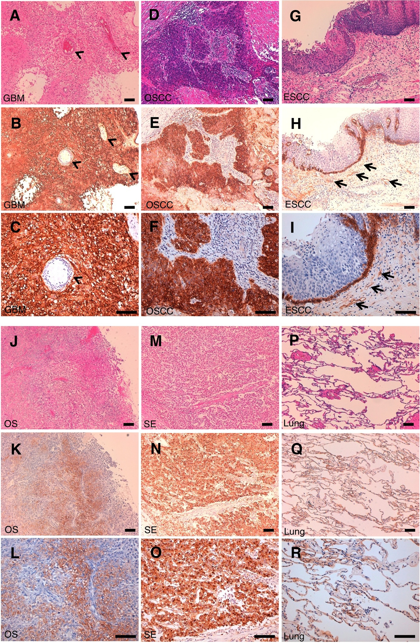
Immunohistochemical analysis by LpMab-13. Glioblastoma
Epitope mapping of LpMab-13
Because LpMab-13 reacted with 38–51 amino acids of hPDPN (data not shown), we further produced several point mutants of those residues. NZ-1 was previously produced by immunizing rats with 38–51 amino acids of hPDPN.(8) To determine the LpMab-13 epitope, we first performed Western blot analysis. LpMab-13 reaction was lost in several point mutations: A42G, G45A, D48A, and D49A (Fig. 3A). In contrast, NZ-1 did not react with M43A and D49A. These results were confirmed by flow cytometry (Fig. 3B). Although alanine scanning is usually performed to determine the binding epitope, conversion to other amino acids has been utilized in some reports(33–35) when alanine might be included in the epitope. Therefore, we further changed Ala42 and Ala46 to other amino acids such as Ser and Val (Fig. 4). LpMab-13 lost the reaction with A42S in the same way with A42G although it reacted with A42V. In contrast, LpMab-13 reacted with A46S and A46V in the same way with A46G. LpMab-7, a positive control against hPDPN, reacted with all point mutants (Figs. 3 and 4). These results indicate that the minimum epitope of LpMab-13 was identified as Ala42–Asp49 of hPDPN, the epitope of which is very similar with a rat anti-hPDPN MAb NZ-1.
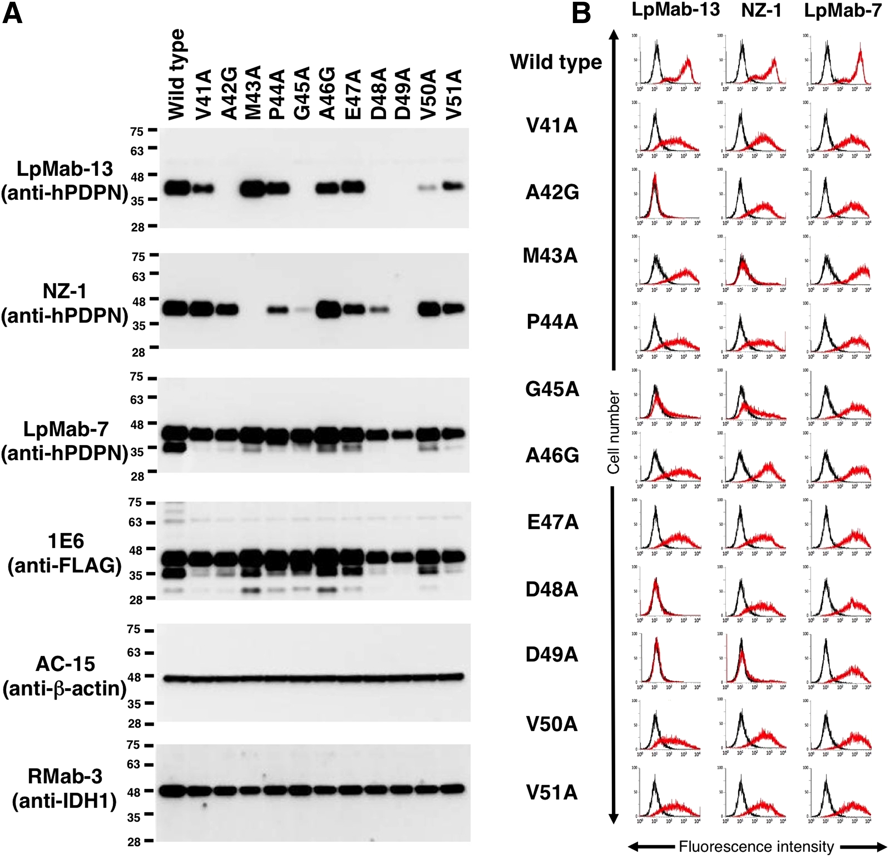
The epitope mapping of LpMab-13.
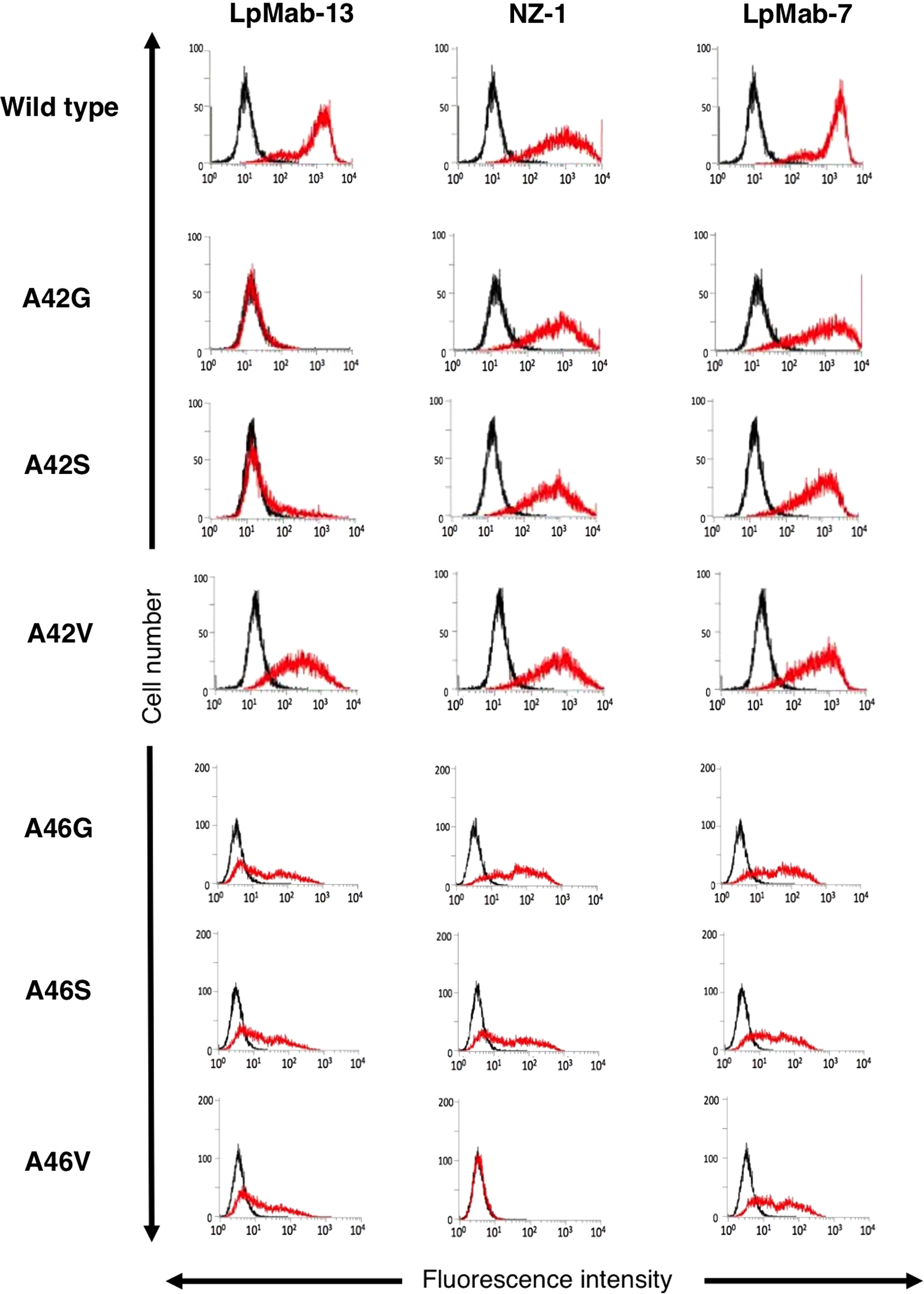
The epitope mapping of LpMab-13. CHO transfectants of each hPDPN mutation were treated with LpMab-13 (1 μg/mL; red), NZ-1 (1 μg/mL; red), LpMab-7 (1 μg/mL; red), or control PBS (black) for 30 minutes at 4°C, followed by treatment with anti-mouse or anti-rat IgG-Oregon green. Fluorescence data were collected using a Cell Analyzer EC800.
Discussion
We previously produced an anti-hPDPN MAb, NZ-1.(8) NZ-1 possesses not only high specificity and sensitivity but also high-binding affinity against hPDPN to be applied for radioimmunotherapy or immunotoxin therapy.(13,36) Another study confirmed the high-binding affinity of NZ-1.(37) NZ-1 is a suitable candidate for therapy against malignant gliomas, because it was highly internalized into glioma cell lines, and also well accumulated into tumors in vivo.(13) Moreover, NZ-1 inhibited the tumor cell-induced platelet aggregation and tumor metastasis by its neutralizing activity.(2) Furthermore, NZ-1 possesses antibody-dependent cellular cytotoxicity and complement-dependent cytotoxicity against hPDPN-expressing tumor cells.(38) Although a rat anti-hPDPN MAb NZ-1 possesses perfect application in all experiments such as Western blot, flow cytometry, immunoprecipitation, immunocytochemical analysis, and immunohistochemical analysis,(8,37) the other mouse anti-hPDPN MAbs such as D2-40(39) or 18H5(40) have been utilized more frequently. Usually, mouse MAbs are preferred for the reason that anti-mouse secondary antibodies are more available for almost all experiments especially for immunohistochemical analysis. For that reason, we recently produced anti-rat PDPN MAbs(41) and anti-rabbit PDPN MAbs(42,43) using mice. In this study, we tried to develop mouse anti-hPDPN MAbs, the epitope of which are similar with rat NZ-1 MAb.
We recently established the CasMab technology, which can produce cancer-specific MAbs although one protein in cancer cells and normal cells possesses the same amino acid sequence.(23) We used LN229/hPDPN cells, not purified recombinant proteins, for immunization to develop novel anti-PDPN MAbs. LpMab-13 detected many PDPN-expressing cancer cell lines such as glioblastoma, lung squamous cell carcinoma, OSCC, and malignant mesothelioma. LpMab-13 does not need antigen retrieval procedure to detect hPDPN in immunohistochemical analysis, although LpMab-2 and LpMab-3, which were also produced by CasMab technology, need antigen retrieval procedure.(23,30) Furthermore, LpMab-9 is not useful for immunohistochemistry, whereas it is very sensitive in flow cytometry or Western blot analysis,(25) indicating that the CasMab technology is not always helpful to produce mAbs for immunohistochemical analysis. Although the epitopes of D2-40 and 18H5 are very similar with those of LpMab-13,(36) D2-40 and 18H5 also need antigen retrieval procedure. NZ-1 is also useful for immunohistochemical analysis without antigen retrieval; therefore, the epitope of LpMab-13 and NZ-1 is critical for this superiority.
In conclusion, LpMab-13 could be useful for investigating the expression and function of hPDPN in cancers and normal tissues. Furthermore, different epitope-possessing anti-hPDPN MAbs should be established as powerful tools for uncovering the function of hPDPN in the future.
Footnotes
Acknowledgments
The authors thank Takuro Nakamura, Noriko Saidoh, Hazuki Kanno, and Kanae Yoshida for excellent technical assistance. This work was supported, in part, by the Platform for Drug Discovery, Informatics, and Structural Life Science (PDIS) from Japan Agency for Medical Research and Development, AMED (Y.K.); by the Basic Science and Platform Technology Program for Innovative Biological Medicine from AMED (Y.K.); by the Regional Innovation Strategy Support Program from the Ministry of Education, Culture, Sports, Science and Technology (MEXT) of Japan (Y.K.); by JSPS KAKENHI Grant Number 26440019 (M.K.K.), Grant Number 25462242 (Y.K.), and Grant Number 16K10748 (Y.K.); and by Takeda Science Foundation (S.O.).
Author Disclosure Statement
No competing financial interests exist.