
Abstract
Pancreatic ductal adenocarcinoma is an aggressive tumor with a poor prognosis. Biomarkers that can detect the tumor in its early stages when it may be amenable to curative resection might improve prognosis. To discover novel markers expressed in primary pancreatic cancer, we generated a panel of monoclonal antibodies against pancreatic ductal adenocarcinoma cell line BxPC3 using a rat medial iliac lymph node method. The antigen recognized by 1B5A5 was expressed on the cell surface and secreted into the conditioned medium of BxPC3 cells, and characterized as glycoproteins with molecular mass between 60 and 90 kDa. A wide range of molecular weights of 1B5A5 antigen in several pancreatic cancer cell lines were observed. Immunohistochemistry using a human multiple organ tumor tissue array showed an enhanced expression of 1B5A5 antigen in pancreas, lung, stomach, breast, urinary bladder, colon, and cervix uteri cancers. Immunoprecipitation followed by proteomic analyses identified CEACAM6 as a 1B5A5 antigen. In addition, western blot analysis results indicated that the 1B5A5 epitope is located within an amino-terminal domain of CEACAM6. These results raised the possibility that our approach could lead to discovery of novel biomarkers for the early stage of cancers in a relatively short period of time.
Introduction
P
The most widely used biomarker for pancreatic cancer is carbohydrate antigen 19-9 (CA19-9), a sialyl Lewis-a antigen found on the surface of proteins. Although CA19-9 is elevated mainly in late-stage pancreatic cancer, it is also elevated in benign diseases of the pancreas and in other malignancies of gastrointestinal tract.(3) Koprowski et al. first identified CA19-9 by hybridoma technology using the colorectal cancer cell line SW1116 as an immunogen.(4) Since the advent of hybridoma technology 40 years ago, research on monoclonal antibodies has developed enormously. The expanding demand of high-quality antibodies with better specificities has resulted in a significant improvement in traditional hybridoma production methods.(5) We have previously demonstrated that a medial iliac lymph node method provides us with substantial amounts of high-quality monoclonal antibodies in a relatively short period of time.(6,7)
The management of cancer through exploitation of properties distinguishing neoplastic and normal cells has been an attractive concept; however, a careful selection of cancer cell lines as immunogens is critical to the discovery of useful biomarkers. It has been shown that the pancreatic ductal adenocarcinoma cell line BxPC3 rapidly developed tumors in the pancreas with rare metastasis to the liver in orthotopic models.(8) In this study, we made an attempt to find novel biomarkers for primary pancreatic cancer by a rat medial iliac lymph node method using BxPC3 cancer cells as immunogens. The results of the present study suggested that our approach could lead to discovery of novel biomarkers for the early stage of cancers in a relatively short period of time.
Materials and Methods
Cell culture, plasmids, and reagents
Human cancer cell lines were obtained from the Japanese Collection of Research Bioresources Cell Bank (Osaka, Japan) and maintained in accordance with the supplier's protocol. FLAG-tagged carcinoembryonic antigen-related cell adhesion molecule 6 (CEACAM6) and its deletion mutant expression plasmids were obtained by subcloning the respective sequence into a pFLAG-CMV-5.1 expression vector (Sigma-Aldrich, St. Louis, MO). Rabbit anti-CEACAM6 monoclonal antibody (OriGene Technologies, Rockville, MD), goat anti-beta actin polyclonal antibodies (Santa Cruz Biotechnology, Dallas, TX), and control rat IgG (BioLegend, San Diego, CA) were also obtained. Dynabeads sheep anti-rat IgG (Invitrogen, Carlsbad, CA), enzymatic protein deglycosylation kit (Sigma-Aldrich) and high-density multiple organ tumor tissue array with normal tissue as control (US Biomax, Rockville, MD) were purchased.
Generation of monoclonal antibodies
Rat anti-BxPC3 monoclonal antibodies were generated based on the rat medial iliac lymph node method.(9) Briefly, a 10-week-old female WKY/Izm rat was injected to the rear footpads with 100 μL of emulsions containing the total lysates of BxPC3 (1.01 × 10⁶ cells) and Freund's complete adjuvant. Two weeks after the first immunization, an additional immunization (7.52 × 10⁵ cells of BxPC3) was performed without adjuvant into the tail base of the rat. Five days after the additional immunization, cells from the iliac lymph nodes of the immunized rat were fused with mouse myeloma Sp2/0-Ag14 cells at a ratio of 5:1 in 50% polyethylene glycol. The resulting hybridoma cells were plated onto 96-well plates and cultured in HAT selection medium. Monoclonal antibodies were purified from the desired hybridoma supernatants.(6)
Immunofluorescence
Pancreatic cancer cells were first fixed with 4% paraformaldehyde. After blocking, the cells were incubated with primary antibody at a concentration of 10 μg/mL. Localization of antigens was visualized using appropriate secondary antibody conjugated Alexa Fluor 488 (Molecular Probes, Eugene, OR).
Immunohistochemistry
A high-density multiple organ tumor and normal tissue microarray (MC5003ac), containing 18 tumor types (20 cases per type) and normal corresponding controls (five cases per type), were purchased from US Biomax. After deparaffinization and rehydration with xylene and ethanol, sections were incubated in 0.3% hydrogen peroxide in methanol for 30 minutes to quench endogenous peroxidase activity and then in blocking solution (Block Ace; Dainippon Pharmaceutical, Osaka, Japan) for 15 minutes at room temperature. Sections were incubated with primary antibody (1B5A5 and normal rat IgG) at a concentration of 10 μg/mL in DISCOVERY Antibody Diluent (Ventana Medical Systems/Roche, Tucson, AZ) at room temperature for 2 hours and then processed using the VECTASTAIN Elite ABC Rat IgG Kit (Funakoshi, Tokyo, Japan) with secondary antibody. The peroxidase reaction product was detected using DAB+, Liquid (Dako Japan, Kyoto, Japan). Images were captured using a brightfield slide scanner VS120 (Olympus, Tokyo, Japan).
Immunoprecipitation and microsequencing
One milligram of BxPC3 whole cell lysates or plasma membrane extracts was prepared, and immunoprecipitation was performed using 1B5A5 followed by Dynabeads sheep anti-rat IgG as previously described.(10) Proteins bound to beads were resolved using 10% sodium dodecyl sulfate polyacrylamide gel electrophoresis (SDS-PAGE) and then visualized by silver staining. Desired bands were excised, in-gel digested with trypsin, and solvent extracted, and the resulting MS/MS spectra were searched using the Mascot search engine.(6)
Western blot
Whole cell extracts were resolved using 10% SDS-PAGE, transferred to a PVDF membrane, and blocked with 5% skim milk for 30 minutes at room temperature. After incubation with primary antibodies, the proteins of interest on immunoblots were detected using an enhanced chemiluminescence detection system.(6)
Results
Since cell surface molecules have been used as markers for isolating, purifying, and targeting cancer cells, we first screened hybridoma supernatants that stain plasma membrane of BxPC3 by immunofluorescence and obtained eight monoclonal antibodies from positive clones (data not shown). However, the results from immunoprecipitation followed by western blot analyses showed that the antigen of a monoclonal antibody, referred to herein as 1B5A5 (rat IgG2b κ), was identical to that of others (data not shown). Next, the expression levels and the distributions of 1B5A5 antigen on human pancreatic cancer cell lines were determined by western blot and immunofluorescence, respectively. KP-3L, an adenosquamous carcinoma of the pancreatic duct, was obtained from liver metastasis of KP-3 in nude mice. A ductal adenocarcinoma, BxPC3, rapidly developed tumors in the pancreas and spread regionally to the spleen with rare metastasis to the liver, whereas MIA PaCa-2 grew more slowly in the pancreas, but rapidly metastasized to distant sites, including portal lymph nodes and liver in orthotopic models.(8) CEA-producing pancreatic islet cell carcinoma, QGP-1, has been shown to migrate from rounded irregular cell aggregates with no hormone secretion.(11) SUIT-2 derived from a metastatic liver tumor of human pancreatic carcinoma has been established, which histopathologically exhibited moderately differentiated tubular adenocarcinoma, and produces and releases at least two tumor markers, CEA and CA19-9, in the culture medium.(12) Forty micrograms of whole cell lysates were prepared, and expression levels of 1B5A5 antigen in six cell lines were compared by western blotting (Fig. 1A). A major band corresponding to molecular mass between 60 and 90 kDa and a faint band of about 125 kDa were detected in the lysates of BxPC3. Only one band corresponding to 125 kDa was observed in the lysates of KP-3 cells; however, KP-3L cells produced several 1B5A5 antigens, ranging from 70 to 180 kDa. Interestingly, the expression of 1B5A5 antigen was not observed in the lysates of MIA PaCa-2. In contrast, QGP-1, a pancreatic islet cell carcinoma, produced high amounts of antigen with molecular mass between 40 and 125 kDa. The pattern of SUIT-2 lysates was similar with that of KP-3L (Fig. 1A). We then determined the distributions of 1B5A5 antigen on pancreatic cancer cell lines by immunofluorescence. Since a marked intensive signal of 1B5A5 was observed in QGP-1, the camera exposure time of QGP-1 used was one-tenth that of others (data not shown). As shown in Figure 1B, 1B5A5 antigen was expressed on the plasma membrane in all five cell lines, regardless of several bands being detected by western blotting. Since 1B5A5 reacted with the cell lysates as diffuse bands by western blotting and the plasma membrane of pancreatic cancer cells by immunofluorescence, we tested the possibility that the 1B5A5 antigen was composed of secreted glycoproteins. As shown in Figure 1C, 1B5A5 antigen in the conditioned medium of BxPC3 was remarkably elevated in a time-dependent manner. In addition, treatment of the whole cell lysates from BxPC3 with a protein deglycosylation enzyme mixture (PNGase, O-glycosidase, α-2(3,6,8,9)-neuraminidase, β-1-4 galactosidase, and β-N-acetylglucosaminidase) that digests N- and O-linked oligosaccharides obviously moved the bands of 1B5A5 antigen forward (Fig. 1D). These results suggested that 1B5A5 antigen is composed of pancreatic cancer cell type-specific secreted glycoproteins.
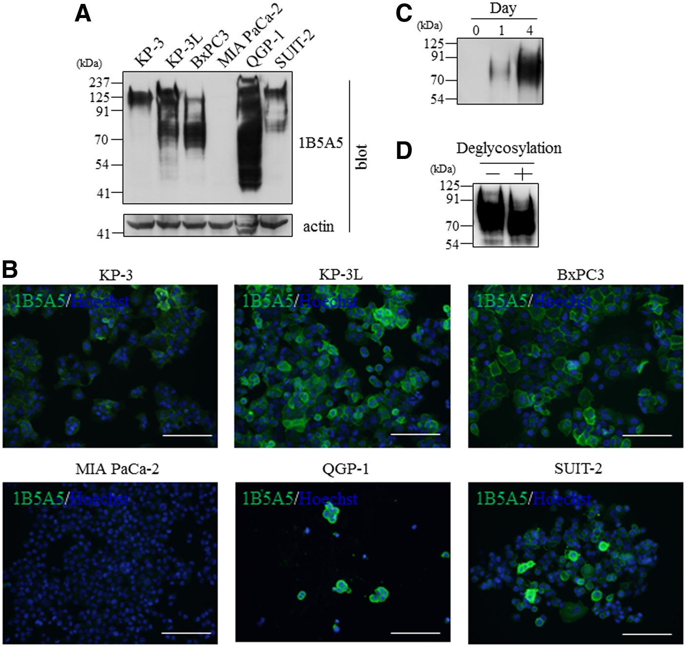
1B5A5 antigen is composed of pancreatic cancer cell type-specific secreted glycoproteins.
Immunohistochemical examination suggested that positive reaction was detected in some tumors (Fig. 2A, B, and Table 1). As expected, some adenocarcinomas (5 out of 19 cases) of the pancreas were positive for 1B5A5 antigen. In the pancreas, we could not determine the spontaneous expression of 1B5A5 antigen because nonspecific reaction was strongly observed in the acinar cells (Fig. 2A). In addition, 3 of 18 cases of urothelial carcinoma of the urinary bladder (17%), 3 of 19 of invasive ductal carcinoma of the breast (16%), and 4 of 18 of adenocarcinoma of the stomach (22%) also showed 1B5A5 antigen positive, whereas all normal tissues of these organs were 1B5A5 antigen negative (Fig. 2A). In contrast to these above organs, positive reaction was also observed in some tissues of the normal lung (two out of four cases), colon (one out of five cases), and cervical uteri (one out of five cases). Positive reaction of the normal lung tissue was weakly observed on the surface of alveolar epithelial cell (Fig. 2B). The surface of epithelial cells of the colon and squamous cells of the cervical uteri were also stained (Fig. 2B). Three of eight adenocarcinomas (38%), four of nine squamous cell carcinomas of the lung, 3 of 20 adenocarcinomas of the colon, and 2 of 20 squamous cell carcinomas of the cervical uteri were strongly stained (Fig. 2B). There was no relationship between tumor malignancy and positive reaction for 1B5A5 antigen (data not shown).
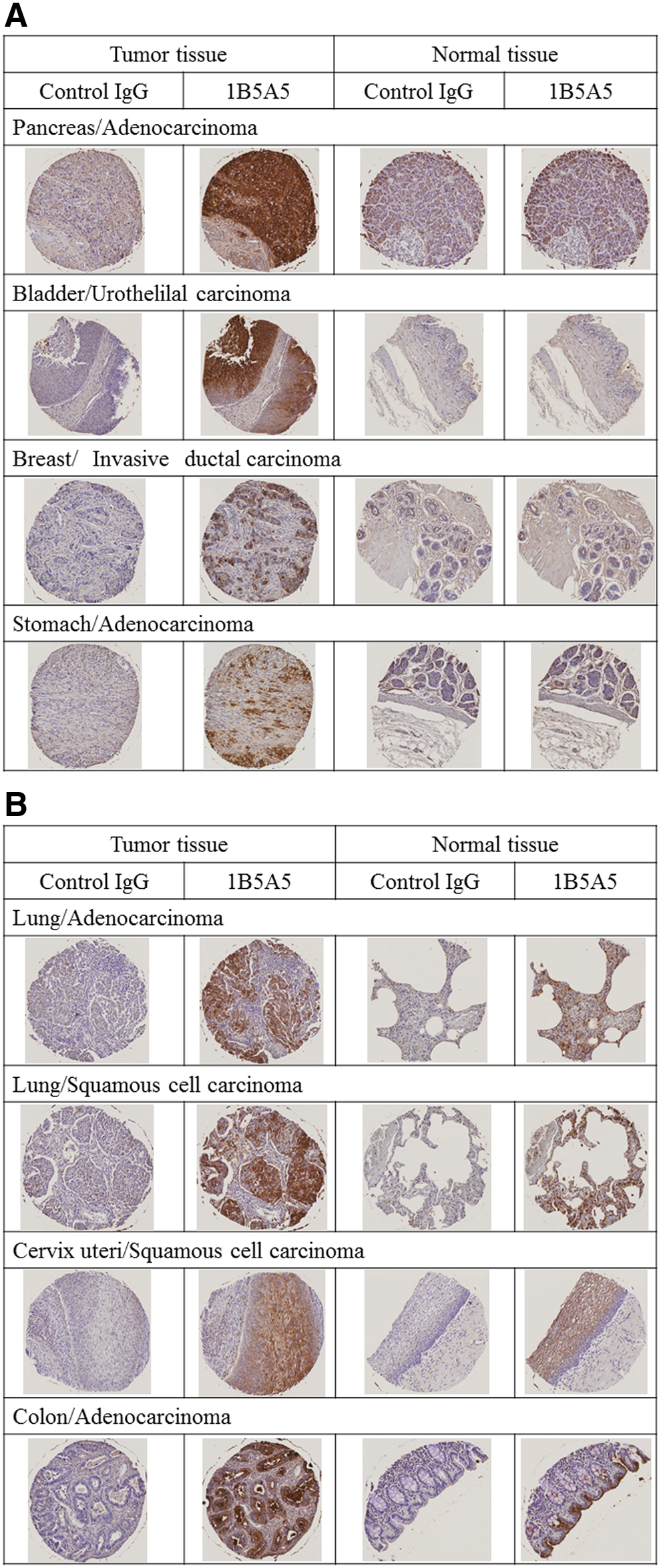
1B5A5 antigen is expressed in representative cases of tumor and normal tissue cores.
N, number of samples examined; ND, undetermined because of nonspecific antibody reaction.
The results described above led us to identify the 1B5A5 antigen. After incubation of the plasma membrane extracts of BxPC3 with 1B5A5, immunoprecipitation was performed. The results of two independent experiments showed that a band with molecular mass of about 80 kDa was consistently detected by western blotting (left) and silver staining (right) (Fig. 3A). The relevant band was excised and analyzed by mass spectrometry, and CEACAM6 was identified as a candidate protein of 1B5A5 antigen (Mascot score >70; the observed m/z 758.9397 and 629.8182 were matched with aa50-aa62 and aa63-aa72 of CEACAM6, respectively). To determine whether 1B5A5 recognizes CEACAM6, we performed immunoprecipitation followed by western blot using 1B5A5 and anti-CEACAM6 antibodies. As shown in Figure 3B, it was evident that a rabbit anti-CEACAM6 monoclonal antibody detected a band with molecular mass between 60 and 90 kDa in immunopurified 1B5A5 precipitates. CEACAM6 protein belongs to the immunoglobulin superfamily and exhibits one variable-like domain, identified as the N-domain, followed by constant C2-like domains termed A and B (Fig. 3C, upper). In addition, CEACAM6 contains a signal peptide at the N-domain and interacts with plasma membrane through a glycosylphosphatidylinositol linkage after cleavage of the pro-peptide at the B-domain. To identify the binding region of 1B5A5 to CEACAM6, we constructed the wild type (WT) and two deletion mutant expression plasmids (N + A and A+B), transfected them into HeLa cells, and performed western blot using the whole cell lysates. Although 1B5A5 recognized both WT and N + A of CEACAM6, the deletion of N-domain abrogated the binding to 1B5A5 (Fig. 3C, lower). These results indicated that the 1B5A5 epitope is located within the N-domain of CEACAM6.

1B5A5 interacts with N-domain of CEACAM6.
Discussion
Pancreatic cancer is the fourth-leading cause of cancer-related deaths in Japan, with a 5-year postdiagnosis survival rate of 7%, and one of the most highly aggressive and lethal of all solid tumors. At present, serum CA19-9 is the only validated tumor marker in widespread clinical use, but precise knowledge of its role in pancreatic cancer diagnosis, staging, determining resectability, response to chemotherapy, and prognosis remains limited.(13) CA19-9 was first discovered by hybridoma technology using a colorectal cancer cell line. In this study, we aimed to discover novel tumor markers for the early stage of pancreatic ductal adenocarcinoma by a rat medial iliac lymph node method using BxPC3 cells as antigens.
During malignant transformation, glycosylation is heavily altered compared with normal tissue due to differential expression of glycosyltransferases, glycosidases, and monosaccharide transporters. Although the CEACAM family members (CEACAMs) were discovered decades ago and many specific monoclonal antibodies have been raised against these proteins, their very similar structures preclude proper assignment of their expression patterns in both normal and tumor tissues.(14) Except for very similar N-domains, characteristic membrane attachments, and the number of glycosylation sites, what differentiates these family members lies in the number of C2-like domains.(15) The availability of mammalian cells transfected with most of the CEACAM gene family members and generation of mutant proteins facilitated proper definition of their respective functions.(16) The present studies showed that 1B5A5 detected a faint band of 125 kDa in addition to a diffuse band with molecular mass between 60 and 90 kDa in the lysates of BxPC3. In contrast, QGP-1 produced 1B5A5 antigen with molecular mass between 40 and 125 kDa. Indeed, 1B5A5 detected the band of 60 kDa as CEACAM6 and recognized the N-domain of CEACAM6, as shown by the gene transfection experiments (Fig. 3C). These results suggest that 1B5A5 reacts with the CEACAMs through the highly conserved N-domain. In contrast, our results do not exclude the possibility that 1B5A5 specifically binds to CEACAM6 because of a wide variety of glycoforms, and some splicing mutants of CEACAMs have been reported.(17,18) Further studies to identify the precise epitope of 1B5A5 would resolve this issue.
Numerous studies have shown that the expressions of CEACAM6 are augmented in many cancers and are now considered valid clinical biomarkers and promising therapeutic targets in melanoma, lung, colorectal, and pancreatic cancers.(15) In this study, we have revealed that 1B5A5-positive reaction was recognized in primary cancers of the pancreas, lung, stomach, breast, urinary bladder, colon, and cervix uteri (Fig. 2A, B), but not observed in the prostate or thyroid tumors (Table 1). These results agree with previous results obtained in other research.(19,20) In contrast, these previous studies suggested that CEACAM6 expression was strongly recognized in ovarian tumors, but no positive reaction for 1B5A5 was observed in our study. The cause of these differences was unclear. While 1B5A5 did not label novel biomarkers, future studies could aid in prediction of the cancer risk. A drug-conjugated murine monoclonal antibody against CEACAM6 led to tumor growth inhibition of pancreatic ductal adenocarcinoma cells.(21) Furthermore, administration of humanized anti-CEACAM6 antibody attenuated tumor growth and angiogenesis and exhibited significant apoptosis in a murine pancreatic ductal adenocarcinoma xenograft model.(22) Thus, another topic we have highlighted is the pharmacological effects of 1B5A5.
In summary, we generated monoclonal antibodies against BxPC3 cells as a model of primary pancreatic ductal adenocarcinoma. Our results enable exploitation of hybridoma technology to discover novel biomarkers for the early stage of cancers in a relatively short period of time.
Footnotes
Author Disclosure Statement
K.H., M.K., N.S., and K.S. are employees of Sumitomo Chemical Co., Ltd. For all other authors, no competing financial interests exist.