
Abstract
Human podoplanin (hPDPN) is expressed in lymphatic vessels, pulmonary type-I alveolar cells, and renal glomerulus. The hPDPN/C-type lectin-like receptor-2 (CLEC-2) interaction is involved in platelet aggregation and cancer metastasis. High expression of hPDPN in cancer cells or cancer-associated fibroblasts (CAFs) leads to a poor prognosis for cancer patients. In our previous research, we reported on several anti-hPDPN monoclonal antibodies (mAbs), including LpMab-2, LpMab-3, LpMab-7, LpMab-9, LpMab-12, LpMab-13, and LpMab-17 of mouse IgG1 subclass, which were produced using CasMab technology. Here we produced a novel anti-hPDPN mAb LpMab-19 of mouse IgG2b subclass. Flow cytometry revealed that the epitope of LpMab-19 includes O-glycan, which is attached to Thr76 of hPDPN. We further identified the minimum epitope of LpMab-19 as Thr76–Arg79 of hPDPN. Immunohistochemistry revealed that LpMab-19 is useful for detecting not only normal cells, including lymphatic vessels, but also glioblastoma and oral squamous cell carcinoma cells. LpMab-19 could be useful for investigating the physiological function of O-glycosylated hPDPN.
Introduction
T
We recently established the CasMab technology, which produces cancer-specific mAbs, although one protein in the cancer and normal cells has the same amino acid sequence.(11) We used LN229/hPDPN cells, not purified recombinant proteins, for immunization to develop novel anti-hPDPN mAbs. Although we have produced many anti-hPDPN mAbs using CasMab technology,(11–17,19,20) almost all mAbs, including LpMab-2, LpMab-3, LpMab-7, LpMab-9, LpMab-12, LpMab-13, and LpMab-17, were determined to belong to the IgG1 subclass. Only the clone LpMab-10 was classified as IgG3 subclass; however, mouse IgG3 subclass is often aggregated.(29) Furthermore, almost all anti-hPDPN mAbs react with PLAG domains (PLAG1–PLAG3) of hPDPN.(21,22,30–33) A novel PLAG domain of hPDPN was recently reported as PLAG4 (81-EDLPT-85), and anti-PLAG4 mAbs were produced.(34) We need to produce further anti-hPDPN mAbs, which target different epitopes of hPDPN, to fully understand the pathophysiological function of hPDPN.
Here we produced a novel anti-hPDPN mAb, LpMab-19 (mouse IgG2b, kappa), the epitope of which is a O-glycosylated glycopeptide of hPDPN.
Materials and Methods
Cell lines
LN229, HEK-293T, Chinese hamster ovary (CHO)-K1, and P3U1 were obtained from the American Type Culture Collection (ATCC, Manassas, VA). LN319 was donated by Prof. Kazuhiko Mishima (Saitama Medical University, Saitama, Japan).(35) CHO-S was purchased from Thermo Fisher Scientific, Inc. (Waltham, MA). LN229 was previously transfected with hPDPN plasmids (LN229/hPDPN) using Lipofectamine 2000 (Thermo Fisher Scientific, Inc.) according to the manufacturer's instructions.(11) The HEK-293T/hPDPN-knockout (KO) cell line (PDIS-2) and the LN319/hPDPN-KO cell line (PDIS-6) were generated by transfection using CRISPR/Cas plasmids (Target ID: HS0000333287) that target PDPN (Sigma-Aldrich, St. Louis, MO). PDIS-2 and PDIS-6 cells were screened using the NZ-1 mAb.(12,15,18–20)
CHO-K1 and P3U1 were cultured in RPMI 1640 medium, including
Production of deletion mutants
The amplified hPDPN complementary DNA (cDNA) was subcloned into a pCAG-Ble(Zeo) vector (Wako Pure Chemical Industries Ltd. [Osaka, Japan]) with an MAP tag, which was detected by PMab-1(36,37) and added at the N-terminus using In-Fusion HD cloning kit (Clontech Laboratories Inc., Mountain View, CA). Deletion mutants of hPDPN were produced using several primers. Sense primers used were as follows: AGAAGACAAAAAGCTTGCCAGCACAGGCCAGCC for dN23, AGAAGACAAAAAGCTTGAAGGCGGCGTTGCCAT for dN37, AGAAGACAAAAAGCTTGCCGAAGATGATGTGGTG for dN46, AGAAGACAAAAAGCTTACCAGCGAAGACCGCTA for dN55, AGAAGACAAAAAGCTTACAACTCTGGTGGCAACA for dN64, AGAAGACAAAAAGCTTGTAACAGGCATTCGCATC for dN75, AGAAGACAAAAAGCTTACTTCAGAAAGCACAGTCC for dN85, AGAAGACAAAAAGCTTCAAAGTCCAAGCGCCAC for dN95, AGAAGACAAAAAGCTTGCCACCAGTCACTCCAC for dN105.
The following antisense primer was used: TCTAGAGTCGCGGCCGCTTACTTGTCGTCATCGT.
CHO-K1 cells were transfected with the plasmids using a Lipofectamin LTX (Thermo Fisher Scientific, Inc.). Deletion mutants were cultured in RPMI 1640 medium including
Production of point mutants
The amplified hPDPN cDNA was subcloned into a pcDNA3 vector (Thermo Fisher Scientific, Inc.), and a FLAG epitope tag was added at the C-terminus. Amino acids in hPDPN were substituted with alanine or glycine using a QuikChange Lightning site-directed mutagenesis kit (Agilent Technologies, Inc., Santa Clara, CA) with oligonucleotides containing the desired mutations. CHO-S or CHO-K1 cells were transfected with the plasmids using a Gene Pulser Xcell electroporation system (Bio-Rad Laboratories, Inc., Berkeley, CA). Point mutants of CHO-K1 and CHO-S were cultured in RPMI 1640 medium including
Hybridoma production
Four-week-old female BALB/c mice (CLEA Japan, Tokyo, Japan) were immunized by intraperitoneal (i.p.) injection of 1 × 10⁸ LN229/hPDPN cells together with Imject Alum (Thermo Fisher Scientific, Inc.).(11) After several additional immunizations, a booster i.p. injection was given 2 days before mice were euthanized by cervical dislocation, and spleen cells were harvested. The spleen cells were fused with P3U1 cells using PEG1500 (Roche Diagnostics, Indianapolis, IN). Hybridomas were grown in RPMI 1640 medium including
Flow cytometry
Cell lines were harvested by brief exposure to 0.25% trypsin/1 mM EDTA (Nacalai Tesque, Inc.). After washing with 0.1% BSA in PBS, cells were treated with primary mAbs for 30 minutes at 4°C, followed by treatment with Oregon Green 488 goat antimouse IgG or antirat IgG (Thermo Fisher Scientific, Inc.). Fluorescence data were collected using a Cell Analyzer EC800 (Sony Corp., Tokyo, Japan).
Immunohistochemical analyses
The use of human cancer tissues was reviewed and approved by Sendai Medical Center Review Board.(19) Written informed consent was obtained for the human cancer tissue samples used in this study. Four-micrometer-thick histologic sections were deparaffinized in xylene and rehydrated. After antigen retrieval procedure (autoclave using citrate buffer, pH 6.0; Agilent Technologies, Inc.), sections were incubated with 1 μg/mL of LpMab-19 for 1 hour at room temperature followed by treatment with Envision+ kit (Agilent Technologies, Inc.) for 30 minutes. Color was developed using 3,3-diaminobenzidine tetrahydrochloride (Agilent Technologies, Inc.) for 5 minutes, and then the sections were counterstained with hematoxylin (Wako Pure Chemical Industries Ltd.).
Results and Discussion
The expression of hPDPN has been reported in many cancers, such as oral cancer, lung cancer, esophageal cancer, malignant mesothelioma, malignant glioma, testicular tumor, bladder cancer, and osteosarcoma.(8,21,23,26,38–46) hPDPN is a PLAG factor, which is involved in cancer metastasis.(8–10) hPDPN-expressing cancer-associated fibroblasts are associated with poor prognosis of several cancers.(47–52) We previously identified C-type lectin-like receptor-2 (CLEC-2) as an endogenous receptor of hPDPN,(28,53) and recently performed comparative crystallographic studies on hPDPN in association with CLEC-2.(54) The interaction with CLEC-2 was mainly observed at Glu47 and Asp48 in the PLAG3 domain and the α2–6-linked sialic acid at Thr52 of hPDPN. Clone LpMab-12, our recently produced mAb, specifically detects this sialylated Thr52.(18)
Herein we used the CasMab technology for production of anti-hPDPN mAbs.(11) LN229/hPDPN cells were immunized into mice, and culture supernatants were screened using ELISA for binding to recombinant hPDPN, which was purified from LN229/hPDPN cells.(11) Finally, LpMab-19 (mouse IgG2b, kappa), the first IgG2b of mouse anti-hPDPN mAbs using CasMab technology, was produced after limiting dilution.
LpMab-19 reacted with LN229/hPDPN, and did not with LN229 (hPDPN-negative cell) in flow cytometry (Fig. 1A). LpMab-19 recognized endogenous hPDPN, which is expressed in LN319 (a glioblastoma cell line), whereas it did not react with LN319/hPDPN-KO cell (PDIS-6) (Fig. 1A). In addition, LpMab-19 detected hPDPN of HEK-293T (an epithelial cell line of kidney), but lost the reaction with HEK-293T/hPDPN-KO cell (PDIS-2) (Fig. 1A). As a positive control, an anti-hPDPN mAb LpMab-13 was used (Fig. 1B). LpMab-13 detected LN229/hPDPN, LN319, and HEK-293T, whereas it lost the reaction with PDIS-6 and PDIS-2.
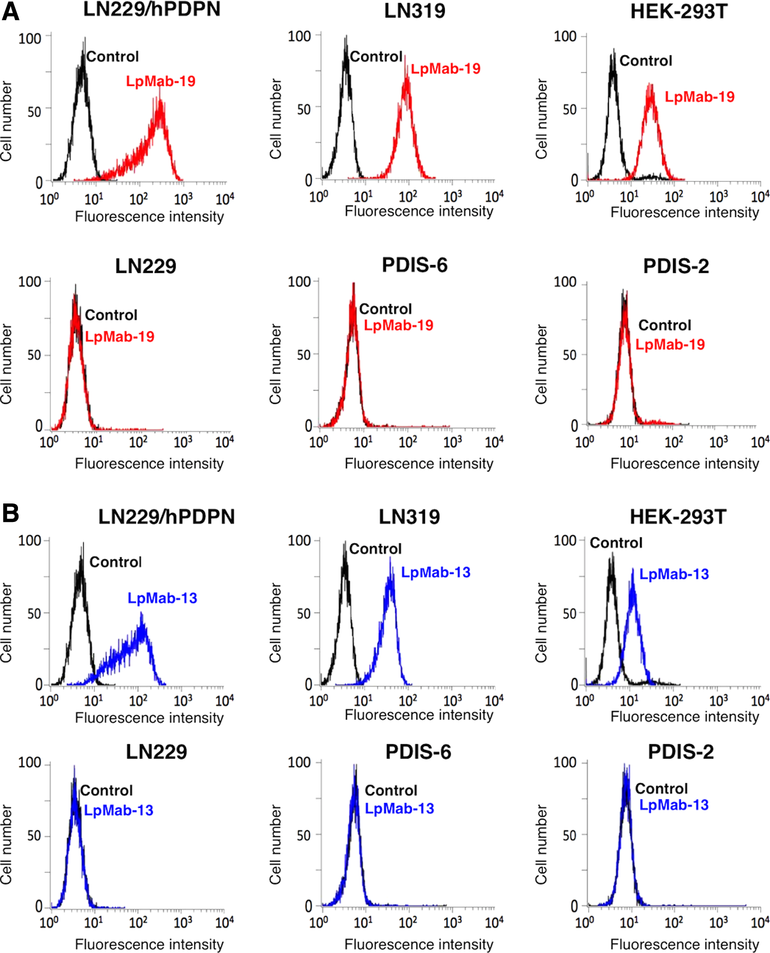
In flow cytometry, LpMab-19 specifically detects hPDPN.
We herein produced several deletion mutants of hPDPN, which were expressed in CHO-K1 cell lines. LpMab-19 detected the delta N-terminus (dN)23 that starts from Ala23. Similarly, LpMab-19 detected dN37, dN46, dN55, dN64, and dN75. In contrast, LpMab-19 lost the reaction with dN85, dN95, and dN105, indicating that the N-terminus of the LpMab-19 epitope exists between the 75th and 85th amino acids (Fig. 2A). All deletion mutants of hPDPN possess the N-terminal MAP tag and were detected by the anti-MAP tag mAb (clone: PMab-1) (Fig. 2B).

Epitope mapping of LpMab-19 using deletion mutants of hPDPN in flow cytometry. Each hPDPN deletion mutant (dN23, dN37, dN46, dN55, dN64, dN75, dN85, dN95, and dN105) was treated with LpMab-19
Next, we produced stable CHO-S cell lines expressing point mutants of hPDPN: T65A, T66A, T70A, S71A, S74A, T76A, T85A, S86A, and S88A (Fig. 3A). The epitope of LpMab-19 was thought to include glycans because LpMab-19 did not react with synthetic peptides of hPDPN (data not shown). LpMab-19 lost the reaction with the T76A mutant, whereas the other point mutants were detected by LpMab-19 (Fig. 3A). All point mutants targeting Ser/Thr residues were detected by LpMab-13 (Fig. 3B). Therefore, the epitope of LpMab-19 includes sialic acid on O-glycan, which is attached to Thr76.
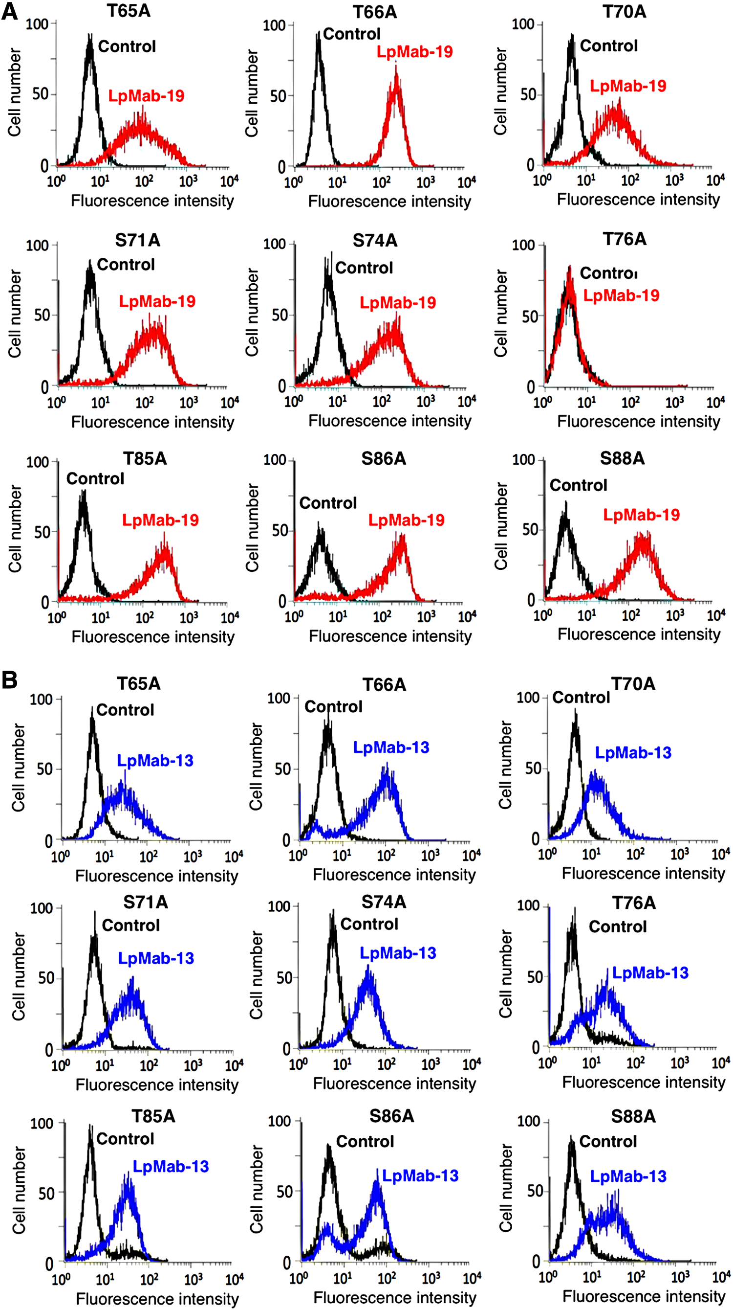
Epitope mapping of LpMab-19 using point mutants of Ser/Thr residues of hPDPN in flow cytometry. Stable CHO-S transfectants expressing hPDPN point mutants (T65A, T66A, T70A, S71A, S74A, T76A, T85A, S86A, and S88A) were treated with LpMab-19
We further determined the LpMab-19 epitope using alanine scanning around Thr76 of hPDPN. Nine hPDPN point mutants against Ser74–Thr85 were transiently expressed in CHO-K1 cells. As shown in Figure 4A, LpMab-19 did not detect T76A, I78A, and R79A. In contrast, LpMab-13 recognized all point mutants (Fig. 4B), indicating that the minimum epitope of LpMab-19 was Thr76–Arg79.
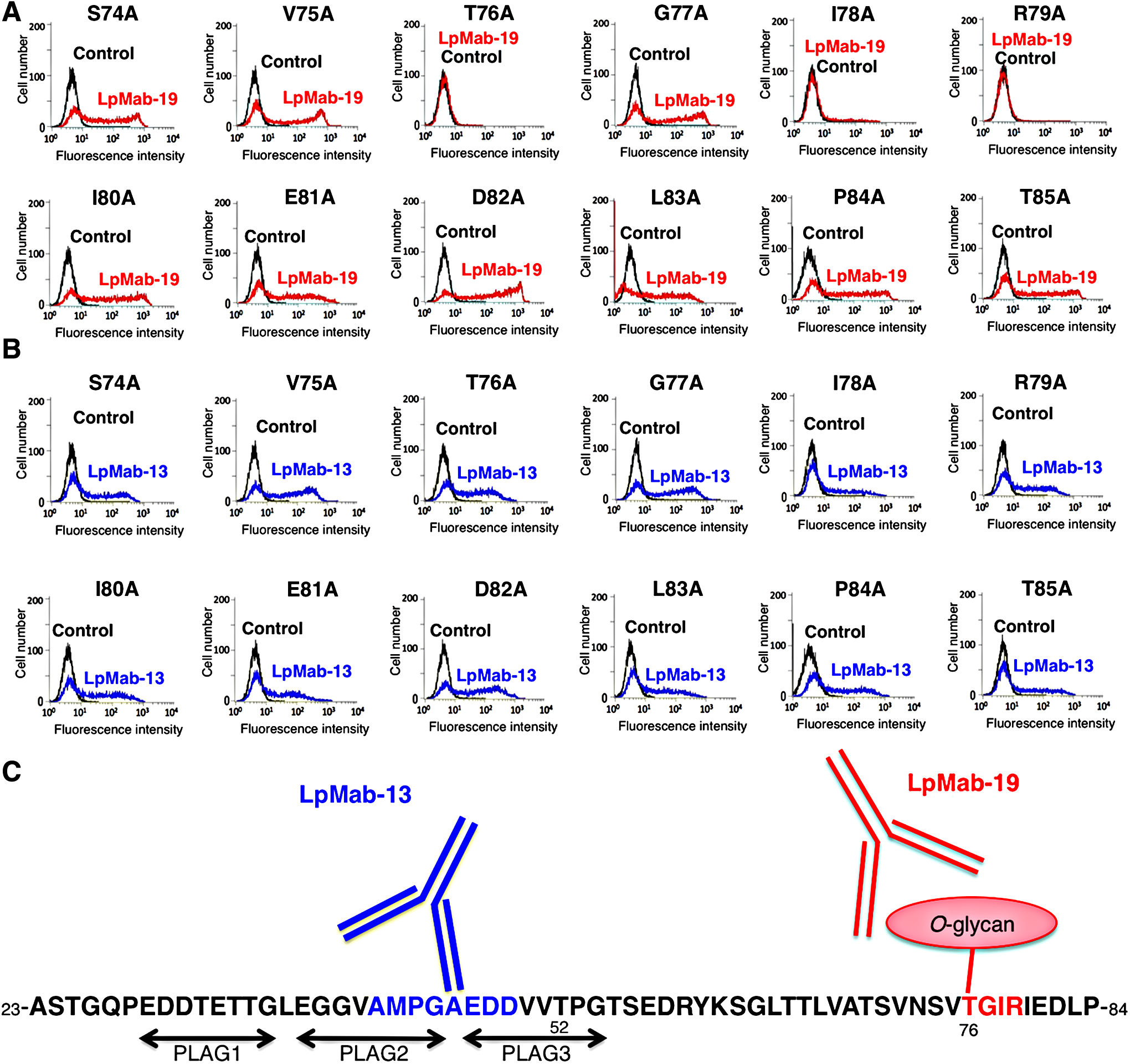
Epitope mapping of LpMab-19 using point mutants of hPDPN in flow cytometry. Nine hPDPN point mutants against Ser74-Thr85 were transiently expressed in CHO-K1 cells. Cells were treated with LpMab-19
It has been reported that hPDPN is expressed in many cancers, such as malignant brain tumor, mesothelioma, oral cancer, lung cancer, esophageal cancer, testicular tumor, and osteosarcoma.(13,38,44) Here we compared the reactivity of LpMab-19 and LpMab-13 in an immunohistochemical analysis using oral squamous cell carcinoma. LpMab-19 stained cancer cells (Fig. 5A, B) and LECs (Fig. 5E, F) in a membrane/cytoplasmic-staining pattern without an antigen retrieval procedure. LpMab-19 did not react with vascular endothelial cells (Fig. 5E, F). The staining pattern of LpMab-19 is similar to LpMab-13, indicating that LpMab-19 is very useful for immunohistochemistry.

Immunohistochemical analysis by LpMab-19 against oral squamous cell carcinoma. Sections were incubated with 1 μg/mL of LpMab-19
Therefore, LpMab-19 could be useful for investigating the expression and function of hPDPN in cancers and normal tissues. In future, different epitope-possessing anti-hPDPN mAbs should be produced for uncovering the function of hPDPN.
Footnotes
Acknowledgments
We thank Takuro Nakamura, Noriko Saidoh, and Kanae Yoshida for their excellent technical assistance. We also thank Yuki Fujii and Ryusuke Honma for their suggestion and advice. This work was supported, in part, by the Platform for Drug Discovery, Informatics, and Structural Life Science (PDIS) from Japan Agency for Medical Research and development, AMED (Y.K.), by the Regional Innovation Strategy Support Program from the Ministry of Education, Culture, Sports, Science and Technology (MEXT) of Japan (Y.K.), by JSPS KAKENHI Grant Number 26440019 (M.K.K.) and Grant Number 16K10748 (Y.K.), and by the Basic Science and Platform Technology Program for Innovative Biological Medicine from AMED (Y.K.). This work was performed, in part, under the Cooperative Research Program of Institute for Protein Research, Osaka University, CR-15–05 and CR-16–05 (Y.K.).
Author Disclosure Statement
No competing financial interests exist.