
Abstract
Podoplanin, a type I transmembrane glycoprotein, is a specific marker of lymphatic endothelial cells (LECs). Recently, we developed PMab-38, an anti-dog podoplanin monoclonal antibody that did not stain canine LECs. In this study, we newly developed PMab-48 against dog podoplanin. Immunohistochemical analysis revealed that PMab-48 reacts not only with canine squamous cell carcinoma cells but also with LECs of the normal colon. Therefore, PMab-48 may be useful in investigating the function of dog podoplanin in LECs.
Introduction
D
Materials and Methods
Cell lines, canine tissues, and animals
P3U1 was purchased from the American Type Culture Collection (ATCC, Manassas, VA), and it was cultured in RPMI 1640 medium (Nacalai Tesque, Inc., Kyoto, Japan) supplemented with 10% heat-inactivated fetal bovine serum (Thermo Fisher Scientific, Inc., Waltham, MA), 100 U/mL of penicillin, 100 μg/mL of streptomycin, and 25 μg/mL of amphotericin B (Nacalai Tesque, Inc.) at 37°C in a humidified atmosphere of 5% CO2 and 95% air. Canine tissues were obtained from North lab (Hokkaido, Japan).(12–14) Female BALB/c mice (4-weeks old) were purchased from CLEA Japan (Tokyo, Japan). Animals were housed under specific pathogen-free conditions. The Animal Care and Use Committee of Tohoku University approved the animal experiments described herein.
Hybridoma production
BALB/c mice were immunized by an intraperitoneal (i.p.) injection of 100 μg of a synthetic peptide (dpp2640: VRPDDIIPGVEDSVVC), conjugated with KLH together with Imject Alum (Thermo Fisher Scientific, Inc.). After several additional immunizations of 100 μg, a booster injection of 100 μg was given i.p. 2 days before spleen cells were harvested. The spleen cells were fused with P3U1 cells by using PEG1500 (Roche Diagnostics, Indianapolis, IN). The hybridomas were grown in RPMI medium with hypoxanthine, aminopterin, and thymidine selection medium supplement (Thermo Fisher Scientific, Inc.).
Enzyme-linked immunosorbent assay
The culture supernatants were screened by using enzyme-linked immunosorbent assay (ELISA) for binding to dpp2640, which is immobilized on Nunc Maxisorp 96-well immunoplates (Thermo Fisher Scientific, Inc.) at 1 μg/mL for 30 minutes. After blocking with 1% bovine serum albumin in 0.05% Tween20/phosphate-buffered saline (Nacalai Tesque, Inc.), the plates were incubated with culture supernatant followed by 1:3000 diluted peroxidase-conjugated anti-mouse IgG (Agilent Technologies, Inc., Santa Clara, CA). The enzymatic reaction was conducted with a 1-Step Ultra TMB-ELISA (Thermo Fisher Scientific, Inc.). The optical density was measured at 655 nm by using an iMark microplate reader (Bio-Rad Laboratories, Inc., Hercules, CA).
Immunohistochemical analyses
Four-micrometer-thick histologic sections were deparaffinized in xylene and rehydrated, and they were autoclaved in citrate buffer (pH 6.0; Agilent Technologies, Inc.) for 20 minutes. Sections were incubated with 50 μg/mL of primary monoclonal antibodies for 1 hour at room temperature followed by treatment with Envision+ kit for 30 minutes (Agilent Technologies, Inc.). Color was developed by using 3,3-diaminobenzidine tetrahydrochloride (Agilent Technologies, Inc.) for 2 minutes, and then the sections were counterstained with hematoxylin (Wako Pure Chemical Industries, Ltd., Osaka, Japan).
Results and Discussion
We immunized mice with a synthetic peptide (dpp2640) corresponding to amino acids 26–40 of dog podoplanin (Fig. 1A); screening was performed by using ELISA. The ELISA-positive wells were screened a second time by flow cytometry. Finally, the positive wells were selected in an immunohistochemical analysis. After limiting dilution, one of the clones, PMab-48 (IgG1, kappa) was established. PMab-48 reacted with Chinese hamster ovary (CHO)/dog podoplanin, not with CHO/cat podoplanin, CHO/bovine podoplanin, CHO/rabbit podoplanin, CHO/mouse podoplanin, CHO/rat podoplanin, CHO/human podoplanin, and CHO/K1 (Supplementary Fig. S1), indicating that PMab-48 is specific against dog podoplanin.
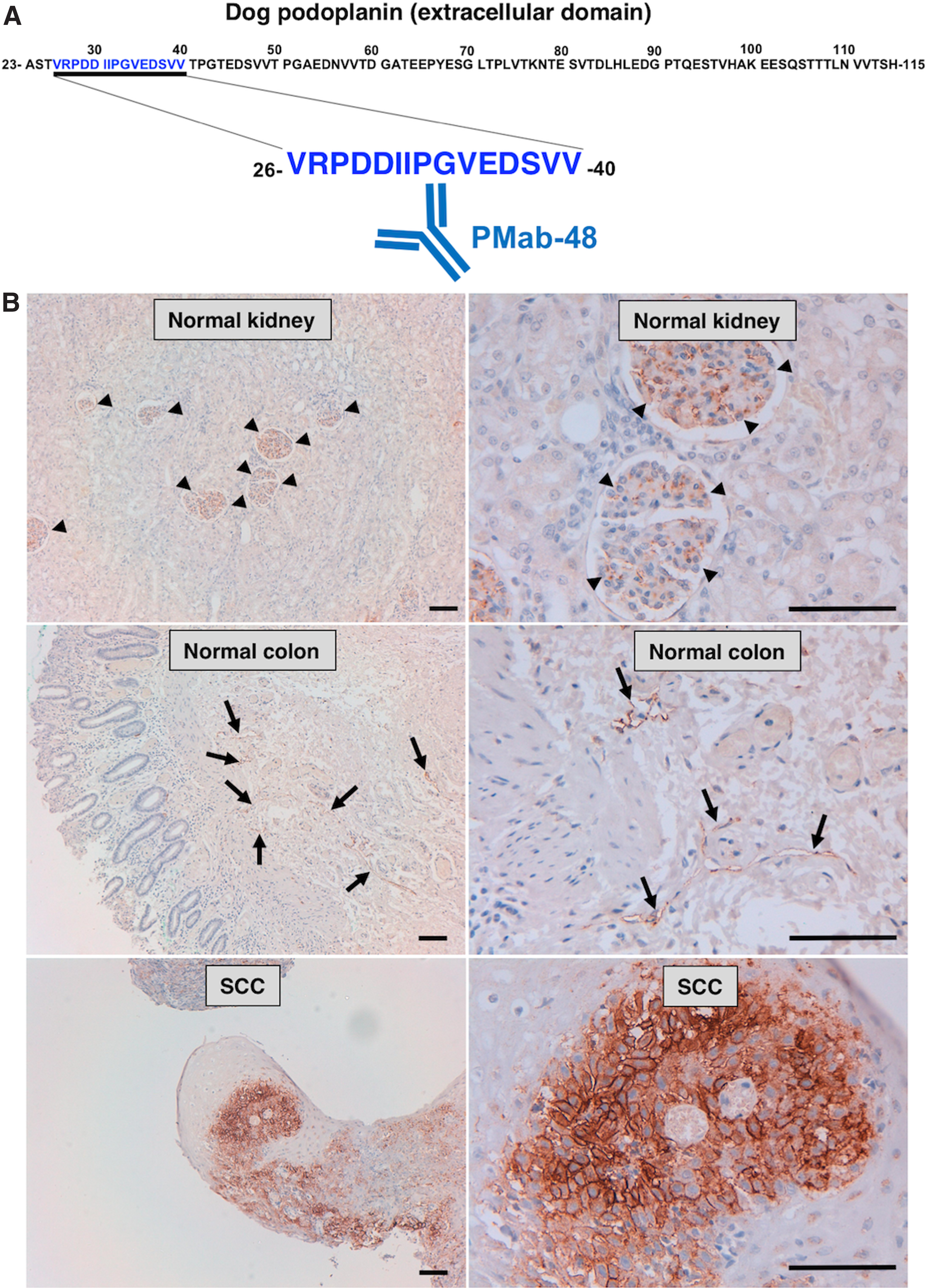
Immunohistochemical analysis against canine tissues by PMab-48.
PMab-48 reacted with podocytes of renal glomerulus and LECs (Fig. 1B and Supplementary Fig. S2); podoplanin expression has been reported in such cells in other species.(3) Conversely, the previously established PMab-38 did not react with LECs.(12) In addition, PMab-48 strongly stained canine squamous cell carcinomas (SCCs) (Fig. 1B and Supplementary Fig. S2). In this study, canine tissues were stained by PMab-48 at a concentration of 10–50 μg/mL. PMab-48 stained SCCs strongly even at a concentration of 10 μg/mL (Supplementary Fig. S2); conversely, a higher concentration (50 μg/mL) was better for staining LECs and glomerulus cells (Fig. 1B).
These results indicate that PMab-48 is advantageous for detecting LECs and may be useful in investigating the function of podoplanin in LECs.
Footnotes
Acknowledgments
This work was supported in part by the Basic Science and Platform Technology Program for Innovative Biological Medicine from Japan Agency for Medical Research and Development, AMED (Y.K.) and by the Platform Project for Supporting Drug Discovery and Life Science Research (Basis for Supporting Innovative Drug Discovery and Life Science Research [BINDS]) from AMED (Y.K.).
Author Disclosure Statement
Y.K. received research funding from Ono Pharmaceutical Co., Ltd. All other authors have no conflicts of interest.
References
Supplementary Material
Please find the following supplemental material available below.
For Open Access articles published under a Creative Commons License, all supplemental material carries the same license as the article it is associated with.
For non-Open Access articles published, all supplemental material carries a non-exclusive license, and permission requests for re-use of supplemental material or any part of supplemental material shall be sent directly to the copyright owner as specified in the copyright notice associated with the article.